
Consensus configurational bias Monte Carlo method and system for pharmacophore structure determination

Inactive Publication Date: 2002-01-22
CURAGEN CORP
View PDF17 Cites 30 Cited by
  • Summary
  • Abstract
  • Description
  • Claims
  • Application Information

AI Technical Summary

Benefits of technology

An important advantage provided by the invention is the ability to make a pharmacophore structure determination without relying on any knowledge of the structure of the target molecule or target. Where the target molecule is a protein, conventional prior art methods have sought to sequence and determine the structure of the protein containing the target, hoping thereby to determine active sites by examination of the structure. A further important advantage of the invention is that this structure determination can be made by use of a relatively small number of actual physical position measurements. In contrast, conventional methods using X-ray crystallography and liquid NMR require determination of positions of all atoms in the molecule ("binder") that specifically binds to the target, and the target. An additional advantage provided by the invention is that, in a preferred embodiment wherein REDOR structural measurements provide input information, the accuracy of the pharmacophore structure determination can be at least approximately 0.25-0.50 .ANG. or better. This accuracy is provided by the combination of an efficient, Monte Carlo technique for structure determination with a few highly accurate distance determinations.

Problems solved by technology

However, achieving desired pharmacological interventions in a predictable manner remains as elusive as ever.
Rational approaches to drug design have met with only limited success.
However, none of these methods appears to be sufficiently accurate for drug design purposes according to current rational approaches.
Drawbacks of x-ray methods include difficulties in obtaining high-quality crystals, expense and time associated with the crystallization process, and difficulties in resolving whether or not the structure of the crystalline forms is representative of the in vivo conformation (Clore et al., 1991, J. Mol. Biol. 221:47; Shaanan et al., 1992, Science 227:961-964).
However, the complexity of the analysis of the various mutual correlations is time consuming, and the correlations (primarily from the nuclear Overhausser effect) provide no better accuracy than X-ray methods.
Protein structures determined by any of these current methods do not predict success in subsequent drug design.
Resolution obtainable either by measurement or computation, generally 0.5-2 .ANG., has often been found to be inadequate for effective direct drug design, or for selection of a lead compound from organic compound libraries.
This accuracy appears to be beyond the capabilities of many current methodologies.
Prior research has identified tools which, although promising, cannot be used in a coordinated manner for drug design.
However, since REDOR measures only a few selected distances, it is not usable in drug design methods which depend on the initial determination of the complete structure of the protein containing the target of interest.
Purely ab initio computational approaches to high resolution structure analysis, such as quantum statistical mechanics and molecular dynamics, require prohibitive computing resources.
Proper quantum statistical mechanics required for an exact evaluation of full protein partition functions is not currently computationally feasible, as it would involve many thousands of atoms including the target, the protein, and the aqueous environment.
Computing the atomic dynamics of all the perhaps thousands atoms of a protein is an extreme computational burden.
In any case, most of the information determined is wasted, since only the structure of the protein binding target are of interest in drug design.
This extreme waste of computer resources results in time consuming, low resolution structure determinations.
To summarize, existing rational drug design methods based on identification of target structure fail to reliably yield drug molecules due to experimental structure determination difficulties and computational difficulties associated with predicting drug structures with ill-defined Hamiltonians.
Unfortunately, there is a need to determine the antibody structures needed for lead compound selection more rapidly and accurately.
While this field has been exploding in the last few years and showing great potential, it is severely limited by its use in isolation without the benefit of integrated structural analysis needed both to derive the high resolution structures of binding peptides and also to direct the construction of additional structured libraries.
Drug design is not aided by having library members recognizing the protein target of interest but without any understanding of why the recognition occurs.
Unfortunately, rational drug design according to current approaches (target structure-based) remains an inefficient, laborious process with a disproportionately high lead-compound failure rate.

Method used


Image

  • Consensus configurational bias Monte Carlo method and system for pharmacophore structure determination
  • Consensus configurational bias Monte Carlo method and system for pharmacophore structure determination
  • Consensus configurational bias Monte Carlo method and system for pharmacophore structure determination

Examples


Embodiment Construction

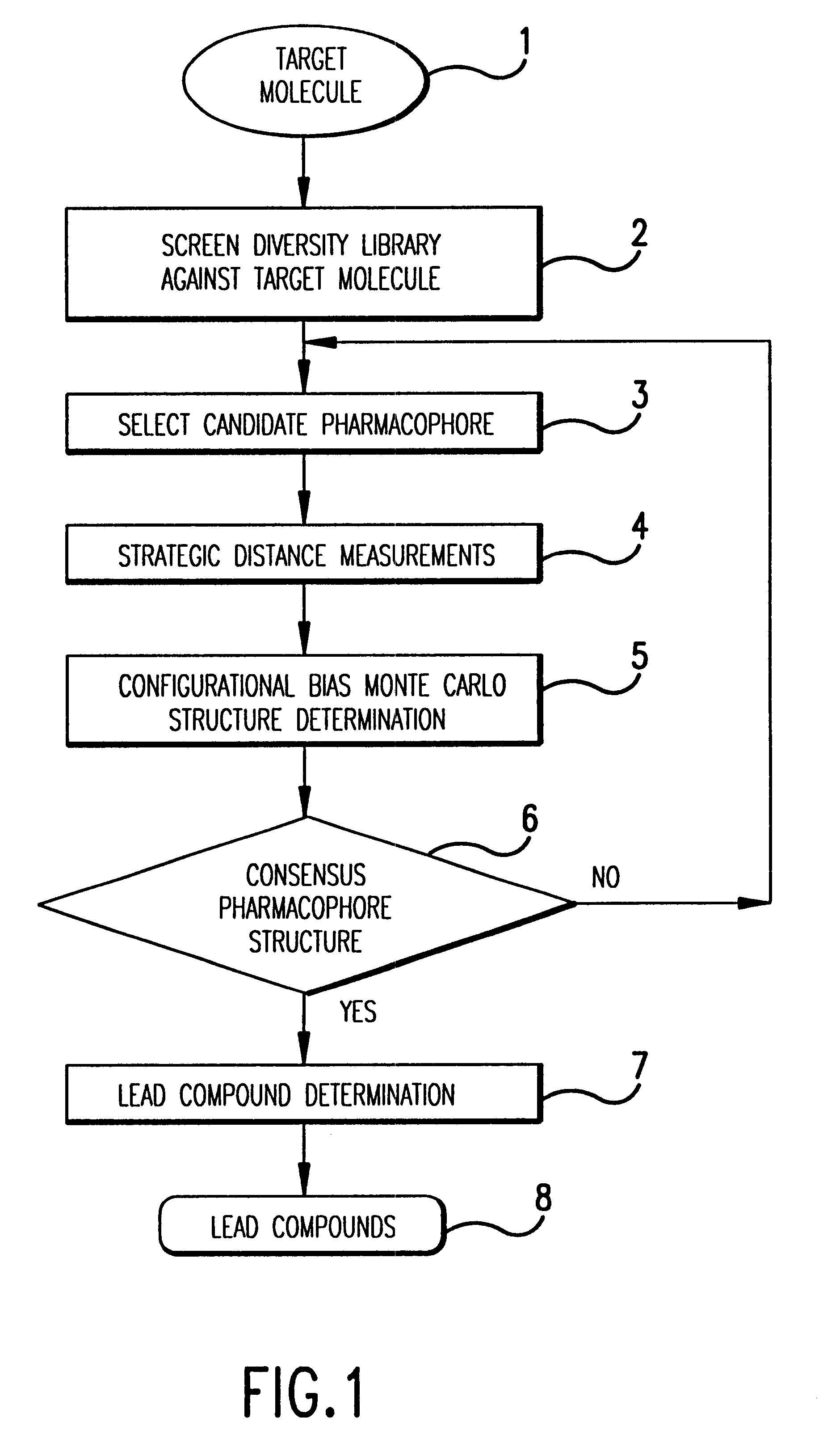
For clarity of disclosure, and not by way of limitation, the detailed description of the invention is described as a series of steps. A broad view of the exemplary steps of which the invention is comprised is presented in FIG. 1, a brief overview of which is presented in the text that follows.

The invention method preferably begins with a target molecule (or molecular complex) 1 having a binding target of biological or pharmacological interest. Specific binding of a molecule to the target is predicted to affect its biological activity and may provide biological effects of interest. For example, these effects might include amelioration of a disease process or alteration of a physiological response. Lead compounds 8 output from the invention are able to specifically bind to target molecule 1 and can serve as starting points for the design of a drug able to specifically bind to the target.

Diversity library screening, step 2, allows the selection from among library members of a plurality...


PUM

PropertyMeasurementUnit
Fractionaaaaaaaaaa
Fractionaaaaaaaaaa
Temperatureaaaaaaaaaa

Abstract

In a specific embodiment, this invention includes a method for determining an accurate, consensus pharmacophore structure shared by compounds that bind selectively to a target molecule. Optionally, the method begins with screening a diversity library against the target molecule of interest to pick the selectively binding members. Next the structure of the selected members is examined and a candidate pharmacophore responsible for the binding to the target molecule is determined. Next, preferably by REDOR nuclear magnetic resonance, several highly accurate interatomic distances are determined in certain of the selected members which are related to the candidate pharmacophore. A highly accurate consensus, configurational bias, Monte Carlo method determination of the structure of the candidate pharmacophore is made using the structure of the selected members and incorporating as constraints the shared candidate pharmacophore and the several measured distances. This determination is adapted to efficiently examine only relatively low energy configurations while respecting any structural constraints present in the organic diversity library. If the diversity library contains short peptides, the determination respects the known degrees of freedom of peptides as well as any internal constraints, such as those imposed by disulfide bridges. Finally, the highly accurate pharmacophore so determined is used to select lead organics for drug development targeted at the initial target molecule.

Description

This specification includes a microfiche appendix containing a listing of the computer programs of this invention, this appendix comprising 2 microfiche of 101 total frames.A portion of the disclosure of this patent document contains material which is subject to copyright protection. The copyright owner has no objection to the facsimile reproduction by any one of the patent disclosure, as it appears in the Patent and Trademark Office patent files and records, but otherwise reserves all copyright rights whatsoever.1. FIELD OF THE INVENTIONThe field of this invention is computer assisted methods of drug design. More particularly the field of this invention is computer implemented smart Monte Carlo methods which utilize NMR and binders to a target of interest as inputs to determine highly accurate molecular structures that must be possessed by a drug in order to achieve an effect of interest. Illustrative U.S. Patents are U.S. Pat. No. 5,331,573 to Balaji et al., U.S. Pat. No. 5,307,28...

Claims


Application Information

Patent Timeline
IPC IPC(8): C07B61/00C07K1/00G01N33/543G01N33/542G01N33/536G01N33/68G01N33/574G06F17/50
CPCC07K1/00G01N33/542G01N33/54326G01N33/6803G01N33/5748B01J2219/007C40B40/10Y10T436/24Y10S977/906Y10S977/953Y10S977/839Y10S977/808
Inventor DEEM, MICHAEL W.ROTHBERG, JONATHAN MARCWENT, GREGORY T.
Owner CURAGEN CORP
Learn More