

Process to study changes in gene expression in granulocytic cells
a technology of granulocytic cells and gene expression, applied in the field of process to study the changes in gene expression in granulocytic cells, can solve the problem that the length needs only be sufficien
- Summary
- Abstract
- Description
- Claims
- Application Information
AI Technical Summary
Benefits of technology
Problems solved by technology
Method used
Image

Examples
example 2
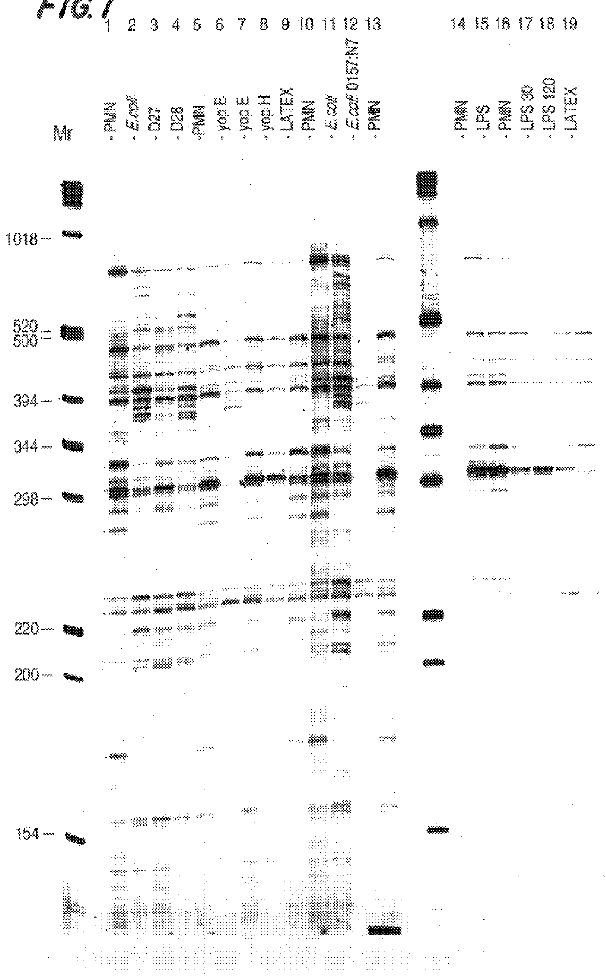
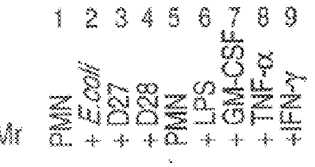
Production of gene expression profiles generated from cDNAs made with RNA isolated from neutrophils exposed to virulent and avirulent bacteria and neutrophils exposed to cytokines.
Neutrophils were isolated from normal donor peripheral blood following the LPS-free method as set forth in Example 1.
Neutrophils were incubated with virulent and avirulent E. coli or Y. pestis, LPS at 1 ng / ml, GM-CSF at 100 units / ml, TNFa at 1000 units / ml, or .gamma.IFN at 100 units / ml. The bacterial cells, LPS or cytokines were added to approximately 3.38.times.10.sup.8 cells in 100 ml of RPMI containing 6% H1 autologous serum. Incubation proceeded for 2 to 4 hours, preferably 2 hours, with gentle rotation in disposable polycarbonate Erlenmeyer flasks at 37.degree. C. After incubation, the cells were spun down and washed once with HBSS.
After incubation of the neutrophils, RNA was extracted and the cDNA profiles prepared as described in Example 1. FIG. 2 is an autoradiogram of the expression profiles gener...
example 3
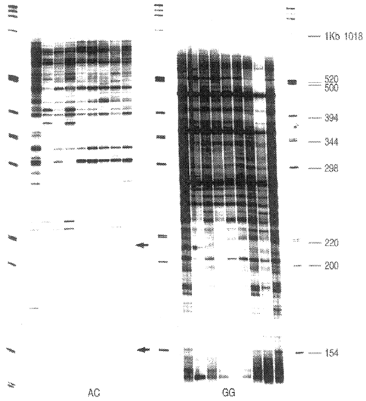
Production of gene expression profiles generated from cDNAs made with RNA isolated from neutrophils exposed to bacteria using all 12 possible anchoring oligo d(T) n1, n2.
Neutrophils were isolated from normal donor peripheral blood following the LPS-free method.
Neutrophils were incubated with E. coli or Y. pestis.
After incubation of the neutrophils, RNA was extracted and the cDNA profiles prepared as described in Example 1. FIG. 3 is an autoradiogram of the expression profiles generated from cDNAs made with RNA isolated from control (untreated) neutrophils (lane 1), neutrophils incubated with avirulent E. coli K12 (lane 2), virulent Y. pestis (lane 3), avirulent Y. pestis (lane 4). The anchoring oligo d(T)18 n1and n2 positions are indicated at the top of the figure. The cDNAs were digested with BglII.
FIG. 4 represents a summary of genes which are differentially expressed in neutrophils upon exposure to virulent and avirulent E. coli and Y. pestis. Expression patterns are determined b...
example 4
Production of expression profiles generated from cDNAs made with RNA isolated from neutrophils isolated from a subject with a sterile inflammatory disease.
Neutrophils are isolated from normal donor peripheral blood following the LPS-free method or from subjects exhibiting the symptoms of a sterile inflammatory disease. RNA is extracted and the gene expression profiles prepared as described in Example 1.
To determine the identity of genes (cDNAs) which are differentially expressed in the neutrophils isolated from a subject exhibiting the symptoms of a sterile inflammatory disease, the cDNA profiles prepared from neutrophils from said subject are compared to profiles prepared from neutrophils isolated from the normal donor. Bands which exhibit altered intensities when compared between the gene expression profiles prepared from neutrophils from said subject and profiles prepared from neutrophils isolated from the normal donor are then extracted from the display gel as previously describ...
PUM
| Property | Measurement | Unit |
|---|---|---|
| total volume | aaaaa | aaaaa |
| concentration | aaaaa | aaaaa |
| concentration | aaaaa | aaaaa |
Abstract
Description
Claims
Application Information