Electrically induced vessel vasodilation

a technology of electric induction and vasodilation, which is applied in the field of electric induction of vessel vasodilation, can solve the problems of systemic administration failure to achieve effective concentrations of drugs at the targeted area, drug investigation also has been investigated, and achieves the effect of increasing the vasodilation of the vessel, and sufficient strength and duration

Inactive Publication Date: 2005-03-08
ONCOSEC MEDICAL
View PDF16 Cites 405 Cited by
  • Summary
  • Abstract
  • Description
  • Claims
  • Application Information

AI Technical Summary

Benefits of technology

[0009]In accordance with the present invention, there are provided methods for inducing or increasing vasodilation of a vessel in a subject by applying an electrical impulse to the vessel, having sufficient strength and duration to induce or increase vasodilation of the vessel. Methods for inducing or increasing the flow of fluid through a vessel in a subject by applying an electrical impulse to the vessel, having sufficient strength and duration to induce or increase the flow of fluid through the vessel, also are provided. For example, a method of the invention employs an electrical impulse applied via electroporation. A method of the invention applies an electrical impulse with an electro-catheter apparatus. Invention methods are useful for treating clinical situations in which it is desired to increase or induce vessel vasodilation or to induce or increase the flow of fluid through the vessel.
[0010]Multiple electrical impulses can be applied in a method of the invention. An electrical impulse can be applied from about 50 to 90 volts per 1.5 mm. An electrical impulse can be applied for about 0.5 ms to 10 ms. Vessels can be denuded prior to, simultaneously with or after applying an electrical impulse in a method of the invention in order to augment the induction of vessel vasodilation or increase in the flow of fluid through the vessel.
[0011]Compositions can be administered to the vessel in the subject prior to, simultaneously with or after the applicati...

Problems solved by technology

A variety of drugs also have been investigated to prevent luminal renarrowing in experimental animal and clinical settings, but without much success.
A primary reason for this may be the failure of systemic administration to achieve effective concentrations of drugs at the targeted area.
Delivery capacity with hydrogel balloon is limited and, during placement, the catheter can lose substantial amount of the drug or agent that is ...

Method used


Image

  • Electrically induced vessel vasodilation
  • Electrically induced vessel vasodilation
  • Electrically induced vessel vasodilation

Examples


example i

[0100]This example describes applying an electrical impulse to a vessel in a subject.

[0101]Animal Preparation and Surgical Approach:

[0102]This study conformed to the care and use of laboratory animals and standard euthanasia procedures policies by the NIH guide set forth in the Institutional Animal Care and Use Committee, and to the position of the American Heart Association on Research Animal Use. New Zealand White rabbits weighing 3.2-4.2 kg of both sexes (N=21) were utilized in the studies. After withdrawal of food for 12-18 hrs, but not water, the animals were sedated using 2 mg / Kg intramuscular xylazine (Miles Inc., KS) and 50 mg / Kg ketamine (Fort Dodge Lab, IA). Sedated animals were anesthetized with 30 mg / Kg α-chloralose (Fisher Scientific, NJ) throughout the ear vein and endotracheally intubated. Once anesthetized, the animal was strapped supine on the surgical table and ventilated with a volume controlled Harvard 665 ventilator (Harvard Apparatus, Natick, Ma.). Corneal and ...

example ii

[0120]This example shows that an electrical impulse applied to a vessel of an animal increases the vessel luminal area.

[0121]Luminal area measurements of the arterial tissue sections were made by NIH image analysis software. Measurement of areas enclosed by the lumen in pulsed artery samples and non-pulsed (control) samples were performed in multiple sections in a given experiment. Statistical testing was performed with “instat” software (Graphpad, San Diego, Calif.). Results were considered statistically significant when the probability of error was p—0.05.

[0122]A total of 120 serial tissue sections obtained from eight animals were examined. Fifty-seven sections were from electrically pulsed arteries and sixty-three sections were from non-electrically pulsed arteries. All experiments but one showed expansion of the vessel lumen after electropulsing at approximately 65 volts and a pulse length of 9 ms. The luminal vessel layer of the pulsed artery showed an average increase of 76% i...

example iii

[0125]This example shows that the application of an electrical impulse to a vessel of an animal can deliver a composition into the vessel.

[0126]Fluoresceinated heparin (F-heparin; 167 units / mg of activity) was used as a probe. To ensure that the covalent bonded fluorescein had not been dissociated by electroporation, in vitro experiments were performed in an electroporation cuvette containing F-heparin. The solution was pulsed using the same electrical parameters as used in the in vivo studies. The sample was then loaded in a LKB ultropak column (TSK G4000 sw) connected to an Anspec HPLC pump with a Shimadzu (SPD-6AV) UV-VIS spectrophotometric detector for analysis. The hard copy of the HPLC profile was obtained using Hitachi D-2500 chromato-integrator. Individual samples were also analyzed in a luminescence spectrometer (Perkin-Elmer LB-5, CA) at 490 nm excitation and emission at 520 nm.

[0127]Luminescence spectrometry and HPLC of electroporated F-heparin showed no differences from ...


PUM


Abstract

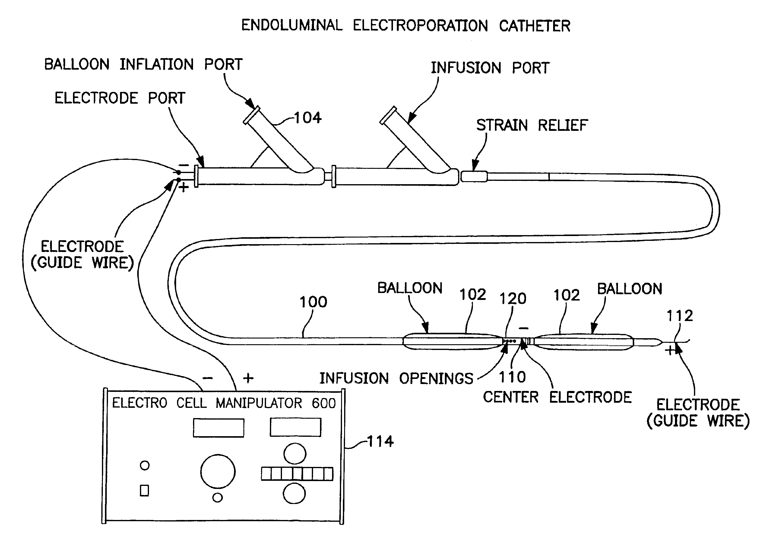
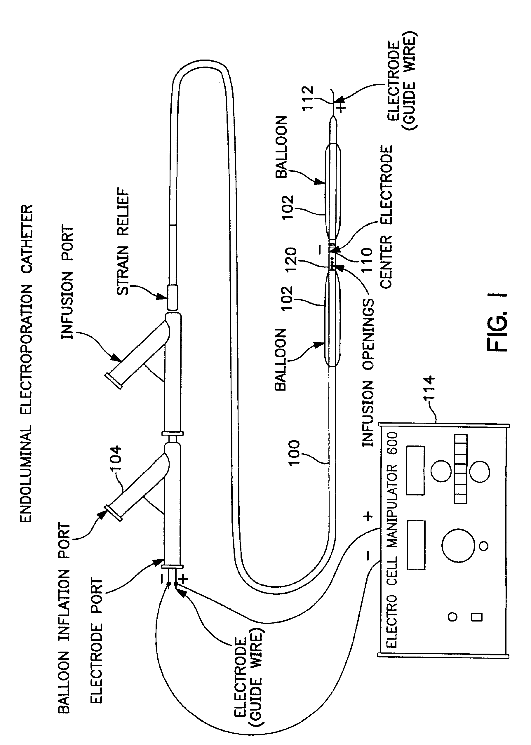
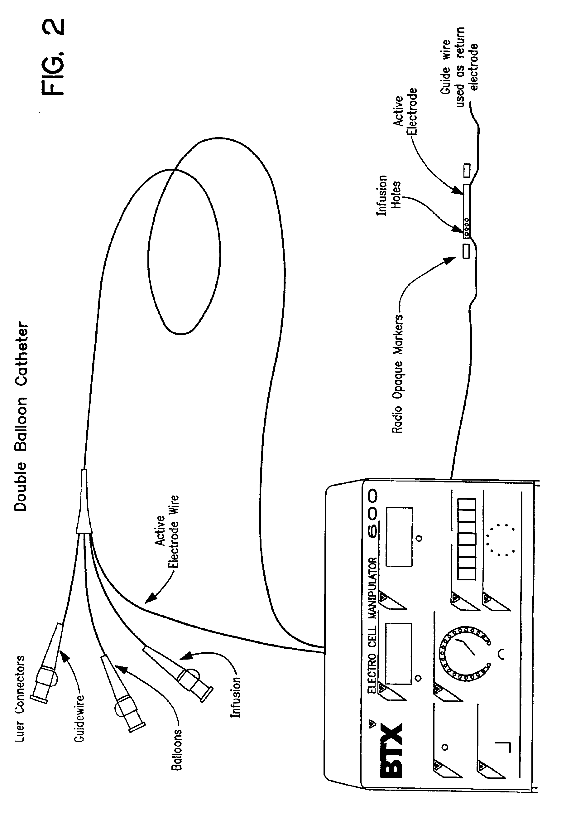
The invention provides methods for inducing or increasing the vasodilation of a vessel. The invention further provides methods for inducing or increasing the flow of fluid through a vessel. An electrical impulse is applied to the vessel in order to induce or increase vessel vasodilation or to induce or increase the flow of fluid through the vessel. In a particular embodiment, a novel double-balloon catheter system incorporating electroporation technology has been designed and is used to apply the electrical impulse endoluminally.

Description

RELATED APPLICATIONS[0002]Under 37 U.S.C. §119(e)(1), this application claims the benefit of prior U.S. provisional application 60 / 084,857, filed May 8, 1998 and U.S. application Ser. No. 09 / 307,216, filed May 7, 1999 now U.S. Pat. No. 6,347,247.FIELD OF THE INVENTION[0003]The present invention relates to the field of vessel vasodilation and, more particularly, to methods for inducing or increasing the vasodilation of a vessel and methods for inducing or increasing the flow of fluid through a vessel by applying an electrical impulse to the vessel.BACKGROUND OF THE INVENTION[0004]Despite procedural success rates greater than 95% achieved by percutaneous transluminal coronary angioplasty (PTCA), luminal renarrowing of blood vessels after balloon angioplasty occurs in 30% to 60% of all cases within 3 to 6 months. Smooth muscle cell proliferation and extracellular matrix remodeling appear to play pivotal roles in the luminal renarrowing process and negate the beneficial effect of vascul...

Claims


Application Information

Patent Timeline
IPC IPC(8): A61N1/32A61N1/30
CPCA61N1/325A61N1/327Y10S977/905Y10S977/883Y10S977/904
Inventor DEV, NAGENDU B.DEV, SUKHENDU B.HOFMANN, GUNTER A.
Owner ONCOSEC MEDICAL
Why Eureka
Try Eureka