Method and device for detecting substances in body fluids by Raman spectroscopy

a raman spectroscopy and substance technology, applied in the field of non-invasive detection or determination, can solve the problems of inability to investigate blood samples, inability to take blood samples in each event, and inability to take blood samples in hospitals or gp surgeries, etc., to achieve easy detection and analysis, low cost, and high reproducibility

Inactive Publication Date: 2005-03-15
MULLER DETHLEFS KLAUS
View PDF18 Cites 49 Cited by
  • Summary
  • Abstract
  • Description
  • Claims
  • Application Information

AI Technical Summary

Benefits of technology

The object of the invention is to provide a method and a device to facilitate a non-invasive detection and analysis of substances in body fluids allowing for a high accuracy and reproducibility while requiring only little time for the measurement which is acceptable for the patient.
The underlying principle of the invention is to use the Raman scattering of primary light by a substance to be detected to obtain a signal that is correlated with the concentration of the substance to be detected. In order to exclude or at least minimise perturbations of the measurement of the Raman signal of the substance to be detected, the wavelength spectrum of secondary light is sampled in the region of the Raman spectrum of the substance to be detected for two different primary wavelengths according to the invention. As was found by the inventor the Raman spectrum of the substance to be detected is shifted corresponding to the different primary wavelength, however, the Raman spectrum of the body tissue is more or less identical for both primary light wavelengths. The comparison of the Raman spectra for the first and for the second primary wavelength allows to eliminate the background signal of the body tissue and to gain a signal which is proportional to the concentration of the substance to be detected.
at least one notch filter is used for eliminating Rayleigh scattering;
at least one notch filter is positioned between the projection optics and the photo detector assembly to eliminate Rayleigh scattering;
One of several advantages of the methods according to the invention consists in allowing a continuous instead of a merely discreet monitoring of the values of the blood analysis. Furthermore, permanent injury of the patient's tissue by taking blood samples can be avoided as well as possibly accompanied tissue inflammation and enhanced danger of infection. The more selective methods and embodiments according to the invention, producing signals with a better signal to noise ratio, allow for very good reproducibility and accuracy of the analysis in short measurement intervals.

Problems solved by technology

However, if a frequent, recurring investigation of the blood is required in regular intervals as for diabetes patients, a frequent taking of blood samples of the patient in a hospital or in a GP surgery constitutes an excessive inconvenience.
The general disadvantage of these procedures is that the taking of a blood sample in each event carries an infection risk—albeit a very small one.
If the required instruments (syringes etc.) are not sterile or not available as sterile a blood investigation is not possible.
Even bigger difficulties arise from blood analysis by taking of a blood sample for patients disposed with haemophilia.
In each case the blood analysis by taking of a blood sample implies a more or less painful operation and an effort the patient is not always up to.
The disadvantage of any chemical analysis, however, remains in the very high standards that must be demanded for cleanliness and precision of dosage.
In addition, for all in vitro methods, there persists the danger that blood and / or chemicals are released into the environment and with that pathogens can be distributed under circumstances.
The taking of a blood sample, often several times per day, constitutes a big burden for the patient both in terms of his health and psychological condition as well as by the restriction of his mobility.
However, severe problems stand in the way of an application of Raman spectroscopy for concentration measurements of substances in tissue fluids.
An additional problem results from the spectral superposition of Raman signals of different other substances with the Raman signal of the substance to be detected.
Due to the complex composition of the medium, interfering signals result which can be substantially higher than the measured signals for the substance to be detected.

Method used


Image

  • Method and device for detecting substances in body fluids by Raman spectroscopy
  • Method and device for detecting substances in body fluids by Raman spectroscopy
  • Method and device for detecting substances in body fluids by Raman spectroscopy

Examples


Embodiment Construction

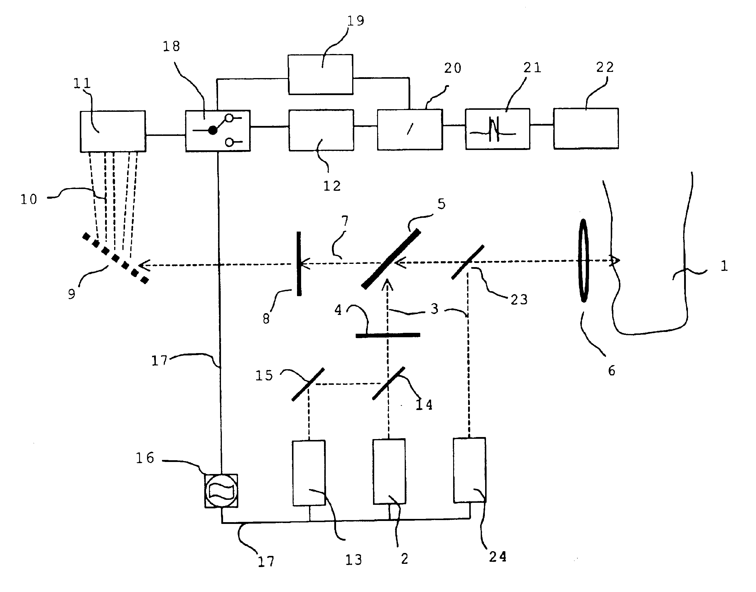
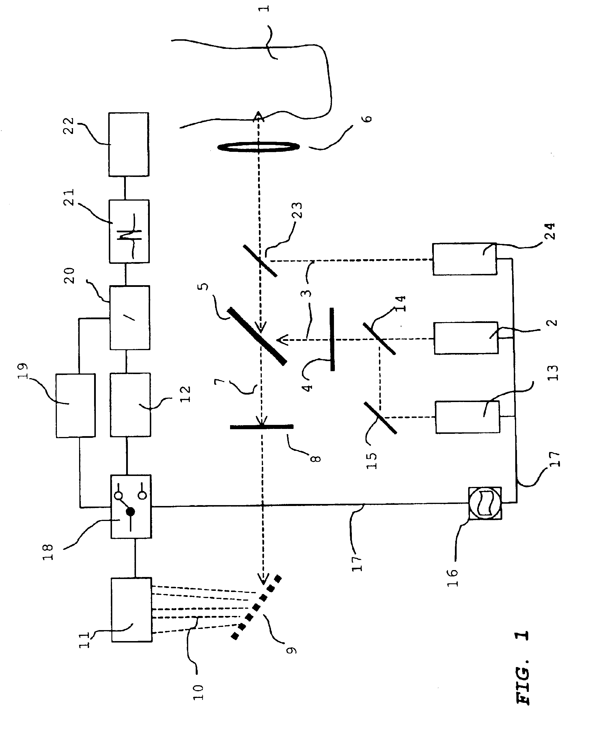
FIG. 1 shows an embodiment of the basic set up for the implementation of an embodiment of the method according to the invention. The device for the non-invasive determination of a concentration of a substance in a body tissue 1 by Raman spectroscopy comprises a first light source 2 that serves for irradiating monochromatic primary light 3 of a first wavelength λ1 into body tissue 1. Primary light 3 passes through a first optical filter 4 that filters out remaining light, possibly present, of a wavelength other than λ1. This band pass filter 4 is particularly of advantage when a non-monochromatic light source 2 is used. The primary light 3 subsequently hits the output beam splitter 5 whose function is explained below. Behind an output beam splitter 5A a projection / collection optics 6 is arranged, projecting primary light 3 onto a spot of body tissue 1. In the shown embodiment the primary light 3 is focussed on an earlobe of a patient which is indicated on the right hand side in FIG. ...


PUM

PropertyMeasurementUnit
wavelength λ1aaaaaaaaaa
wavelength λexcaaaaaaaaaa

Abstract

The invention relates to a method and to a device for the non-invasive detection or determination, by Raman spectroscopy, of the concentration of substances in body fluids. The aim of the invention is to facilitate a non-invasive in vivo detection of substances in body fluids that allows for very accurate, reproducible results of analysis while requiring only little time for measuring which is acceptable for the patient. To this end, the inventive method and device for carrying out a Raman spectroscopy of a body fluid in a body tissue (1) records at least two Raman spectrums under different physical conditions and compares them with each other. The result of comparison allows detection of the substance one is looking for and measurement of the concentration of said substance.

Description

BACKGROUND OF THE INVENTIONThe invention relates to a method and a device for the non-invasive detection or determination, by Raman spectroscopy, of the concentration of substances in body fluids. In particular the invention is concerned with detection or determination of concentrations of glucose, cholesterol, lactate or such in human blood.For the detection of substances dissolved in the blood of a patient in most cases a blood sample is taken from the patient and subsequently analysed. Provided this blood sample is taken as outpatient only once or in relatively large intervals and by skilled personnel this is not an excessive inconvenience for the patient. However, if a frequent, recurring investigation of the blood is required in regular intervals as for diabetes patients, a frequent taking of blood samples of the patient in a hospital or in a GP surgery constitutes an excessive inconvenience. For this reason so called “home monitoring” procedures have been developed by which th...

Claims


Application Information

Patent Timeline
Patent Type & Authority Patents(United States)
IPC IPC(8): A61B5/00G01N21/65G01N21/63A61B5/145A61B5/1455A61B10/00
CPCA61B5/14532G01N21/65A61B5/1455
Inventor MULLER-DETHLEFS, KLAUS
Owner MULLER DETHLEFS KLAUS
Why Eureka
Try Eureka