Method of designing agonists and antagonists to IGF receptor

a technology of agonists and antagonists, applied in chemical methods analysis, instruments, hormone peptides, etc., can solve the problems of limiting the application of this type of therapy and the cost of humanised monoclonal production

Inactive Publication Date: 2006-03-28
WALTER & ELIZA HALL INST OF MEDICAL RES
View PDF2 Cites 34 Cited by
  • Summary
  • Abstract
  • Description
  • Claims
  • Application Information

AI Technical Summary

Benefits of technology

[0038]In another preferred embodiment, the computer-assisted method further includes the step of selecting one or more chemical structures from step (e) which interact with the receptor site of the molecule in a manner which prevents the binding of natural ligands to the receptor site.

Problems solved by technology

The large doses required and the cost of production of humanised monoclonal antibody is likely to limit the application of this type of therapy.

Method used


Image

  • Method of designing agonists and antagonists to IGF receptor
  • Method of designing agonists and antagonists to IGF receptor
  • Method of designing agonists and antagonists to IGF receptor

Examples


example 1

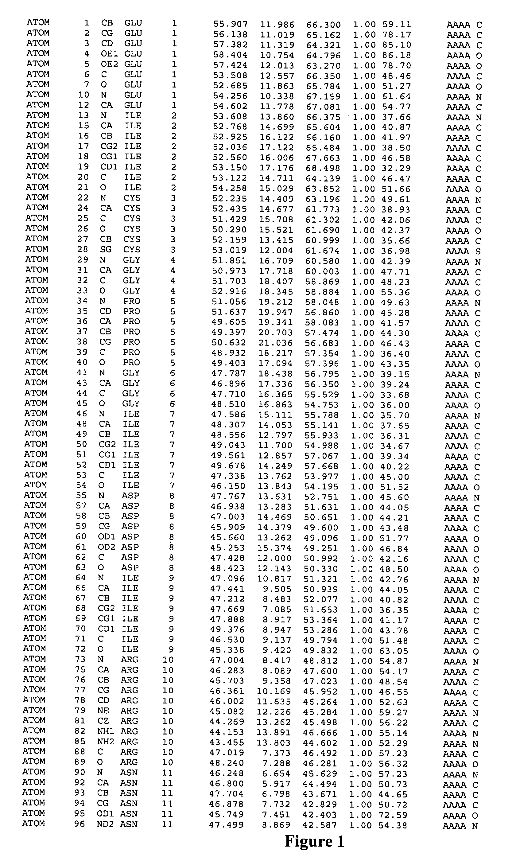
Expression, Purification and Crystallization of the IGF-1R Fragment.

[0089]Several factors hamper macromolecular crystallization including sample selection, purity, stability, solubility (McPherson, A., et al., 1995, Structure 3:759–768); Gilliland, G. L. & Ladner, J. E., 1996, Curr. Opin. Struct. Biol. 6:595–603), and the nature and extent of glycosylation (Davis, S. J., et al., 1993, Protein Eng. 6:229–232). Initial attempts to obtain structural data from soluble IGF-1R ectodomain (residues 1–906) protein, expressed in Lec8 cells (Stanley, P. 1989, Molec. Cellul. Biol. 9:377–383) and purified by affinity chromatography, produced large, well-formed crystals (1.0 mm×0.2 mm×0.2 mm) which gave no discernible X-ray diffraction pattern (unpublished data). Similar difficulties have been encountered with crystals of the structurally-related epidermal growth factor receptor (EGFR) ectodomain, which diffracted to only 6 Å, insufficient for the determination of an atomic resolution structure ...

example 2

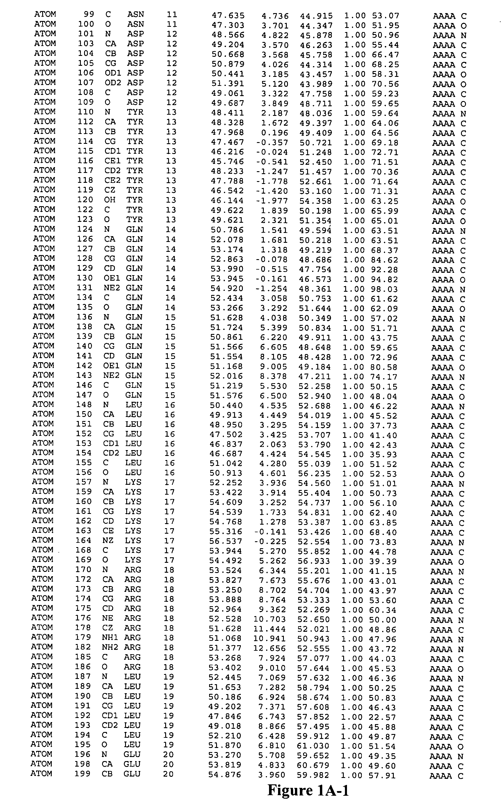
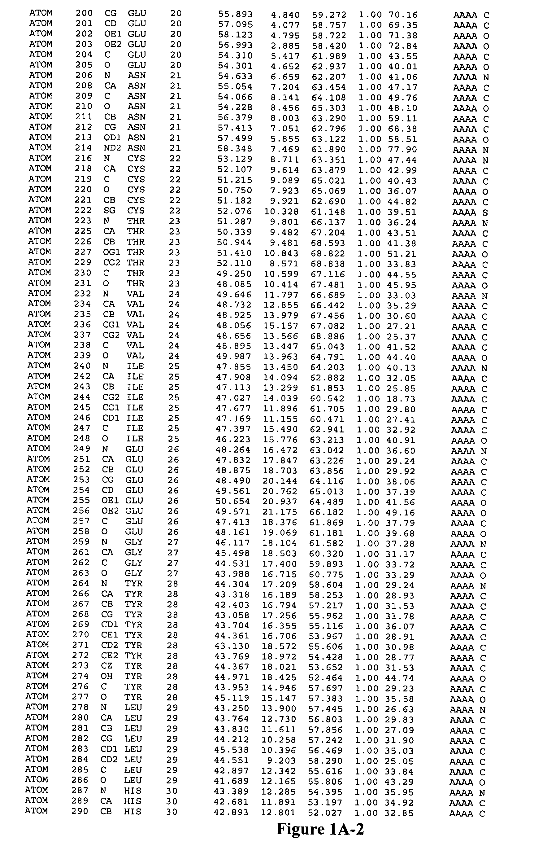
Structure of the IGF-1R / 1-462

[0098]Crystals were cryo-cooled to −170° C. in a mother liquor containing 20% glycerol, 2.2 M ammonium sulfate and 100 mM Tris at pH 8.0. Native and derivative diffraction data were recorded on Rigaku RAXIS IIc or IV area detectors using copper Kα radiation from a Siemens rotating anode generator with Yale / MSC mirroroptics. The space group was P212121 with a=77.39 Å, b=99.72 Å, and c=120.29 Å. Data were reduced using DENZO and SCALEPACK (Otwinowski, Z. & Minor, W., 1996. Mode. Meth. Enzym. 276:307–326). Diffraction was notably anisotropic for all crystals examined.

[0099]Phasing by multiple isomorphous replacement (MIR) was performed with PROTEIN (Steigeman, W. Dissertation (Technical Univ. Munich, 1974) using anomalous scattering for both UO2 and PIP derivatives. Statistics for data collection and phasing are given in Table 1. In the initial MIR map regions of protein and solvent could clearly be seen, but the path of the polypeptide was by no means obvi...

example 3

Prediction of 3D Structure of the Corresponding Domains of IRR and IR Based on Structure of IGF-R Fragment.

[0121]The sequence identities between the different members of the insulin receptor family are sufficient to allow accurate sequence alignments to facilitate 3D structure predictions by homology modelling. The alignments of the ectodomains of human IGF-1R, IR, and IRR are shown in FIG. 9.


PUM

PropertyMeasurementUnit
pHaaaaaaaaaa
volumeaaaaaaaaaa
pHaaaaaaaaaa

Abstract

Identification of the human insulin receptor binding site sequence of certain spatial molecular structures conforming to such binding site and of certain insulinomimetic sequences including the binding site sequence are disclosed.

Description

FIELD OF THE INVENTION[0001]This invention relates to the field of receptor structure and receptor / ligand interactions. In particular it relates to the field of rising receptor structure to predict the structure of related receptors and to the use of the determined structures and predicted structures to select and screen for agonists and antagonists of the polypeptide ligands.BACKGROUND OF THE INVENTION[0002]Insulin is the peptide hormone that regulates glucose uptake and metabolism. The two types of diabetes mellitus are associated either with an inability to produce insulin because of destruction of the pancreatic islet cells (Homo-Delarche, F. & Boitard, C., 1996, Immunol. Today 10: 456–460) or with poor glucose metabolism resulting from either insulin resistance at the target tissues, or from inadequate insulin secretion by the islets or faulty liver function (Taylor, S. I., et al., 1994, Diabetes, 43: 735–740).[0003]Insulin-like growth factors-1 and 2 (IGF-1 and 2) are structur...

Claims


Application Information

Patent Timeline
Patent Type & Authority Patents(United States)
IPC IPC(8): G01N33/48G01N33/50G01N33/53G06F19/00C07B61/00C07K14/65C07K14/705C07K14/71G01N33/15G01N33/566G06F17/50
CPCC07K14/65G01N2333/71G01N2333/65
Inventor BENTLEY, JOHN DAVIDCOSGROVE, LEAH JANEFRENKEL, MAURICE JOHNGARRETT, THOMAS PETER JOHNLAWRENCE, LYNNE JEANLOU, MELZHENLOVRECZ, GEORGE OSCARWARD, COLIN WESLEY
Owner WALTER & ELIZA HALL INST OF MEDICAL RES
Why Eureka
Try Eureka