

Tissue electroperforation for enhanced drug delivery
a technology of enhanced drug delivery and tissue electroperforation, which is applied in the field of tissue electroperforation for enhanced drug delivery, can solve the problems of high molecular weight, difficult-to-deliver” drugs, and difficult-to-deliver” drugs, and achieves the effects of high hydrophilicity and/or high molecular weigh
- Summary
- Abstract
- Description
- Claims
- Application Information
AI Technical Summary
Benefits of technology
Problems solved by technology
Method used
Image


Examples
example 1
Increase in Transepidermal Water Loss (TEWL) After Electroperforation in Piqs
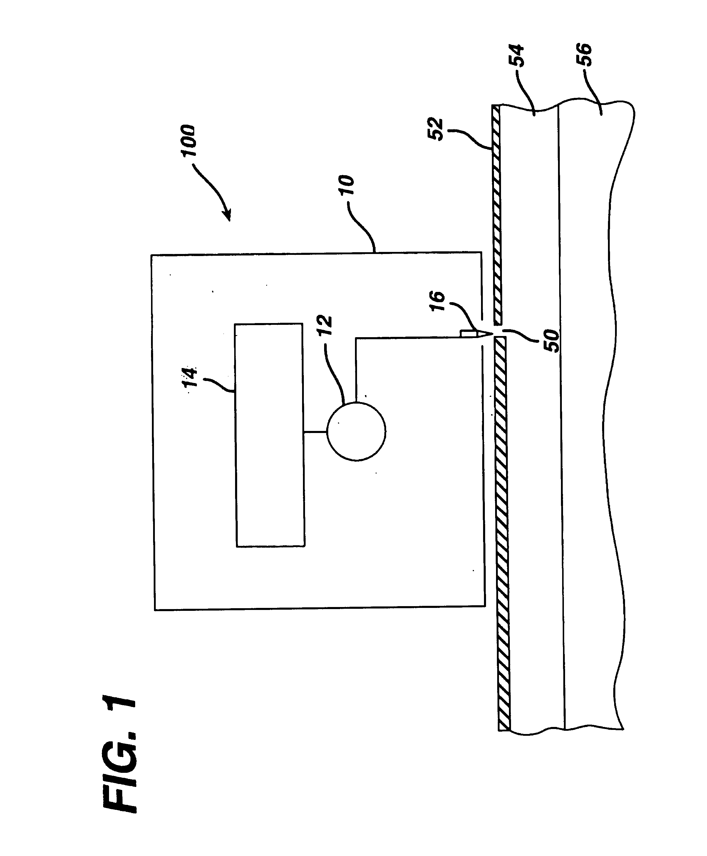
[0091]To evaluate the pore transport pathway created through the stratum corneum of the skin by electroperforation, an in vivo experiment was conducted on the back skin of Yorkshire pigs (female, ˜12 kg) using an electrosurgery apparatus (Surgitron™, Ellman International, Inc., Hewlett, N.Y.). The pigs were immobilized with appropriate anesthetics and analgesics. Electrofulguration current was used with a fine wire electrode (0.26 mm in diameter) and power output setting at between scale 3 to 10. A small pore was created on the surface of the skin by carefully moving the electrode towards the skin until the tip of the electrode almost touched the skin. The electrode was quickly moved away from the skin as soon as an electric arc appeared in the gap between the electrode tip and the skin surface.
[0092]Typical microscopic biopsy results (magnification=220×) of the pig skin treated with electroperforation are ...
example 2
Electroperforation Followed by Passive Diffusion of Insulin for Transdermal Delivery
[0095]The electroperforation procedure described in Example 1 was conducted in two pigs with a pore density of 39 pores / cm2 of the skin and subsequently followed by transdermal insulin delivery with passive diffusion. An insulin-containing chamber was immediately placed onto the electroperforation-treated skin. The chamber was made of flexible polyethylene containing 0.5 ml of insulin injection solution (Pork insulin, Molecular Weight≅6000 daltons, 100 U / ml, Regular Iletin® II, Eli Lilly, Indianapolis, Ind.). The contact area of the insulin solution in the chamber to the electroperforation-treated skin was 2.3 cm2. The chamber was affixed to the pig skin with a veterinary silicone adhesive at the rim of the chamber. Blood glucose of the pigs was monitored by obtaining blood samples of the ear vein, which were analyzed using two blood glucose analyzers separately to assure the accuracy (One Touch® Bas...
example 3
Electroperforation followed by Iontophoresis of Insulin for Transdermal Delivery
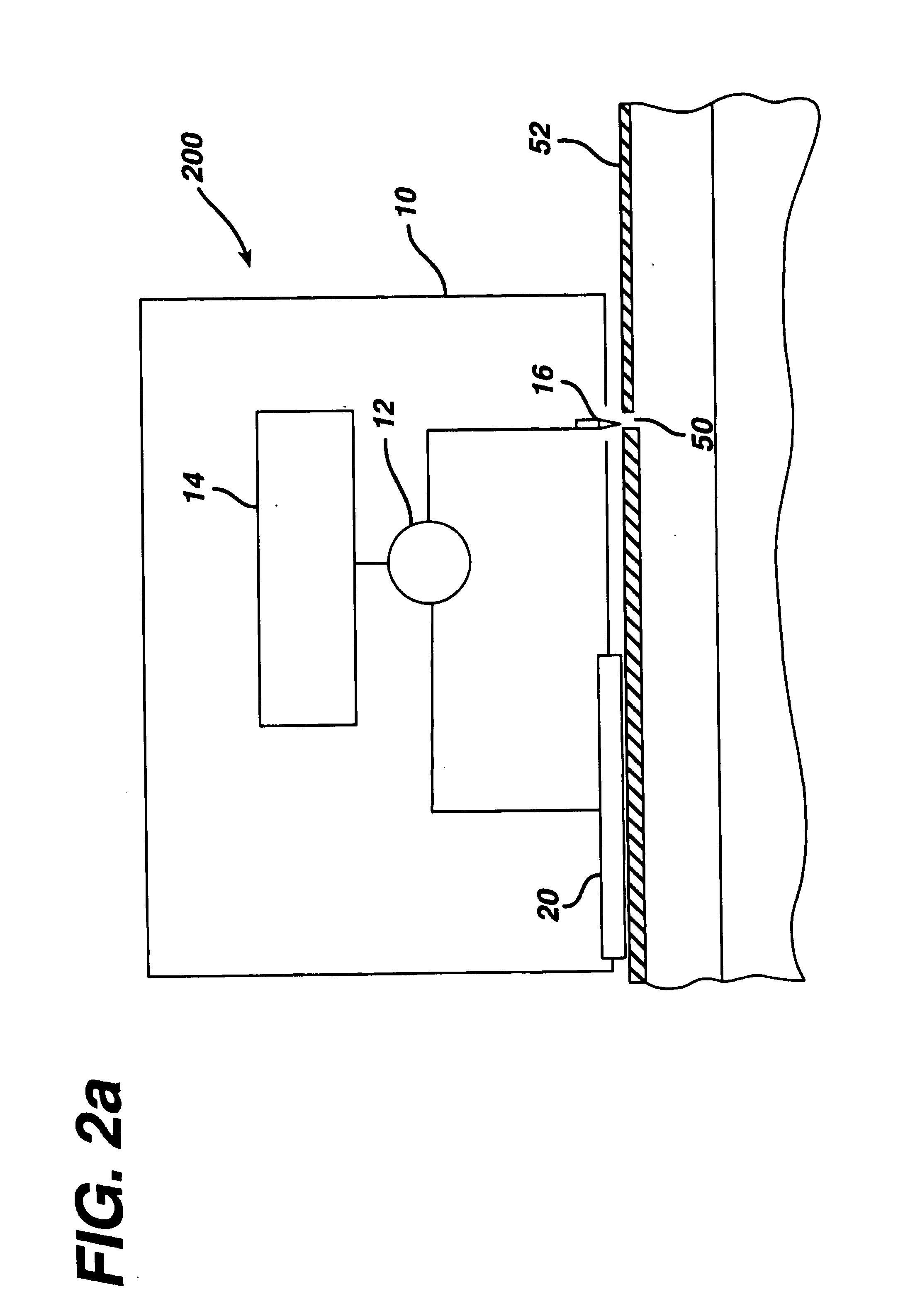
[0096]An electroperforation procedure was conducted in two pigs similar with a pore density of 9 pores / cm2 on the skin and subsequently was followed by transdermal insulin delivery. The purpose of using a lower pore density in this experiment was to examine the effect of pore number (e.g., the extent of the transport pathway available) to transdermal insulin delivery. The same insulin-containing chamber and drug application procedures were used in this experiment as those in the Example 2. However, a steel wire was placed in the insulin-containing chamber to serve as a delivery electrode for iontophoresis. The power source of iontophoresis was a commercial iontophoresis apparatus (Phoresor II™, PM700, Motion Control, Inc., Salt Lake City, Utah). The first 1.5 hours of the delivery experiment was by passive diffusion of insulin only. Iontophoresis of insulin was conducted twice in two 30-minute sections w...
PUM
| Property | Measurement | Unit |
|---|---|---|
| Electric potential / voltage | aaaaa | aaaaa |
| Frequency | aaaaa | aaaaa |
| Frequency | aaaaa | aaaaa |
Abstract
Description
Claims
Application Information