Method for treating onychomycosis

a technology for onychomycosis and treatment methods, applied in the field of treatment methods for onychomycosis, can solve the problems of inability to accurately evaluate the effect of drugs, inability to cure tinea unguium completely, and patient stopping or taking drugs

Active Publication Date: 2007-05-08
KAKEN PHARMA CO LTD
View PDF8 Cites 40 Cited by
  • Summary
  • Abstract
  • Description
  • Claims
  • Application Information

AI Technical Summary

Benefits of technology

[0013]The present invention has been accomplished based on findings that it is desirable that an effect of antimicrobial agent such as particularly antifungal agent is evaluated after removing a drug remaining in the infected site after treatment of an animal or a biosample such as skin with the pathogenic microorganism. An object of the present invention is to provide a novel method for evaluating the effect of the antimicrobial agent and the antimicrobial agent obtained according to the method for evaluating the drug effect. In detail, the present invention provides the method for detecting the viable pathogenic microorganism in the above-mentioned infected site of the animal or the biosample with the pathogenic microorganism after removing the antimicrobial agent which has been administered to the animal or the biosample, and the method for evaluating the effect of antimicrobial agent which can accurately evaluate the effect of the antimicrobial agent without the influence of the antimicrobial agent remaining in the infected site of the animal or the biosample with a pathogenic microorganism. In addition, the present invention provides the antimicrobial agent obtained according to the above-mentioned the method for evaluating the drug effect, and the detecting method of the antimicrobial agent which comprises detecting the existing antimicrobial agent in the infected site of the animal or the biosample with the pathogenic microorganism, to which the antimicrobial agent is administered.
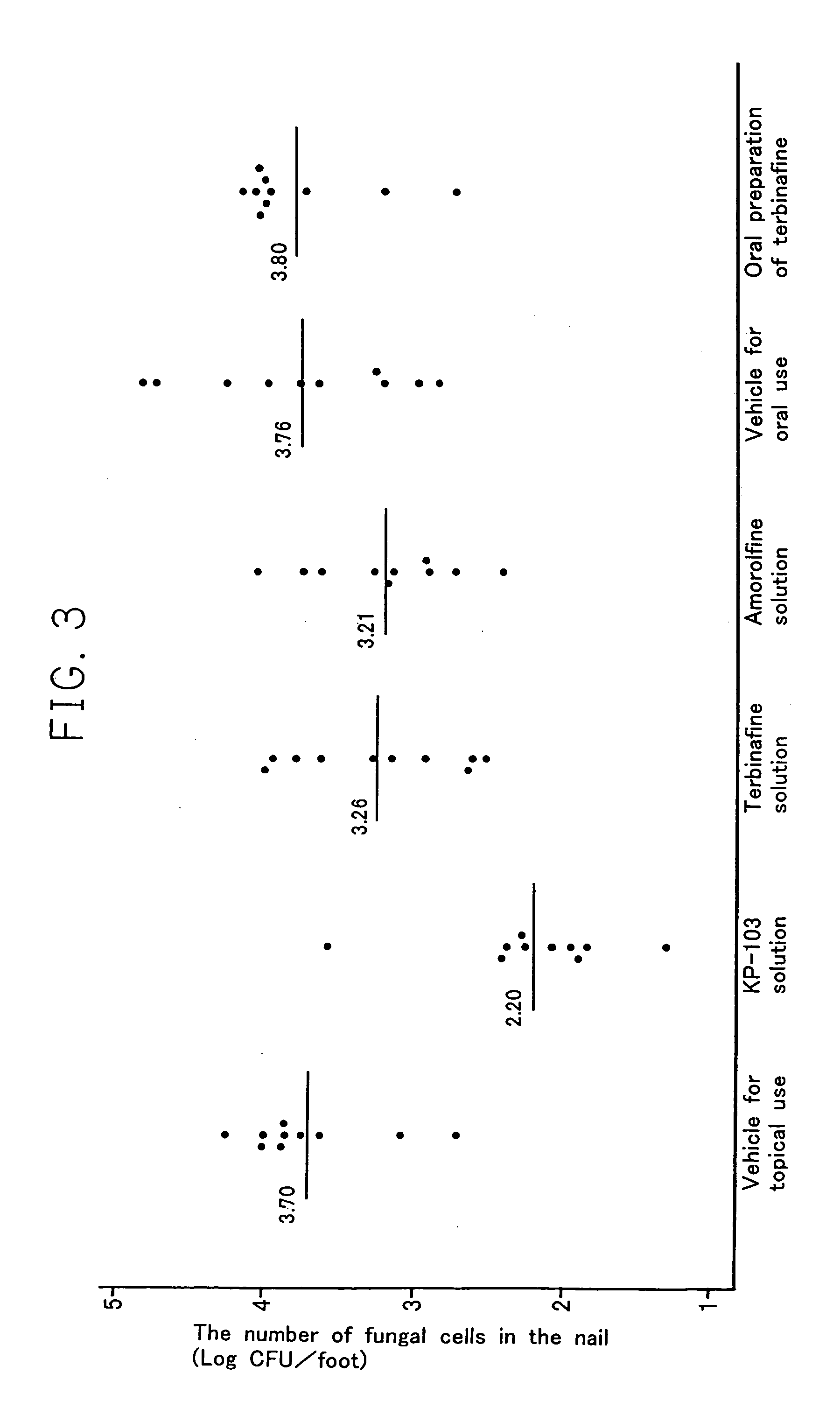
[0016]Additionally, an object of the present invention is to provide the evaluation method of a drug which enables the effect of an antifungal agent to accurately evaluate in a guinea pig model of tinea unguium. Another object of present invention is to provide a therapeutic agent for onychomycosis which exhibits the effect on tinea unguium by topical application and which is capable of curing tinea unguium shorter period than that of the marketed oral preparation due to good permeability, good retention capacity and conservation of high activity in nail plate as well as the potent antifungal activity thereof based on the present invention. Another object of the present invention is to provide the effective therapeutic agent for onychomycosis exhibiting no side effect even if therapeutically effective amounts of it are administered sufficiently.

Problems solved by technology

Therefore, it became to clear that the conventional method has the problem that the drug effect can not be accurately evaluated, because the apparent therapeutic effect need to be evaluated after removing the drug remaining in the skin.
However, there are many cases where the patient stops taking the drug or that takes the drug irregularly, since they have to take the drug for a long period, for example at least a half a year in order to completely cure tinea unguium.
It is thought that this is a main cause of difficulty of curing tinea unguium completely.
Furthermore, by taking the drug for a long period, griseofulvin has the problem of side effects on internal organ (gastrointestinal disorder, hepatotoxicity) and hepatotoxicity is reported as the side effect in terbinafine.
However, in case of the simple application on nail plate with the current antifungal agent for topical use, the antifungal effect on fungus in the nail was not seen, because the drug could not sufficiently permeate the thick keratin in nail plate (Markus Niewerth and Hans C. Korting, Management of Onychomycoses, Drugs, 58: 283–296, 1999).
In addition, the therapeutic effect of a topical preparation of antifungal agent on the experiment model of trichophytosis can not be evaluated using the conventional method as mentioned above.

Method used


Image

  • Method for treating onychomycosis
  • Method for treating onychomycosis
  • Method for treating onychomycosis

Examples


example 1

Determination of drug remaining in skin which has been already evaluated five days after the last treatment according to conventional method.

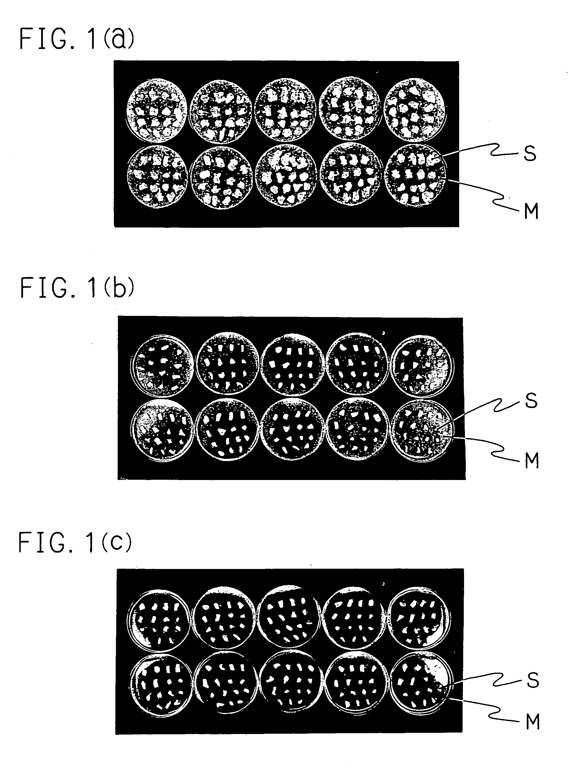
[0068]A model was prepared according to Comparative Example 1. Lanoconazole being a test compound was used for a therapeutic experience as 1% solution with the same vehicle as KP-103. For the infected control group without an application of a drug, the KP-103-treated group and the lanoconazole-treated group, 20 animals were employed, respectively. The two hind feet were excised from each animal five days after the last treatment in the same manner as in Comparative Example 1. A total of 20 light feet were used for an evaluation by the conventional method, and a total of 20 right feet were used in the present evaluation method.

[0069]The skin pieces of light foot were placed on 20 ml of Sabouraud dextrose agar medium containing Trichophyton mentagrophytes KD-04 strain (2×10⁴ cells / ml) and the antibiotic substance described in Comparative Example ...

example 2

Determination of Remaining Drug after Removing Drug from Skin.

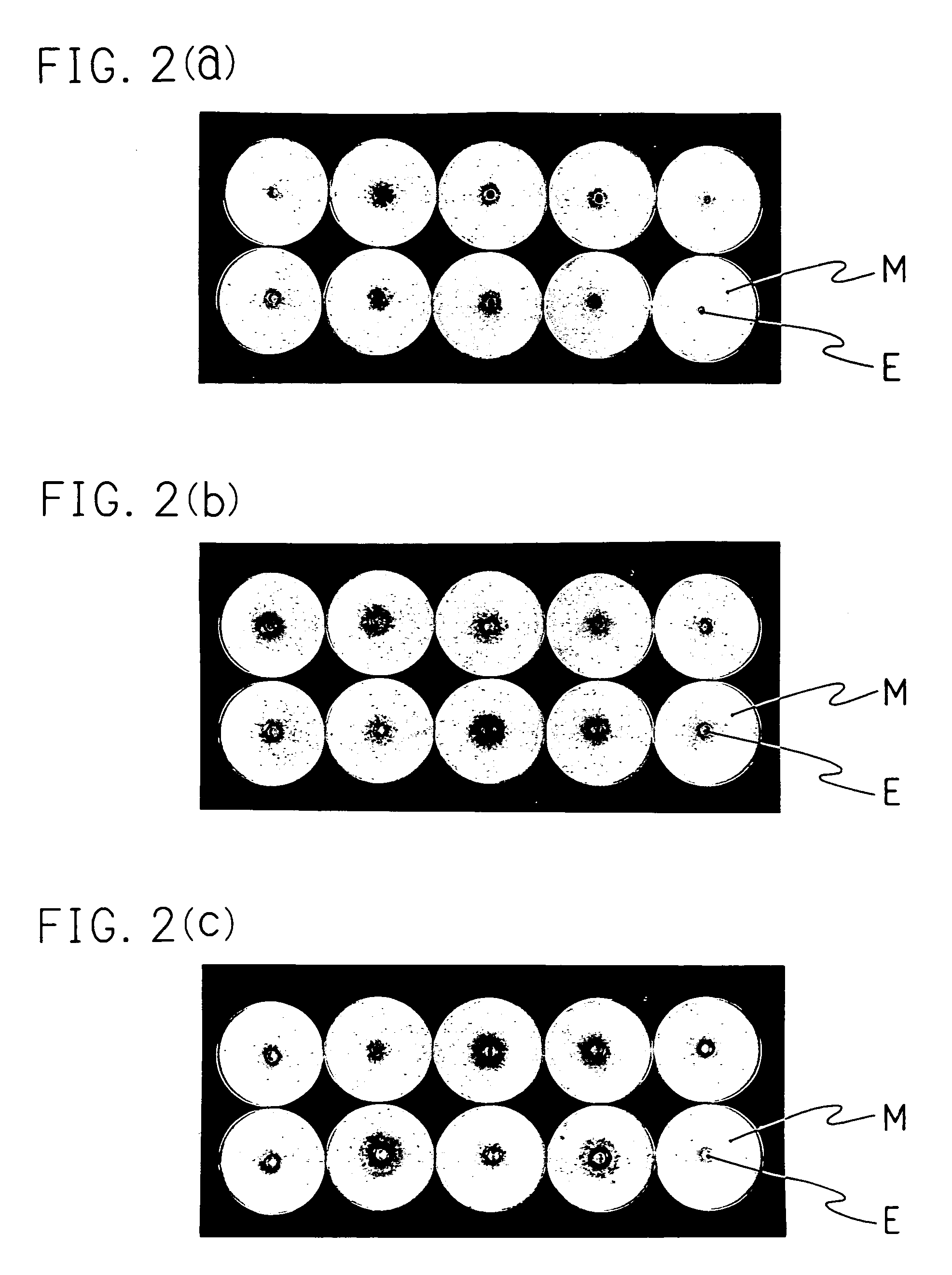
[0072]As Example 1, 20 right feet were excised from each animal five days after the last treatment, and sufficiently wiped with the cotton sweb containing alcohol. The planta was cut off from each foot. The skin mincced by a scissors was put into dialysis membrane (fractional molecular weight: 12,000–14,000, made of cellulose, available from VISKASE SALES Corporation) together with 4 ml of distilled water. Dialysis was carried out under 3 L of distilled water at 4° C. for 2 days. The dialysis water was changed twice a day 4 times in total. The content was transfer into a glass homogenizer. Thereto 4 ml double-concentration phosphate buffered saline containing 4% of trypsin derived from pig pancreas (available from BIOZYME Laboratories Limited) was added and the resulting mixture was homogenized. It was left at 37° C. for one hour and was filtrated with the two-ply gauze. The resulting filtrate was centrifuged. To a precip...

example 3

Detection of Viable Fungus in Skin and Evaluation of Drug Effect

[0076]To two mediums of Sabouraud dextrose agar medium (20 ml) containing the antibiotic substance described in Comparative Example 1 were applied 100 μl of the suspension from one right feet of each animals obtained in Example 2. After the cultivation was carried out at 30° C. for 10 days, the result is described as “fungus-negative” when a colony of fungus was not observed in two agar plates (detection limit: 10 CFU (colony forming unit) / feet). The number of fungus-negative feet was counted. On the other hand, 20 left feet were evaluated in the same manner as in Comparative Example 1. Table 2 shows the result of comparing the therapeutic effect evaluated by the conventional method with that by the present evaluation method.

[0077]

TABLE 2The number of fungus-negativefeet / Total number of infectedfeetConventionalPresent evaluationTest substanceMethodmethodInfected control 0 / 20 0 / 20KP-10319 / 2017 / 20Lanoconazole20 / 20 3 / 20

[00...


PUM

PropertyMeasurementUnit
temperatureaaaaaaaaaa
weightaaaaaaaaaa

Abstract

A novel method for evaluating an effect of an antimicrobial agent which comprises removing the antimicrobial agent remaining in a biological sample or the like to thereby accurately evaluate the effect of the antimicrobial agent without being affected by the remaining antimicrobial agent. A therapeutic agent for onychomycosis which can be obtained according to the evaluation method of the drug effect.

Description

[0001]This application is a divisional under 35 U.S.C. § 120 of U.S. Non-Provisional application Ser. No. 10 / 031,929 filed 25 Jan. 2002, now abandoned, which was a National Stage filing under 35 U.S.C. § 371 of PCT / JP00 / 04617 filed 11 Jul. 2000, which claimed priority to Japanese patent application Ser. No. 11 / 214,369 filed 28 Jul. 1999.TECHNICAL FIELD[0002]The present invention relates to a method for detecting pathogenic microorganism, method for evaluating an effect of an antimicrobial agent on pathogenic microorganism and a method for detecting an antimicrobial agent. The present invention also relates to an antimicrobial agent and a therapeutic agent for onychomycosis, which are obtained according to the above-mentioned method for evaluating the drug effect.BACKGROUND ART[0003]A method for evaluating a drug effect with an animal model is needed in order to explore a novel antimicrobial agent (also hereinafter referred to “drug”). Further, a method enabling a drug effect to be e...

Claims


Application Information

Patent Timeline
Patent Type & Authority Patents(United States)
IPC IPC(8): C12Q1/18A01N43/26A01N43/34A61K31/445A61K31/4196A61K31/454A61P31/10C07D401/12C07D521/00C12Q1/04
CPCA61K31/4196A61K31/454C07D231/12C07D233/56C07D249/08C12Q1/04C12Q1/18G01N33/56911G01N33/56961C07D401/12G01N2500/00A61P31/10
Inventor TATSUMI, YOSHIYUKIYOKOO, MAMORUNAKAMURA, KOSHOARIKA, TADASHI
Owner KAKEN PHARMA CO LTD
Why Eureka
Try Eureka