Signal for packaging of influenza virus vectors
a technology for packaging and influenza viruses, applied in the direction of viruses/bacteriophages, antibody medical ingredients, microorganisms, etc., can solve the problems of intertypic reassortment between, and achieve the effects of immunizing the individual, increasing the amount of endogenous protein in the mammal, and increasing the expression of endogenous protein
- Summary
- Abstract
- Description
- Claims
- Application Information
AI Technical Summary
Problems solved by technology
Method used
Image

Examples
example 1
Materials and Methods
[0100]Viruses and cells. Human H3N2 viruses isolated from a single patient, either in embryonated chicken eggs (A / Tottori / AT1 / AM2AL3 / 94; AM1AL3) of Madin-Darby canine kidney (MDCK) cells (A / Tottori / 872 / K4 / 94; K4), were obtained from T. Ito (Tottori University, Tottori, Japan). Virus stocks were grown either in 10 day-old embryonated chicken eggs (AMZAL3 virus) or on MDCK cells (K4 virus) in minimal essential medium (MEM) supplemented with 0.3% bovine serum albumin and 0.5 mg of trypsin / ml. MDCK cells were maintained in MEM supplemented with 5% newborn calf serum (Sigma, St. Louis, Mo.).
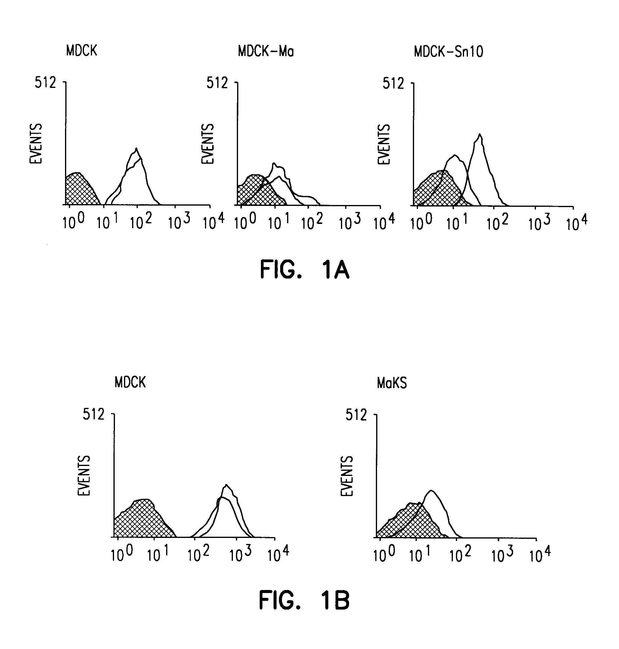
[0101]Generation of lectin-resistant cell lines. MDCK cells grown to 75% confluency were washed three times with phosphate-buffered saline and incubated with Maakia amurensis (MAA) lectin (100 mg / ml; Boehringer Mannheim, Mannheim, Germany) or Sambucus nigra (SNA) lectin (100 mg / ml; Boehringer Mannheim) in MEM containing 0.3% bovine serum albumin. After a 48 hour incubation, the me...
example 2
Materials and Methods
[0123]Cells. 293T human embryonic kidney cells were maintained in Dulbecco's medium supplemented with 10% fetal calf serum (FCS) and Madin-Darby canine kidney (MDCK) cells were maintained in Eagle's medium supplemented with 5% newborn calf serum.
[0124]Plasmid-based reverse genetics. Influenza A viruses were generated using plasmids possessing the cDNA of A / WSN / 33(H1N1) viral genes under the control of an RNA polymerase I promoter and terminator (referred to as Poll plasmids) and pCAGGS / MCS plasmids expressing influenza viral proteins as described in Neumann et al. (1999) (FIG. 4B). Briefly, Poll plasmids and protein expression plasmids were mixed with a transfection reagent, Trans IT LT-1 (Panvera, Madison, Wis.), incubated at room temperature for 10 minutes, and added to 1×10⁶ 293T cells cultured in Opti-MEM (GIBCO / BRL). Forty-eight hours post-transfection, 0.5 μg per ml of trypsin was added to the medium to activate the HA protein, followed by incubation for 1...
example 3
[0144]To obtain a true image of the viral contents, electron microscopy tomography was performed (FIG. 11A). An image of virions with a 50 nm thickness was collected. Then, an analysis of one of the virions was conducted and the 3D images of virion content reconstituted. The structures (rods) inside the particle are colored to distinguish each structure (FIGS. 11B–F, showing views from the top, side and bottom). Most of the views for the rods are cut, however, for one view, in which rods were cut across the middle, the entire molecule is shown. Nevertheless, all of the views show inter-rod interactions.
[0145]Based on the results summarized above, including data which support that each viral segment contains a unique sequence that is important for incorporation into virions, which likely contributes to the formation of the unique morphologic features of the viral contents, vRNA segments are likely selectively incorporated into influenza virions. This information is useful not only to...
PUM
| Property | Measurement | Unit |
|---|---|---|
| emission wavelength | aaaaa | aaaaa |
| excitation wavelength | aaaaa | aaaaa |
| emission wavelength | aaaaa | aaaaa |
Abstract
Description
Claims
Application Information
 - R&D
- Intellectual Property
- Life Sciences
- Materials
- Tech Scout
- Unparalleled Data Quality
- Higher Quality Content
- 60% Fewer Hallucinations
Browse by: Latest US Patents, China's latest patents, Technical Efficacy Thesaurus, Application Domain, Technology Topic, Popular Technical Reports.
© 2025 PatSnap. All rights reserved.Legal|Privacy policy|Modern Slavery Act Transparency Statement|Sitemap|About US| Contact US: help@patsnap.com