

Methods for detecting chromosome aberrations
a technology of aberration and chromosome, applied in the field of methods for detecting chromosomal aberration, can solve the problems of false negative reading, high background, and large genomic nucleic acid probes derived from genomic nucleic acids (>100 bp) may not be well suited for hybridization analysis,
- Summary
- Abstract
- Description
- Claims
- Application Information
AI Technical Summary
Benefits of technology
Problems solved by technology
Method used
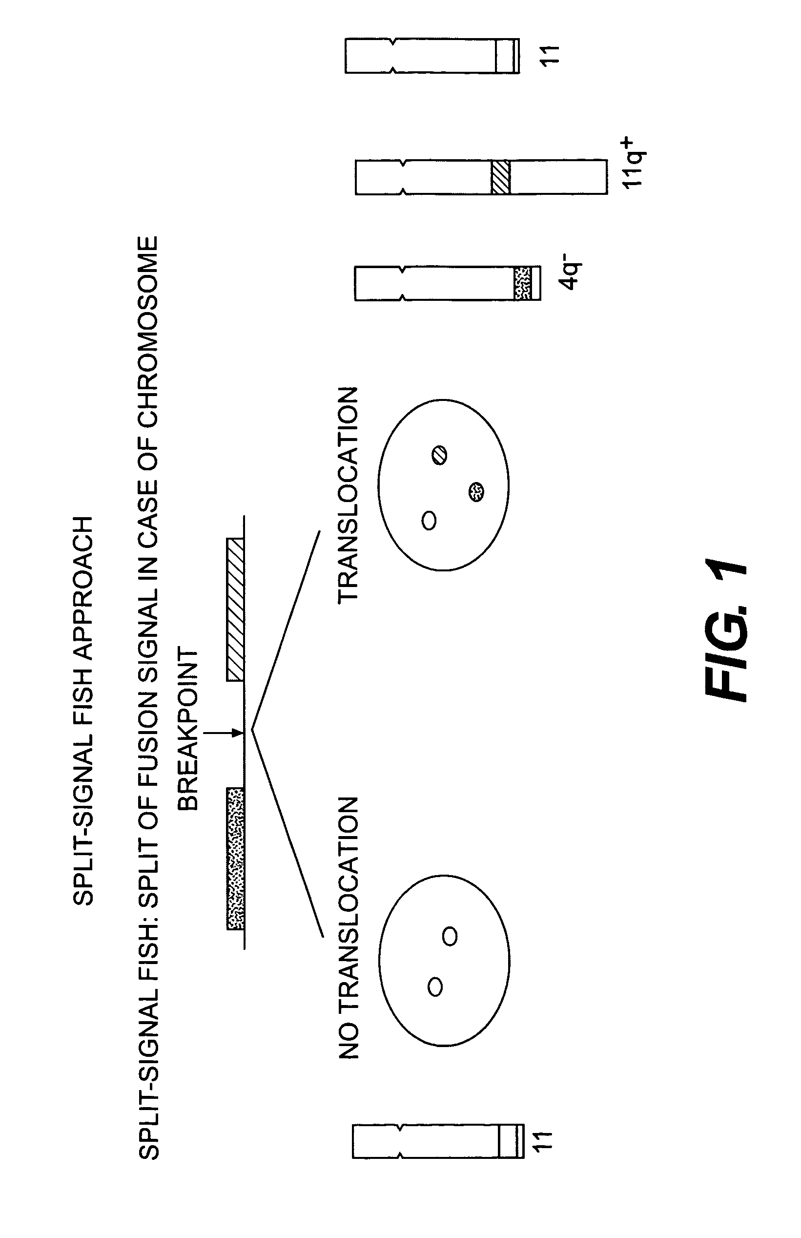
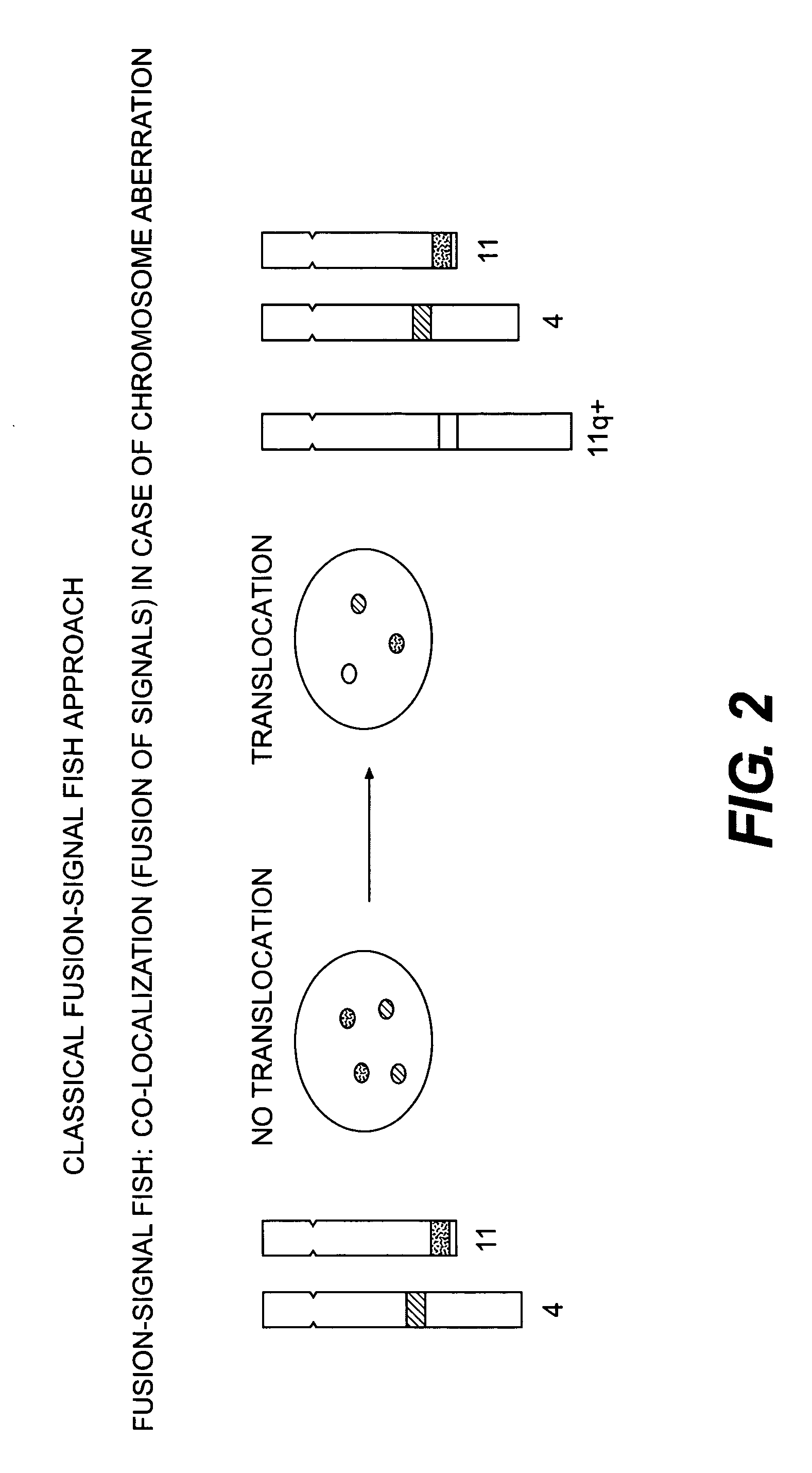
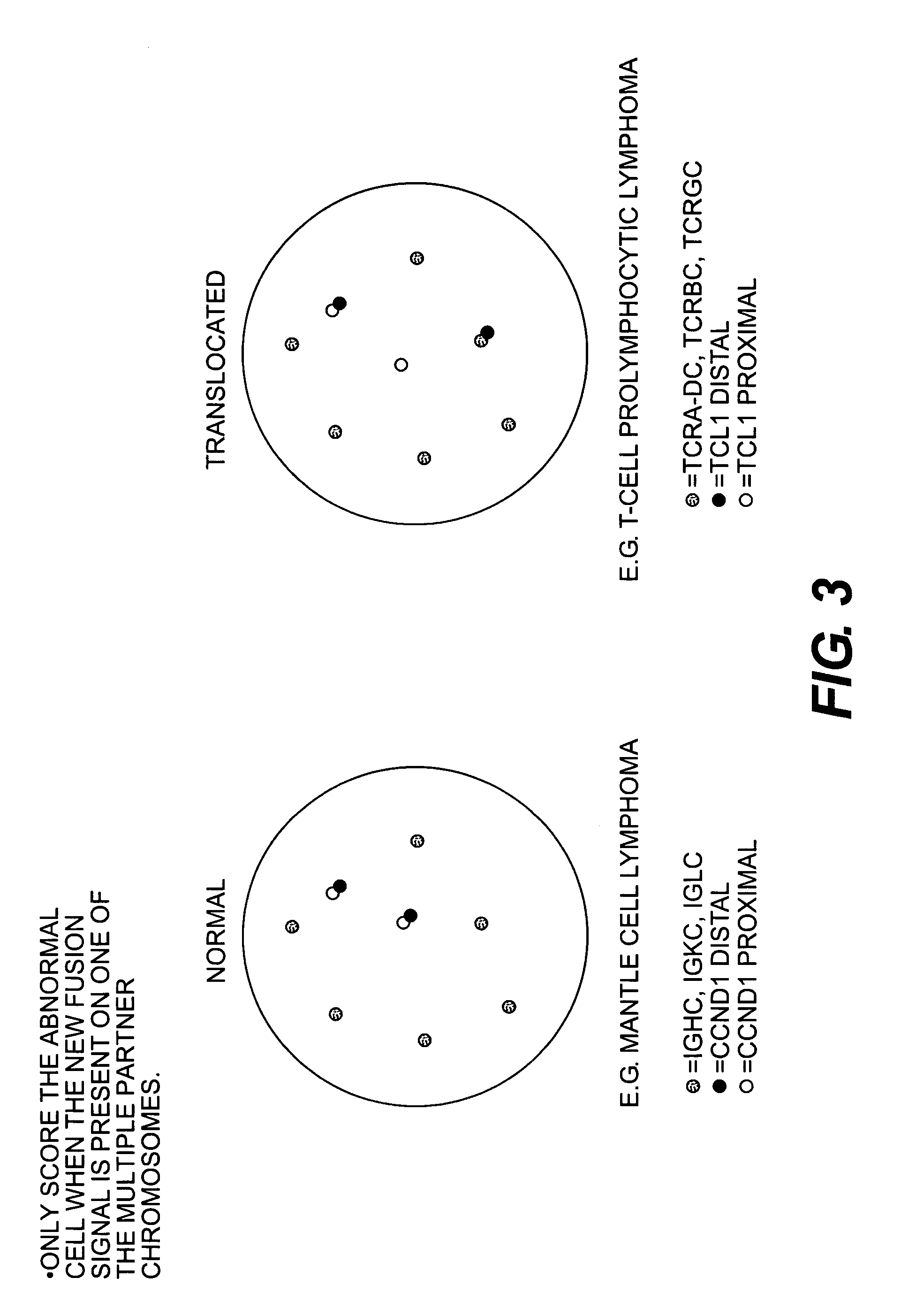
Image

Examples
example 1
[0088]A slide containing metaphase spreads and interphase nuclei was pretreated in TBS (50 mmol / L Tris, 150 mmol / L NaCl, pH 7.6) with 3.7% formaldehyde for 2 minutes at room temperature, as described in Human Cytogenetics, a Practical Approach, Volume 1, 2nd ed., D. E. Rooney and B. H. Czepulkowski editors (1992). The slide was then rinsed twice in phosphate buffered saline (PBS) for 5 minutes per wash at room temperature. After rinsing, the slide was dehydrated in a cold (4° C.) series of ethanol by incubating the slide in 70% ethanol at 4° C. for 2 minutes; incubating the slide in 85% ethanol at 4° C. for 2 minutes; and then incubating the slide in 96% ethanol at 4° C. for 2 minutes, followed by air drying.
[0089]On each target area, 10 μL of DNA Hybridization Buffer (100 ng fluorescein labelled probe, 100 ng Texas Red labelled probe, 5 μM PNA Oligo Mix, 45% formamide, 300 mM NaCl, 5 mM NaPO4, 10% Dextran sulphate) was added and an 18×18 mm coverslip was applied to cover the hybrid...
example 2
[0092]Slides containing metaphase spreads and interphase nuclei from normal cells and test sample cells are pretreated in TBS as described above. The slides are then rinsed twice in PBS for 5 minutes per wash at room temperature. After rinsing, the slide are dehydrated in a cold (4° C.) series of 70%, 85%, and 96% ethanol as described above, followed by air drying.
[0093]On each target area, 10 μL of DNA Hybridization Buffer (100 ng fluorescein labeled probe, 100 ng Texas Red labeled probe, 100 ng AMCA (7-amino-4-methylcoumarin-3-acitic acid) labeled probe, 5 μM PNA Oligo Mix, 45% formamide, 300 mM NaCl, 5 mM NaPO4, 10% Dextran sulphate) is added and an 18×18 mm coverslip is applied to cover the hybridization area on the slides. The edges of the coverslip are sealed with rubber cement before denaturation at 82° C. for 5 minutes. The slides are hybridized overnight at 45° C. After hybridization, the coverslip is removed and the slides are washed for 10 minutes in a stringent wash buff...
example 3
[0096]PDGFRA probes were hybridized to metaphase chromosomes from metaphase spreads from normal tissue and from the cell line ELO-1. A normal PDGFRA locus on chromosome 4 was detected in normal cells as either a yellow dot or by co-localized green / red signals due to the juxtaposition of the red probes and green probes hybridizing to regions upstream and downstream from an anticipated disease-related change in chromosomal structure. An additional chromosome 4 was also detected as a result of an amplification of the derivative chromosome 4.
PUM
| Property | Measurement | Unit |
|---|---|---|
| pH | aaaaa | aaaaa |
| chromosomal structure | aaaaa | aaaaa |
| non-nucleic acid | aaaaa | aaaaa |
Abstract
Description
Claims
Application Information