Decellularisation of matrices

a matrices and matrix technology, applied in the field of decellularisation of matrices, can solve the problems of inherently toxic most of protease inhibitors, serious limitations of methods, and deleterious effects of sds on heart valve extracellular matrix

Inactive Publication Date: 2008-04-08
TISSUE REGENIX +1
View PDF9 Cites 55 Cited by
  • Summary
  • Abstract
  • Description
  • Claims
  • Application Information

AI Technical Summary

Benefits of technology

[0020]We have found Aprotinin particularly effective as a proteolytic inhibitor and of particular utility because of its low toxicity, stability in solution at different pHs and stability at a variety of different temperatures.

Problems solved by technology

However, serious limitations of the method have been reported (Bodnar E et al, Thorac. Cardiovasc. Surgeon.1985 34: 82-85; Courtman D Wet al, J Biomed Mater Res.
These researchers all report that SDS has a deleterious effect on heart valve extracellular matrix (ECM) and in particular on the collagen and elastin fibre components.
A further problem associated with decellularising tissue implant is to minimise the degradation to the ECM during the process.
However, most of the protease inhibitors are inherently toxic which is undesirable if the matrix is to be seeded with living cells and implanted into a human or animal.
Moreover, some of the protease inhibitors used so far, for example PMSF, are extremely unstable in solution having a half life of less than 1 hour, and since decellularisation is a lengthy process i.e. several days, this limits the choice of inhibitors that have sufficient half lives.

Method used


Image

  • Decellularisation of matrices
  • Decellularisation of matrices

Examples


example 1

Porcine Aortic Valves

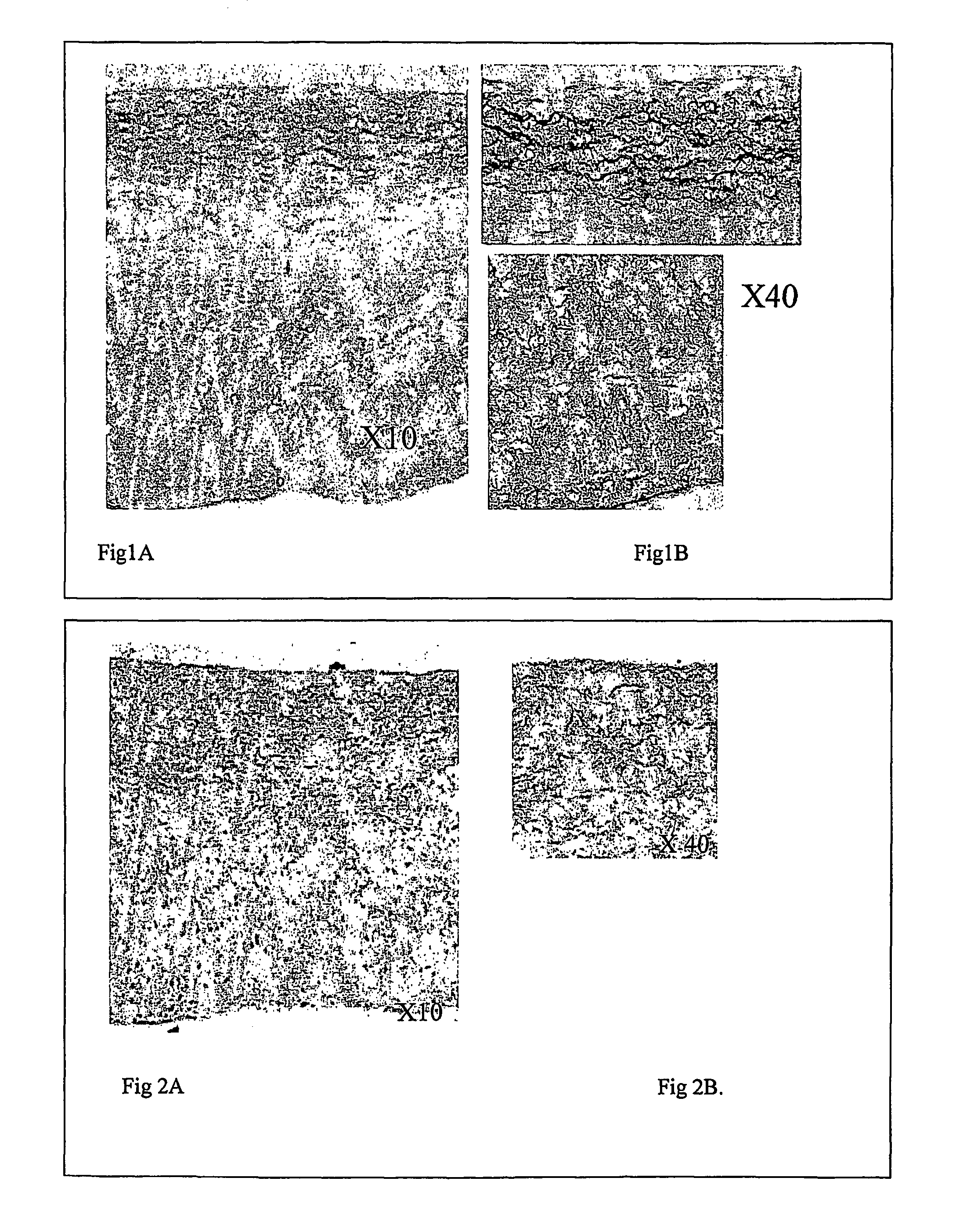
[0058]Porcine hearts were procured from a local abattoir within 2 hours of slaughter and transported on ice to the laboratory. On arrival at the laboratory, aortic valve roots were dissected from the heart and washed in transport solution [Hanks' balanced salt solution (HBSS), 10 KIU / ml Aprotinin, 10 u / ml penicillin, 100 μg / ml streptomycin, 100 U / ml Nystatin, 10 mM HEPES pH7.6). The aortic valves were incubated overnight (14 hours) in hypotonic tris buffer (10 mM tris pH8, 0.1% (w / v) ethylene diamine tetraacetic acid (tDTA), 10 KIU Aprotinin in distilled water DW).

[0059]Subsequently, the aortic valves were incubated for 24 hours with shaking at ambient temperature in (0.05%-0.1%) (w / v) sodium dodecyl sulphate (SDS) or 0.5% sodium deoxycholate in hypotonic tris buffer. They were then washed (×3) with tris buffered saline (0.15M NaCl, 0.05M tris pH 7.6 in DW) containing protease inhibitors (0.1% w / v EDTA and 10 KIU / ml Aprotinin). They were then subjected to a furt...

example 2

Porcine Patella Tendons

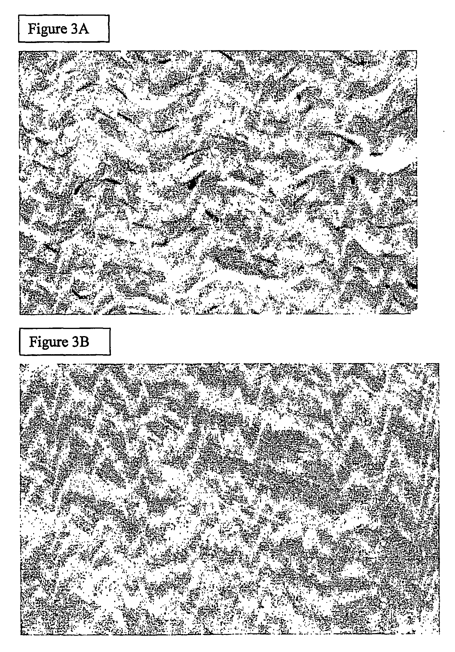
[0061]Porcine patella tendons were dissected and then washed in PBS. The tendons were incubated overnight (24 hours) in hypotonic Tris buffer (10 mM Tris pH 8, 0.1% ethylene diamine tetraacetate (EDTA), 10 KIU Aprotinin in distilled water (DW)]. Tendons were subsequently incubated for a further 24 hours with shaking at ambient temperature in 0.03-0.1% w / v sodium dodecyl sulphate (SDS) or 0.5% sodium deoxycholate in hypotonic Tris buffer. They were then washed (×3) with PBS containing protease inhibitors (0.1% EDTA and 10 KIU / ml Aprotinin).

[0062]With reference to FIG. 3A there is shown a photomicrograph of fresh porcine patella tendon, stained with heamatoxylin and eosin (×400). FIG. 3B shows a photomicrograph of porcine patellar tendon following de-cellularisation treatment as described above and also stained with heamatoxylin and eosin (×400). It is apparent from comparing the Figures that decellularisation has been achieved whilst maintaining the histoarchit...


PUM

PropertyMeasurementUnit
concentrationaaaaaaaaaa
half lifeaaaaaaaaaa

Abstract

A method of preparing matrices or tissue engineered biomaterials for implantation, and in particular to a method of improving decellularisation of matrices or tissue engineered biomaterials prior to implantation. The method employs a single anionic detergent in combination with protease inhibitors.

Description

RELATED APPLICATION[0001]This application claims priority from PCT Application PCT / GB02 / 02341, filed 20 May 2002, which claims priority from British Application No. 0112586.3, filed 24 May 2001. These disclosures are hereby incorporated by reference herein in their entireties.[0002]The present invention relates to a method of preparing matrices or tissue engineered biomaterials for implantation, and in particular to a method of improving decellularisation of matrices or tissue engineered biomaterials prior to implantation.BACKGROUND TO THE INVENTION[0003]A large variety of body implants are known for medical uses such as substitute vascular prostheses, skin dressings and coverings, and for other purposes. The implant material can be synthetic or body tissues from the same species or other species as the species to be implanted. When body tissues and structures are to be implanted, they may be used fresh from the donor but in many cases, it is preferred to have some means of preservi...

Claims


Application Information

Patent Timeline
Patent Type & Authority Patents(United States)
IPC IPC(8): C12N9/00C12N9/48A61L27/36
CPCA61L27/3633A61L27/3687
Inventor FISHER, JOHNBOOTH, CATHERINEINGHAM, EILEEN
Owner TISSUE REGENIX
Why Eureka
Try Eureka