Cytological imaging system and method

a cytological imaging and cytological technology, applied in the field of cytological material analysis, can solve the problems of difficult analysis of traditional stains by an automated system, difficult identification difficulty in analyzing the cytoplasm of traditional stains by the human eye, so as to achieve less heat, less power, and less light and heat.

Inactive Publication Date: 2008-08-12
CYTYC CORP
View PDF106 Cites 3 Cited by
  • Summary
  • Abstract
  • Description
  • Claims
  • Application Information

AI Technical Summary

Benefits of technology

[0022]An additional aspect of the invention relates to counterstaining previously stained material, wherein the counterstain includes a component from the previous stain. The process of counterstaining the previously stained material replenishes any loss of the previous stain component that may have occurred subsequent to the initial staining, for example, through rinsing the material after staining. In one embodiment of this method, the previously stained material is rinsed prior to counterstaining. The rinse can be an alcohol bath.
[0023]Another aspect of the invention relates to an optical instrument lighting system that includes a first light source and a second light source. The first light source has a first wavelength, which may be between about 690 nm and about 750 nm. The first light source is used to verify a stain used on a cytological sample. The second light source has a second wavelength different than the first wavelength, and may be between about 500 nm and about 600 nm. The second light source is used to illuminate the cytological sample for viewing. In various embodiments, the first light source can be a red light emitting diode (LED) and the second light source can be a green LED or an array of up to eight or more green LEDs. The light source can operate on low voltage, such as 5 volts DC. LEDs are bright, stable, and available in a very wide range of illumination wavelengths. Furthermore, LEDs efficiently produce a narrow band of illumination (typically 15 nm), eliminating the need for narrow band filters and allowing all energy (heat) to be put into the desired illumination wavelength. Conventional illumination such as tungsten-halogen bulbs put out a lot of wasted light and heat when narrow band illumination is desired. LEDs generate significantly less heat and require substantially less power than conventional imaging light sources. In fact, standard light sources are often insufficient to obtain the necessary shutter times. In addition, LEDs are relatively tiny.

Problems solved by technology

Traditional stains, including the Papanicolaou stain, are difficult for an automated system to analyze.
The variety of colors in the cytoplasm from traditional stains, which are straightforward for the human eye to distinguish, are not readily analyzed with automated imaging systems because they contrast to varying degrees with the traditional blue hematoxylin stain of the nucleus.
The varying contrast makes automated analysis unreliable.
While standardization of a Papanicolaou-like stain has been proposed for many years, there has been little incentive for laboratories to do so.
This variability affects current imaging technologies which may reject numerous slides either because of problems inherent with a conventional Pap smear preparation, or because of poor staining that produces nuclear-cytoplasmic contrast that is inadequate for image acquisition and analysis.
Many require a high level of sophistication that is costly in terms of hardware and software.
Further, these approaches have not proven accurate and reliable enough to be widely used in clinical cytological and histological diagnoses.
Typically, these devices generate a significant amount of heat, require filters for obtaining correct wavelengths, and are relatively large.

Method used


Image

  • Cytological imaging system and method
  • Cytological imaging system and method
  • Cytological imaging system and method

Examples


Embodiment Construction

[0055]Embodiments of the present invention are described below. It is, however, expressly noted that the present invention is not limited to these embodiments, but rather the intention is that modifications that are apparent to the person skilled in the art and equivalents thereof are also included.

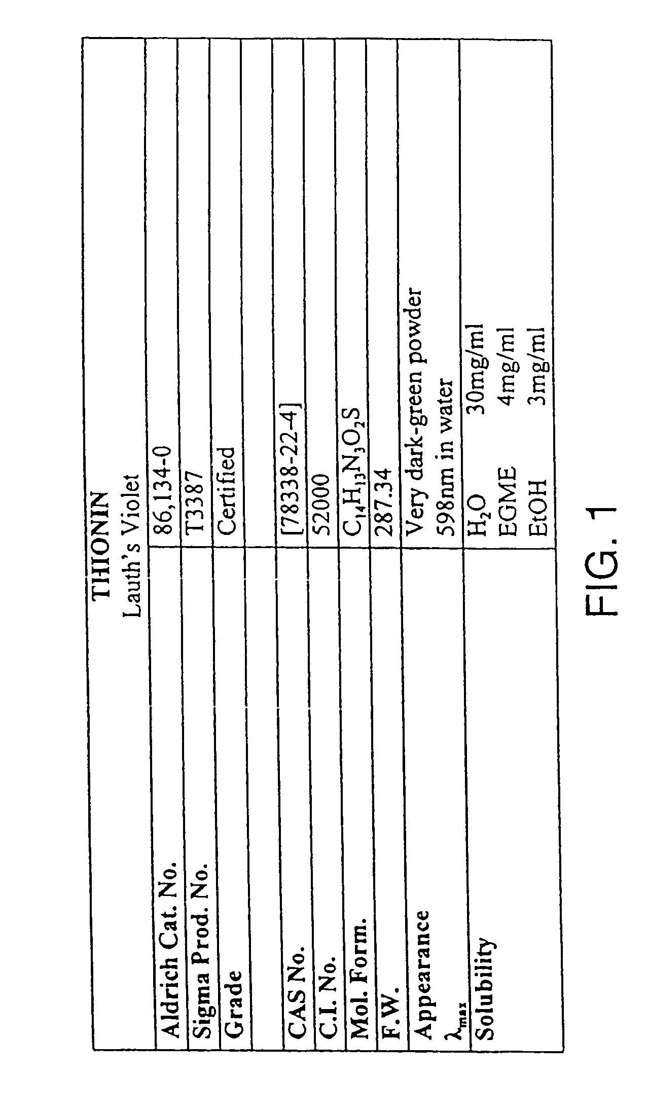
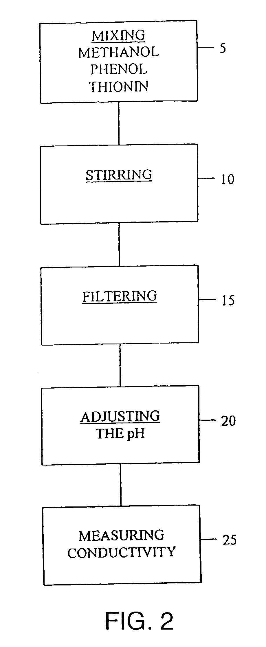
[0056]One embodiment of the stain is a thionin-phenol solution that includes methanol, phenol, and thionin. The stain may also include an acid for adjusting the pH of the stain solution. The purity of the methanol (methyl alcohol) component can be various grades The phenol component is supplied as loose crystals having an ACS grade purity of at least about 95%. The phenol is a chaotrophic agent available through Aldrich Chemical Company, Inc. of Milwaukee, Wisc.; however, an equivalent may be substituted. The thionin component is supplied as a certified dye powder, specifically a BSC-certified, metachromatic, cationic thiazine dye. FIG. 1 depicts the characteristics of one embodiment of t...


PUM

PropertyMeasurementUnit
wavelengthaaaaaaaaaa

Abstract

The present invention relates to the analysis of cytological material. Specifically, the invention relates to stains and methods of producing the stains, methods of staining cells for cytological or histological analysis to contrast the nuclear portion of the cell from the cytoplasmic portion, and systems and methods for illuminating a cytological sample. The analysis can be automated or manual.

Description

CROSS-REFERENCE TO RELATED APPLICATIONS[0001]This application incorporates herein by reference three U.S. patent applications filed of even date herewith entitled Cytological Stain Composition, Cytological Stain Composition Including Verification Characteristic, and Apparatus and Methods for Verifying the Location of Areas of Interest Within a Sample in an Imaging System, and identified by Ser. Nos. 09 / 430,116, 09 / 430,196, and 09 / 430,198, respectively.TECHNICAL FIELD[0002]The present invention relates to the analysis of cytological material. Specifically, the invention relates to stains and methods of producing the stains, methods of staining cells for cytological or histological analysis to contrast the nuclear portion of the cell from the cytoplasmic portion, and systems and methods for illuminating a cytological sample. The analysis can be automated or manual.BACKGROUND INFORMATION[0003]Cytology is the branch of biology dealing with the study of the formation, structure, and func...

Claims


Application Information

Patent Timeline
Patent Type & Authority Patents(United States)
IPC IPC(8): G01N33/48F21V9/00G01N21/00G02B21/06G01N21/75G01N1/30G01N15/14G01N21/31G06T7/00
CPCG01N1/30G01N15/1475G01N21/3151G01N15/1484G01N15/147G01N2015/1488G01N2015/1472
Inventor ZAHNISER, DAVID J.ZELINSKI, MATTHEW S.DOLASH, THOMAS M.MAENLE, GARRICK L.FLEMING, MARKLAUDO, JOHN S.
Owner CYTYC CORP
Why Eureka
Try Eureka