
Non-continuous immunoassay device and immunoassay method using the same

a technology of immunoassay and continuous strip, which is applied in the direction of specific use bioreactors/fermenters, instruments, biomass after-treatment, etc., can solve the problems of ineffective control of the reaction rate of antigen-antibody reaction with the conventional assay strip, the shape of the assay device including the strip b>10/b> is limited to the straight rod shape, and the flow speed of the whole blood components, such as the hemolyzed erythr

Inactive Publication Date: 2008-10-07
SD
View PDF15 Cites 3 Cited by
  • Summary
  • Abstract
  • Description
  • Claims
  • Application Information

AI Technical Summary

Benefits of technology

"The present invention provides an immunoassay device that can control the flow speeds of liquid buffer, conjugate, and analyte, resulting in improved sensitivity and specificity in immunochromatographic assay. The device can also control the migration speed of the mobile phase and the antigen-antibody reaction time. Additionally, the device is non-continuous, meaning it can be produced in a simple manner and the whole pad does not need to be discarded if one pad is damaged during production. The shape of the device can also be modified for easy application and detection of the test result. The detection of the test result is easy, and the background of the detection line is clear even when whole blood is used as the liquid sample."

Problems solved by technology

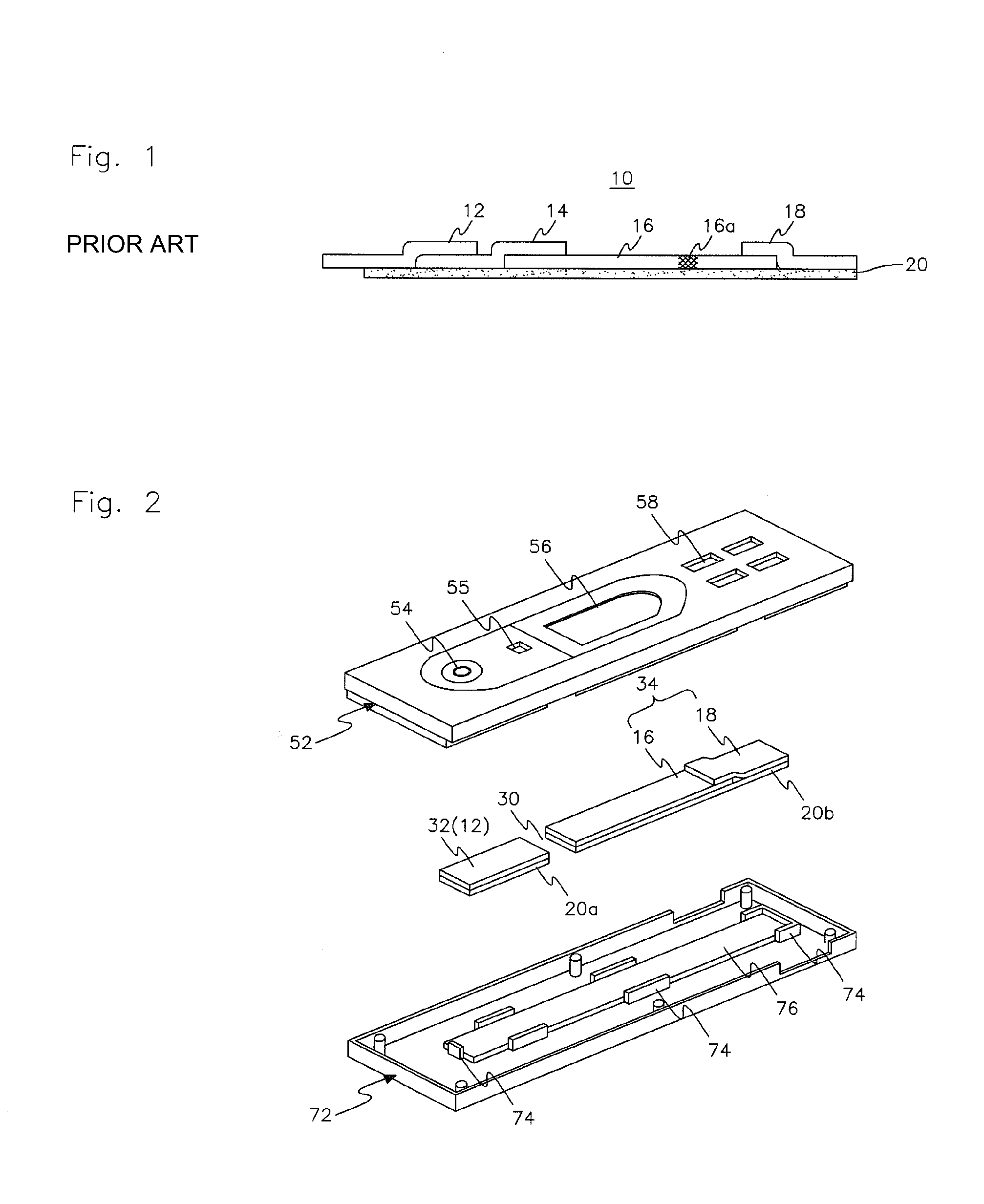
Thus, the flow speed of the liquid sample and the reaction rate of antigen-antibody reaction cannot be effectively controlled with the conventional assay strip 10.
The conventional assay strip 10 has a further disadvantage in that the shape of the assay strip 10 or the shape of the assay device including the strip 10 is limited to the straight rod shape.
In addition, when whole blood is used as the liquid sample, the flow speeds of the whole blood components, such as the hemolyzed erythrocyte, are relatively slow compared to the analyte in the liquid sample.
This makes it difficult to determine the test result.
However, this is not practical since it takes much time for the detection or determination of the test result.
Such problems mainly occur when using the whole blood to examine disease, such as malaria, AIDS, hepatitis C, hepatitis B, syphilis, helicobacter pylori, tumor markers (AFP, PSA, CEA), tuberculosis, SARS, Dengue fever, leprosy, and so on.

Method used


Image

  • Non-continuous immunoassay device and immunoassay method using the same
  • Non-continuous immunoassay device and immunoassay method using the same
  • Non-continuous immunoassay device and immunoassay method using the same

Examples


experimental example 1

Test of Influenza Virus Using Immunoassay Device

[0056](A) Manufacture of nitrocellulose pad. The monoclonal antibodies against the nucleocapsid antigens of influenza virus type A and B were diluted with phosphate buffer solution, and the diluted antibodies were spread over a nitrocellulose pad (width: 25 mm, pore size: 10 to 12 μm to form test line 1 and 2, respectively. An anti-mouse immunoglobulin G antibody was obtained by immunizing a goat with a mouse immunoglobulin G, and the antibody was diluted with phosphate buffer solution. The diluted antibody was spread over the nitrocellulose pad to form a control line, and was dried in 37° C. Thermostat for immobilization. Then, phosphate buffer solution containing 0.05% by weight bovine serum albumin, 4% by weight % sucrose and 0.0625% by weight ionic surfactant was sprayed on the blank space of the nitrocellulose pad, and the pad was dried in 30° C. Thermostat for 60 to 120 minutes. The nitrocellulose pad was attached to a polypropyl...

experimental example 2

Test of Syphilis Using Immunoassay Device

[0059]Except of using syphilis antigen produced by gene recombination instead of the monoclonal antibody against the nucleocapsid antigen of influenza virus, the nitrocellulose pad and the antigen-gold conjugate pad were manufactured by the same method of Experimental example 1. The nitrocellulose pad was attached to a polypropylene backing plate on which an adhesive is coated, then an absorbent pad (U.S.A., Millipore company) was also attached to the backing plate so that the absorbent pad and the nitrocellulose pad were overlapped by 1 mm. In addition, a sample pad for whole blood, an auxiliary pad and the antigen-gold conjugate pad were consecutively attached on a separate polypropylene backing plate on which an adhesive is coated so that the neighboring pads were separated by 1 mm, respectively. The two produced strips were installed on a lower case with a separation distance of 2 mm. Then, an upper case having three connecting members fo...

experimental example 3

Test of HGC Using Immunoassay Device

[0060]Except of using the monoclonal antibody against the alpha HCG antigen instead of the monoclonal antibody against the nucleocapsid antigen of influenza virus, the nitrocellulose pad and the antibody-gold conjugate pad were manufactured by the same method of Experimental example 1. A sample pad, the antibody-gold conjugate pad, and the nitrocellulose pad were consecutively attached on a polypropylene backing plate on which an adhesive is coated so that the neighboring pads were separated by 1 mm, respectively. Then an absorbent pad (obtained from Millipore Corp., U.S.A.) was also attached to the backing plate so that the absorbent pad and the nitrocellulose pad were overlapped by 1 mm. The produced strip was installed on a lower case. Then, an upper case having two connecting members for connecting the sample pad, the antibody-gold conjugate pad and the nitrocellulose pad was assembled with the lower case to produce the immunoassay device acco...


PUM

PropertyMeasurementUnit
pore sizeaaaaaaaaaa
gap distanceaaaaaaaaaa

Abstract

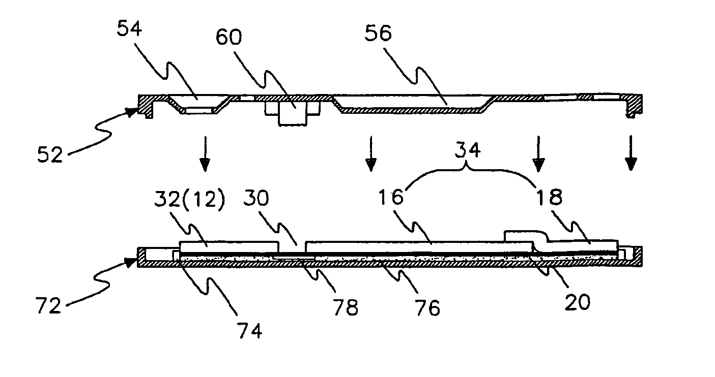
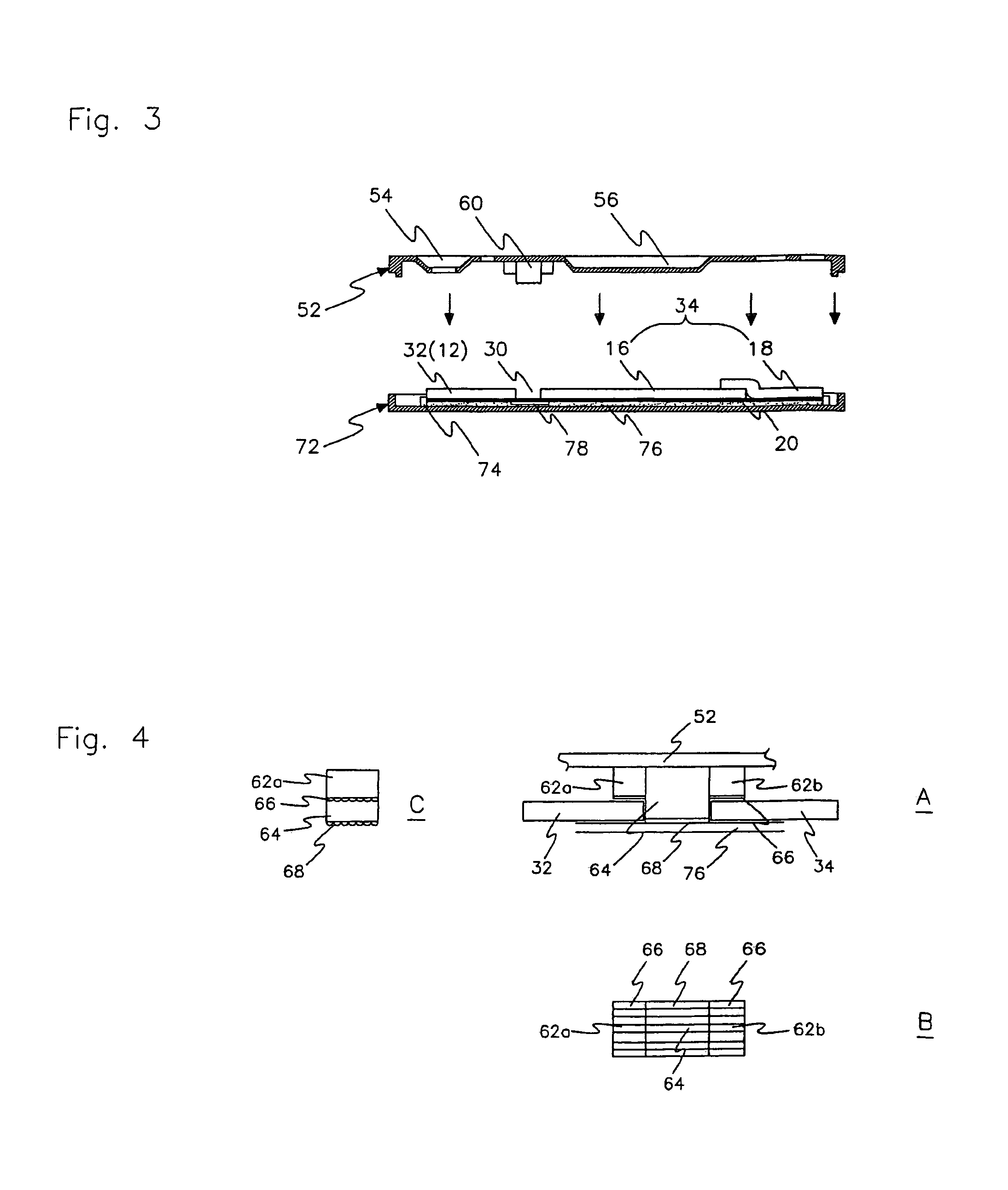
A non-continuous immunoassay device which includes two or more separated pads for immunoassay analysis, and is capable of controlling the migration speed of a mobile phase between the separated pads, and an immunoassay method using the same are disclosed. The immunoassay device includes a first pad receiving a mobile phase; a second pad which is spatially separated from the first pad by a predetermined distance, and to which the mobile phase migrates; an upper case for covering the upper parts of the first pad and the second pad; a lower case for covering the lower parts of the first pad and the second pad; and a connecting member which is formed on at least one of the upper case and the lower case, and located between the first pad and the second pad to form a passage for moving the liquid sample.

Description

[0001]This application claims priority of Korean Patent Application Nos. 2005-9677 filed on Feb. 2, 2005 and 2005-31525 filed on Apr. 15, 2005.FIELD OF THE INVENTION[0002]This invention relates to a non-continuous immunoassay device. More particularly, this invention relates to a non-continuous immunoassay device which includes two or more separated pads for immunoassay analysis, and is capable of controlling the migration speed of a mobile phase between the separated pads, and an immunoassay method using the same.BACKGROUND OF THE INVENTION[0003]The immunochromatographic assay known as a “rapid testing method” has been developed for a simple, qualitative and quantitative analysis of a small amount of analyte. The assay utilizes antigen-antibody reaction for analysis, and is applied in various fields or industries, such as a medical field for diagnosing diseases, agriculture, livestock raising, foods, military, environment and so on. Typically, the immunochromatographic assay is car...

Claims


Application Information

Patent Timeline
Patent Type & Authority Patents(United States)
IPC IPC(8): G01N33/53
CPCG01N33/558Y10S436/815Y10S436/81Y10S436/805G01N33/54388Y02A50/30
Inventor CHO, YOUNG-SHIKLEE, HYO-KEUNCHO, BYUNG-KI
Owner SD
Learn More