P53 dependent apoptosis-associated gene and protein
a p53-dependent, apoptosis-associated technology, applied in the direction of depsipeptides, peptide/protein ingredients, fungi, etc., can solve the problem that the mechanism of p53-induced apoptosis is not sufficient to explain the mechanism of p53-induced apoptosis, and the mechanism of cell growth inhibition or apoptosis is not known
- Summary
- Abstract
- Description
- Claims
- Application Information
AI Technical Summary
Benefits of technology
Problems solved by technology
Method used
Image
Examples
example 1
Isolation of a Novel p53-inducible Transcription Unit
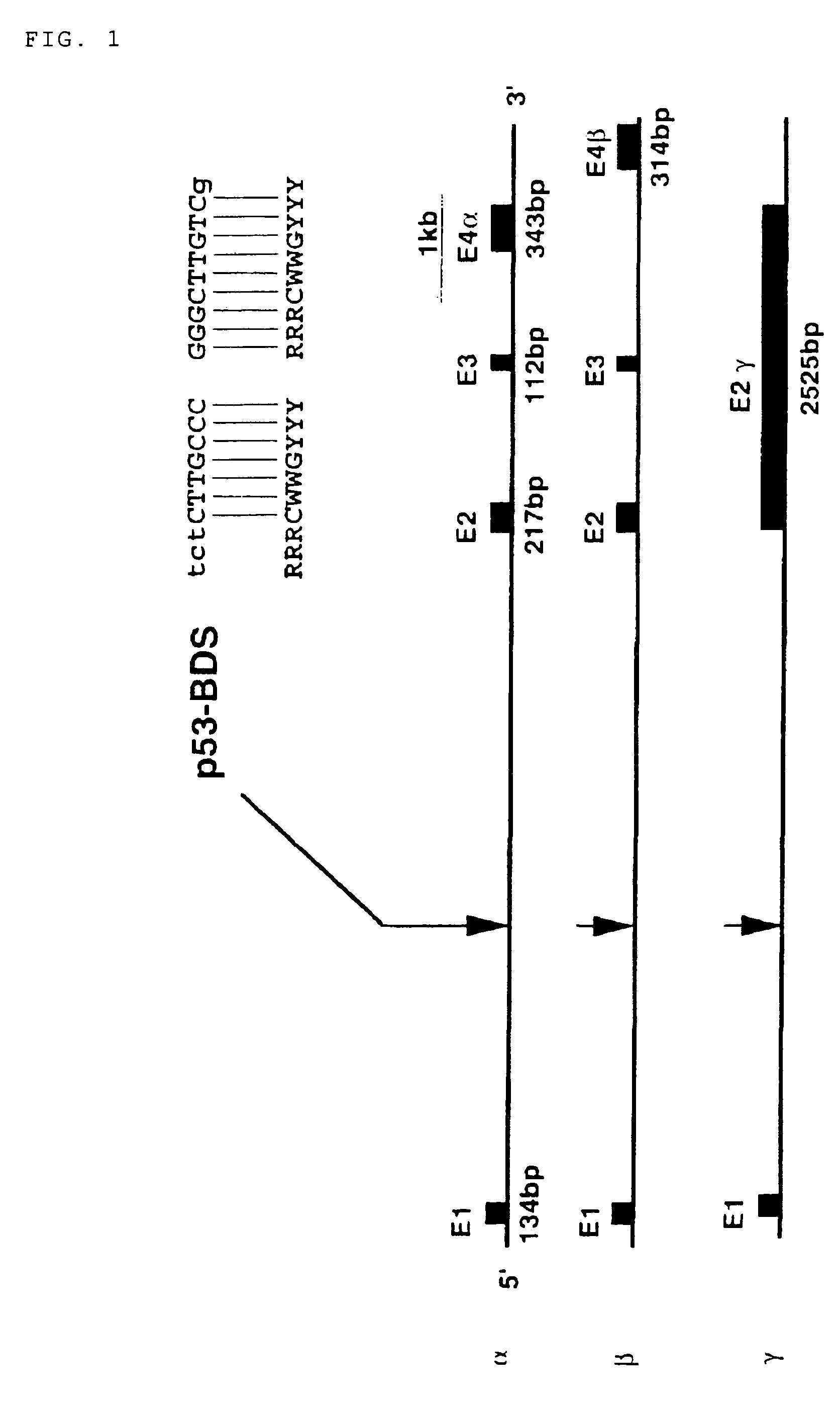
[0136]The present inventors isolated a number of human genomic sequences, whose transcription was presumably activated by p53, using a yeast enhancer trap system (p53-target site; Tokino et al. Hum. Mol. Genet. 3: 1537-1542, 1994). The inventors determined the nucleotide sequence of a 190-bp DNA fragment, corresponding to one of the above-described sequences (clone TP53-41 in Tokino et al., Hum. Mol. Genet. 3: 1537-1542, 1994), and found a putative p53-binding sequence (BDS) within the fragment (FIG. 1). To isolate a larger genomic region comprising the fragment, the 190-bp DNA fragment comprising a p53-target site was radiolabeled, and used as a probe for screening a human cosmid library consisting of 106 colonies. A cosmid designated as TP53-cos38 was isolated by the screening, and then subjected to FISH analysis. FISH analysis was performed using the cosmid clone TP53-cos38 as a probe according to the method described in Inazaw...
example 2
Isolation of the p53AIP1 Gene
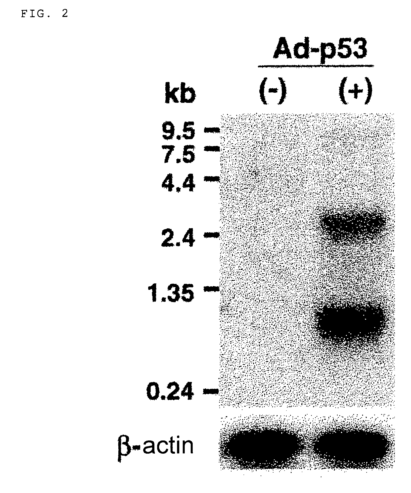
[0139]In order to isolate a full length cDNA of the above-described DNA fragment, glioma cells, U373MG, were infected for 48 hr with a replication-defective adenovirus expressing wild-type p53 (Ad-p53), and then poly(A)+RNA was isolated from resulting cells. Northern blotting analysis using the 474-bp cDNA fragment as a probe was performed as follows.
[0140]Poly(A)+RNA (3 μg) extracted from U373MG cells infected with Ad-p53 was separated by electrophoresis on a 1% agarose gel containing 1×MOPS and 2% formaldehyde, and transferred to a nylon membrane. The RNA blot was hybridized with a random-primed 32P-labeled p53AIP1 cDNA probe or β-actin in 5×SSPE / 10×Denhardt's / 2% SDS / 50% formamide at 50° C., washed in 0.1×SSC / 0.1% SDS at 65° C., and then subjected to autoradiography at −80° C.
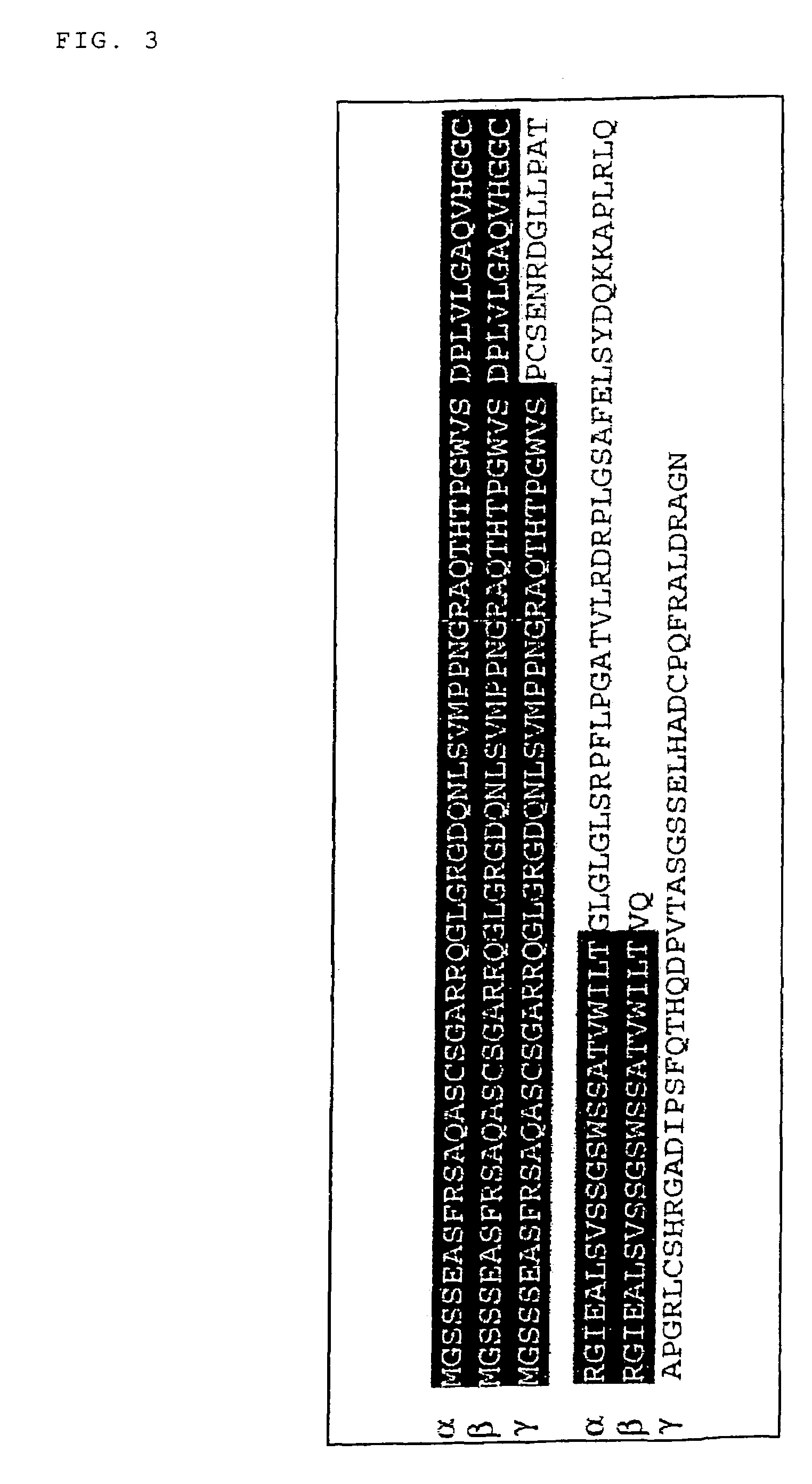
[0141]The analysis detected 0.8-kb and 2.7-kb transcripts whose transcription were strongly induced in U373MG cells by infection with Ad-p53 (FIG. 2). Northern blotting analysis u...
example 3
p53AIP1 as a Novel Target-Molecule for p53
[0143]In order to determine whether p53 was able to bind to oligonucleotides corresponding to the BDS sequence in intron 1 of the p53AIP1 gene, electrophoretic mobility shift assay (EMSA) was performed as described below. First, the following oligonucleotides were designed:
[0144]
(oligomer 1S, SEQ ID NO:9)5′-TCTCTTGCCCGGGCTTGTCG-3′;(oligomer 1A, SEQ ID NO:10)5′-CGACAAGCCCGGGCAAGAGA-3′.
and
[0145]Next, the oligomers 1S and 1A were annealed, and labeled with [γ-32P] DATP. H1299 lung carcinoma cells (obtained from ATCC) were infected-with Ad-p53. Nuclear extracts were prepared from these cells, and incubated with 0.5 μg of sonicated salmon sperm DNA, EMSA buffer (consisting of 0.5×TBE, 20 mM HEPES (pH 7.5), 0.1 M NaCl, 1.5 mM MgCl2, 10 mM dithiothreitol, 20% glycerol, 0.1% NP-40, 1 mM PMSF, 10 μg / ml pepstatin, and 10 μg / ml leupeptin), and the 32P-labeled double-stranded oligomers for 30 min at room temperature. In some cases, the nuclear extracts ...
PUM
 Abstract
Description
Claims
Application Information
 - R&D
- Intellectual Property
- Life Sciences
- Materials
- Tech Scout
- Unparalleled Data Quality
- Higher Quality Content
- 60% Fewer Hallucinations
Browse by: Latest US Patents, China's latest patents, Technical Efficacy Thesaurus, Application Domain, Technology Topic, Popular Technical Reports.
© 2025 PatSnap. All rights reserved.Legal|Privacy policy|Modern Slavery Act Transparency Statement|Sitemap|About US| Contact US: help@patsnap.com


