Electromagnetic sensors for biological tissue applications and methods for their use

a technology of electromagnetic sensors and biological tissues, applied in the field of electromagnetic sensors, can solve the problems of increasing the risk of extravasation, affecting the detection of fluid and other materials in biological tissues, and the needle being pushed through the wall of the vessel, so as to improve the signal coupling and the resulting measurement

Active Publication Date: 2009-09-22
BAYER HEALTHCARE LLC
View PDF72 Cites 274 Cited by
  • Summary
  • Abstract
  • Description
  • Claims
  • Application Information

AI Technical Summary

Benefits of technology

[0015]In accordance with one aspect of the present invention, a sensor element for injecting and/or receiving electromagnetic waves into body tissue comprises a housing with a substrate mounted within the housing. A superstrate is mounted to the substrate and has a base facing the substrate and an outer surface extending away from the substrate and having a transitional periphery interconnecting the base to an outer surface plateau extending over at least a central portion of the superstrate. A generally planar antenna ele

Problems solved by technology

Extravasation can be caused, for example, by rupture or dissection of fragile vasculature, valve disease, inappropriate needle placement, or patient movement resulting in the infusing needle being pulled from the intended vessel or causing the needle to be pushed through the wal

Method used


Image

  • Electromagnetic sensors for biological tissue applications and methods for their use
  • Electromagnetic sensors for biological tissue applications and methods for their use
  • Electromagnetic sensors for biological tissue applications and methods for their use

Examples


experimental examples

[0105]1. Inorganic Phantom Experiments.

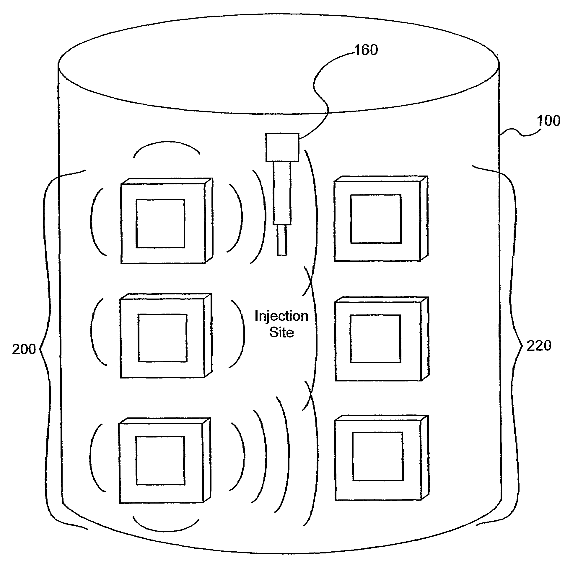
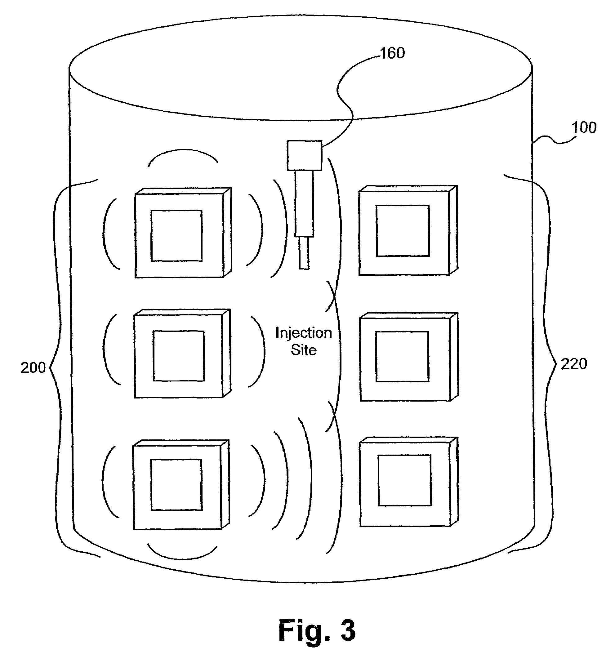
[0106]Several experiments demonstrating the use of the sensors of the present invention were carried out using single and dual microstrip patch antenna configurations and an inorganic phantom to represent human tissue. Such inorganic phantoms provide the opportunity to accurately control both the position and amount of a simulated extravasation in an environment of generally known and simple dielectric properties as compared to human tissue.

[0107]Below approximately 1 GHz, the wavelength becomes too large to provide adequate sensitivity to changes of interest in the tissue and approaching and, beyond 10 GHz, the penetration of the waves into the tissue becomes too small to provide adequate sensing depth. Thus, the antennae of the present invention that were used for this experiment were designed to resonate at an intermediate frequency of approximately 4 Ghz.

[0108]The microstrip antennae used in the studies leading to the present invention were...


PUM


Abstract

Tissue sensors house one or more sensor elements. Each element has a housing mounted substrate and a superstrate with a planar antenna between. A transitional periphery (TP) of a superstrate outer surface interconnects a base to a plateau. At least some of the TP has a generally smooth transition. Plural elements are spaced by the housing. Alternately, the superstrate TP is flat, the housing extends to the outer superstrate surface and a shield surrounds the element. The housing is flush with or recessed below the superstrate and defines a TP between the housing and superstrate. A method converts a reference signal to complex form; plots it in a complex plane as a reference point (RP); converts a measurement signal to complex form; plots it in the complex plane as a measurement point (MP); determine a complex distance between the MP and the RP; and compares complex distance to a threshold.

Description

CROSS-REFERENCE TO RELATED APPLICATIONS[0001]This application claims the benefit of U.S. Provisional Patent Application Ser. No. 60 / 308,012, filed Jul. 26, 2001, the disclosure of which is incorporated herein by reference. This application is related to an application (Express Mail Label No. EL886755793US) filed on even date herewith, entitled DETECTION OF FLUIDS IN TISSUE, also claiming priority to U.S. Provisional Patent Application Ser. No.60 / 308,012, filed Jul. 26, 2001, the disclosure of which is also incorporated herein by reference.BACKGROUND OF THE INVENTION[0002]The present invention relates in general to the detection of fluid and other materials in biological tissues and, more particularly, to electromagnetic sensors and methods of using those sensors for the detection of fluid and other materials in tissues. While the present invention is generally applicable to sensing of bodily or injected fluid levels or the presence of other foreign materials, such as tumors, in tiss...

Claims


Application Information

Patent Timeline
IPC IPC(8): A61B5/117A61B5/103A61B5/05A61B5/053A61M5/168A61N2/04G01N22/00H01Q1/27H01Q1/40H01Q9/04H01Q13/18H01Q21/06H01Q21/20
CPCA61B5/05H01Q21/20A61B5/0537A61B5/411A61B5/4878A61M5/16836G01N22/00H01Q1/273H01Q1/40H01Q9/0407H01Q9/0414H01Q13/18H01Q21/06H01Q21/065A61B5/0507A61B2562/02A61B2562/046A61B2562/143
Inventor BOUTON, CHAD EDWARD
Owner BAYER HEALTHCARE LLC
Why Eureka
Try Eureka