Detection of smooth muscle motor activity

Inactive Publication Date: 2009-09-22
MEDISENSE TECH INT +1
View PDF11 Cites 17 Cited by
  • Summary
  • Abstract
  • Description
  • Claims
  • Application Information

AI Technical Summary

Benefits of technology

[0008]The present invention provides a method for detecting smooth muscle motor activity. The invention is based on the observation that the repolarization time of action potentials in smooth muscle cells is several seconds which is comparable to the period of the slow waves. It was further found that, in accordance with

Problems solved by technology

Moreover, it was also found that overlapping action potentials reinforce one anothe

Method used


Image

  • Detection of smooth muscle motor activity
  • Detection of smooth muscle motor activity
  • Detection of smooth muscle motor activity

Examples


example 1

Validation of the Method and System of the Invention in Dogs

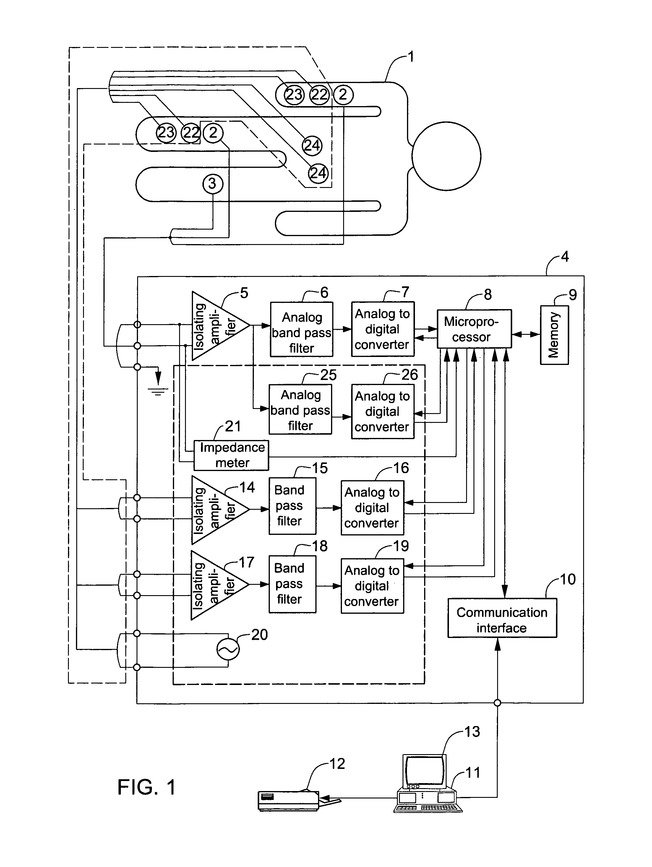
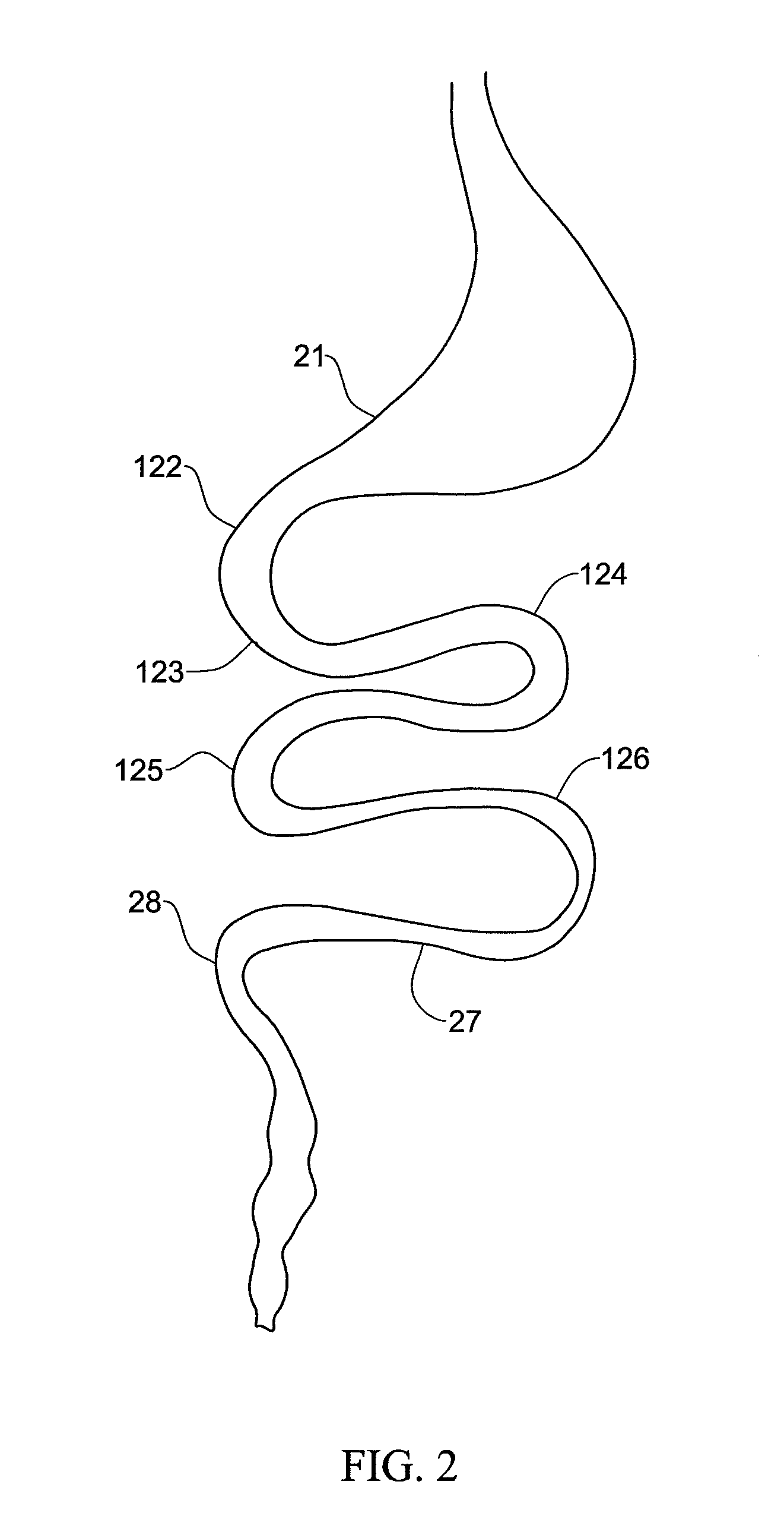
[0052]The method of the invention was validated in 6 healthy dogs by comparing the signal recorded by cutaneous electrodes in accordance with the invention with signals recorded simultaneously by electrodes implanted in various locations in the GI tract of the dogs. Cutaneous Ag—AgCl electrodes (Red Dot™, 3M™ Company, Canada) were used. A recording electrode was disposed on each one of the right limbs and a reference electrode was disposed on the left hind limb of each dog. The electrodes had a noise level of less than 1 mV below 0.04 Hz. In addition, 8 bipolar platinum electrodes (0.2 mm diameter, 2 mm separation) were implanted in the GI tract of the dogs. The locations of the implanted electrodes were as indicated in FIG. 2. Location 21 is the stomach antrum, 122 is the proximal part of the duodenum, 123 is the distal part of duodenum, 124, 125, 126, and 27 are different parts of jejunum, and 28 is the ileum.

[0053]The am...

example 2

Recording the Migrating Myoelectrical Complex in Dogs

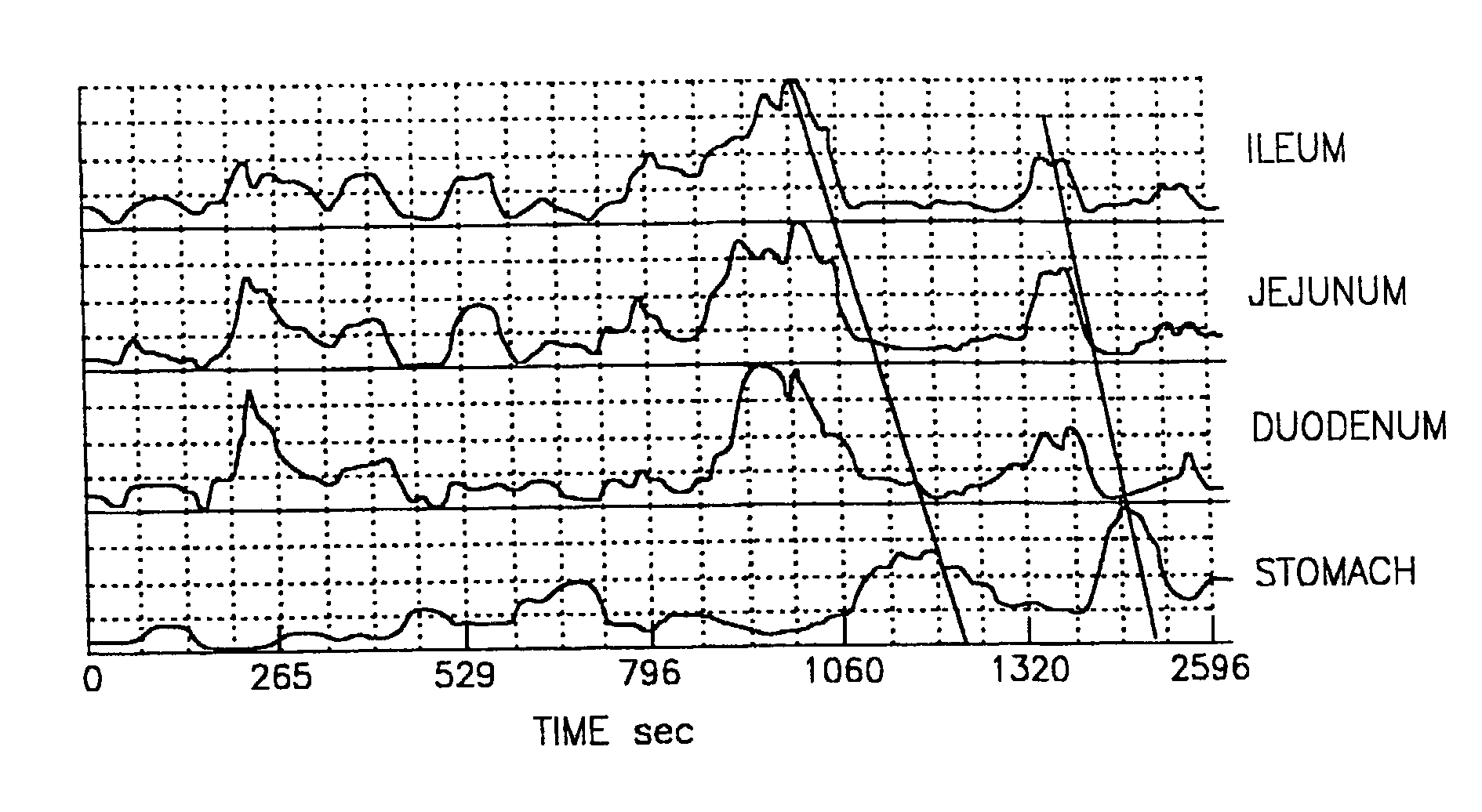
[0059]The system and method of Example 1 were used to detect the migrating myoelctrical complex (MMEC) in dogs. FIG. 5, panels a to f, show the action potentials recorded simultaneously by electrodes implanted in the GI tract of a dog over time. Bursts of action potentials corresponding to muscle contraction are indicated in FIG. 5, panels a-f by brackets 51 to 56, respectively. Transient muscle contraction was detected in the stomach (bracket 51 in FIG. 5a). The region of contraction then migrated to the duodenum (bracket 52 in FIG. 5b), then along the jejunum (brackets 53, 54, 55 in FIGS. 5c-e). In the ileum, bracket 56 indicates the muscle contraction of the previous wave (FIG. 5f). FIG. 5, panels g-j, show simultaneous measurements of the energy E obtained by processing the signals recorded by the cutaneous electrodes. In accordance with the invention, elevated E indicates increased muscle contraction in the organ, and this is...

example 3

Validation of the Method and System of the Invention in Human Subjects

[0060]Cutaneous electrodes were disposed on human subjects as shown in FIG. 1. The cutaneous electrodes used as well as the processing of the signal recorded by these electrodes were as described in Example 1. Evaluation of GI tract motor function by measurement of bowel pressure was simultaneously performed using an open catheter inserted into the jejunum during surgery. FIG. 6a shows the total signal recorded by the cutaneous electrodes over a period of about 40 min, and FIG. 6b shows the simultaneous pressure measurements recorded by the catheter. At the time indicated by the vertical line 51, 0.2 mg / kg body weight prozerin was administered to the subject. This treatment induced a transient increase in the amplitude of the slow waves during the time period 52. Simultaneously, pressure waves indicative of smooth muscle contraction were detected by the catheter (FIG. 6b).

[0061]Power spectra of the signal from the...


PUM


Abstract

A system and method for determining smooth muscle motor activity in body organs. The system comprises electrodes for recording an analog signal in the subject's body, an analog to digital converter that converts the analog signal to a digital signal and a processor. Processing the digital signal includes obtaining power spectra of the digital signal, and identifying one or more frequency peaks in the power spectra. A peak in the power spectrum is identified within a frequency range in which an organ generates slow waves. The energy of the identified frequency peak is measured and an energy above a characteristic threshold is indicative of smooth muscle motor muscle motor activity in the respective organ.

Description

[0001]This application is the national phase under 35 U.S.C. § 371 of PCT International Application No. PCT / IL00 / 00215 which has an International filing date of Apr. 11, 2000, which designated the United States of America and was published in English.FIELD OF THE INVENTION[0002]The present invention is in the field of medical devices. More specifically, the invention relates to devices for examining smooth muscle organs.BACKGROUND OF THE INVENTION[0003]The major smooth muscle organs of the body include the gastrointestinal (GI) tract organs, the urinary bladder, and the uterus. The motor activity associated with these organs is essential in many physiological processes such as digestion, excretion and parturition. Many pathologies arising from impaired smooth muscle motor activity are known. In the GI tract, for example, smooth muscle dysfunction is a common pathology often making life for those suffering from it uncomfortable and painful.[0004]The electrical activity of smooth musc...

Claims


Application Information

Patent Timeline
IPC IPC(8): A61B5/04A61B5/02A61B5/0408A61B5/0488
CPCA61B5/04884A61B5/392
Inventor VASILIEV, VLADISLAVAZAROV, YOKOVNOTOV, OLGA
Owner MEDISENSE TECH INT
Why Eureka
Try Eureka