Eureka AIR delivers breakthrough ideas for toughest innovation challenges, trusted by R&D personnel around the world.

Recombinant modified adenovirus fiber protein

a technology of adenovirus and fiber protein, which is applied in the field of recombinant adenovirus having a modified fiber protein, can solve the problems of limiting the exploitation of this strategy, reducing yield, and hampered flexibility of this “fiber swapping” approach, so as to expand the functionality of the adenovirus system, and the wild-type tropism of the adenovirus

Inactive Publication Date: 2009-11-03
IST DI RICERCHE DI BIOLOGIA MOLECOLARE P ANGELETTI +1
View PDF20 Cites 4 Cited by
  • Summary
  • Abstract
  • Description
  • Claims
  • Application Information

AI Technical Summary

Benefits of technology

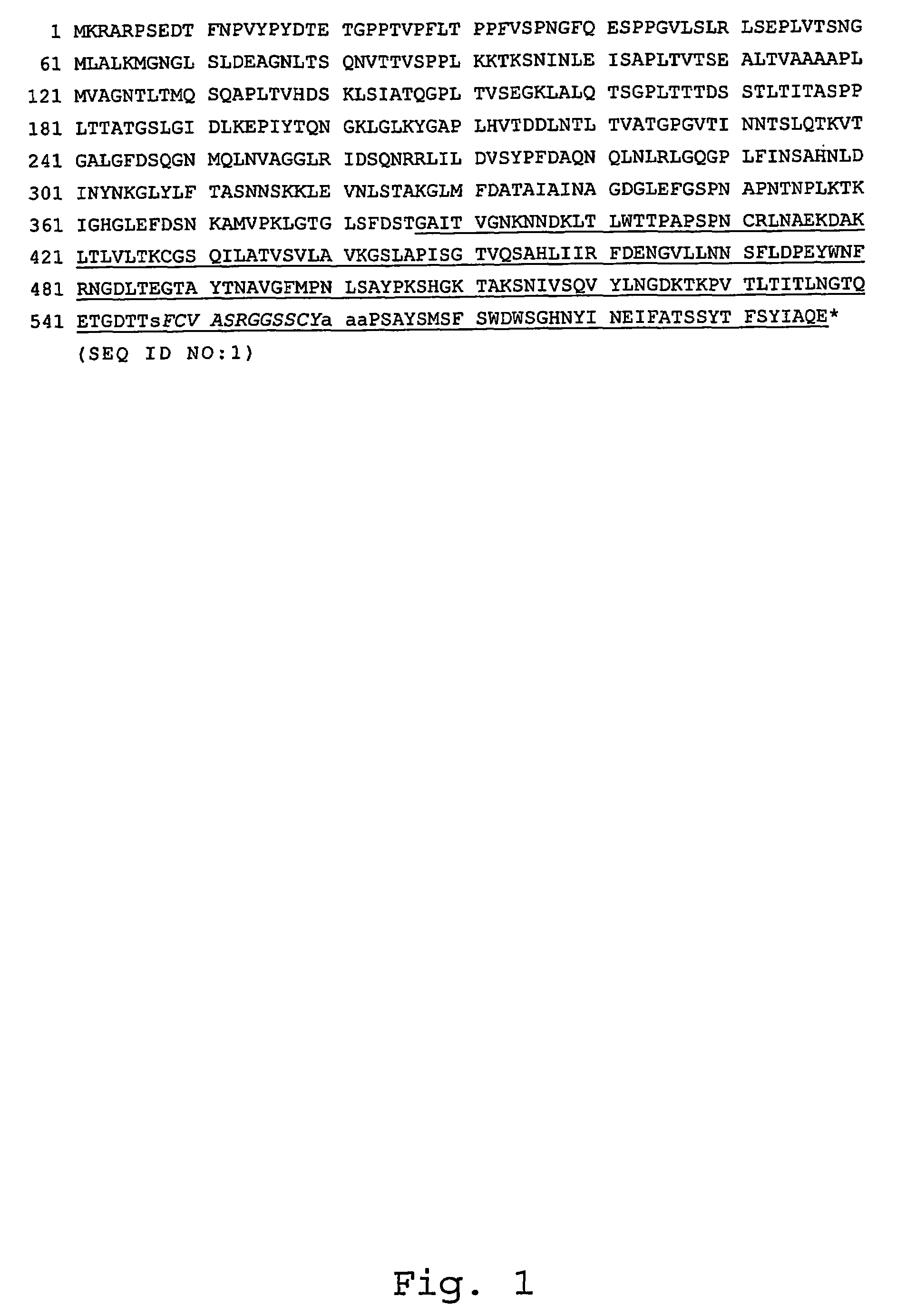
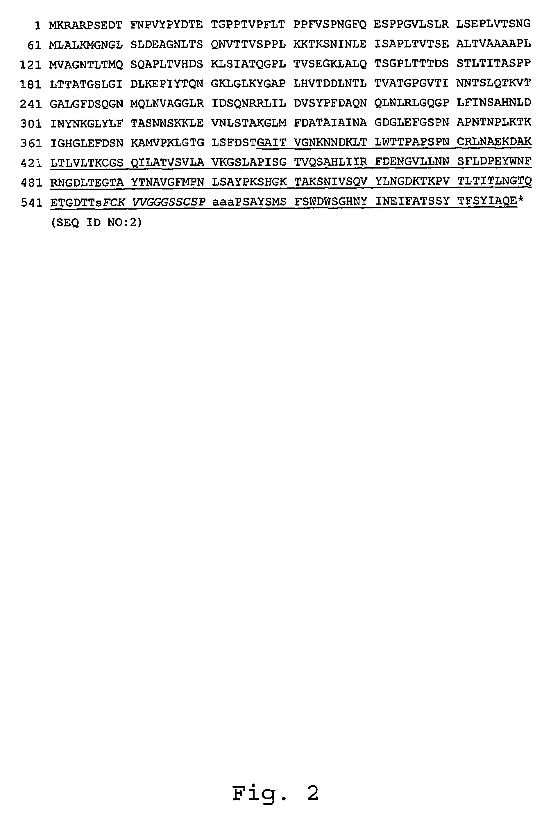
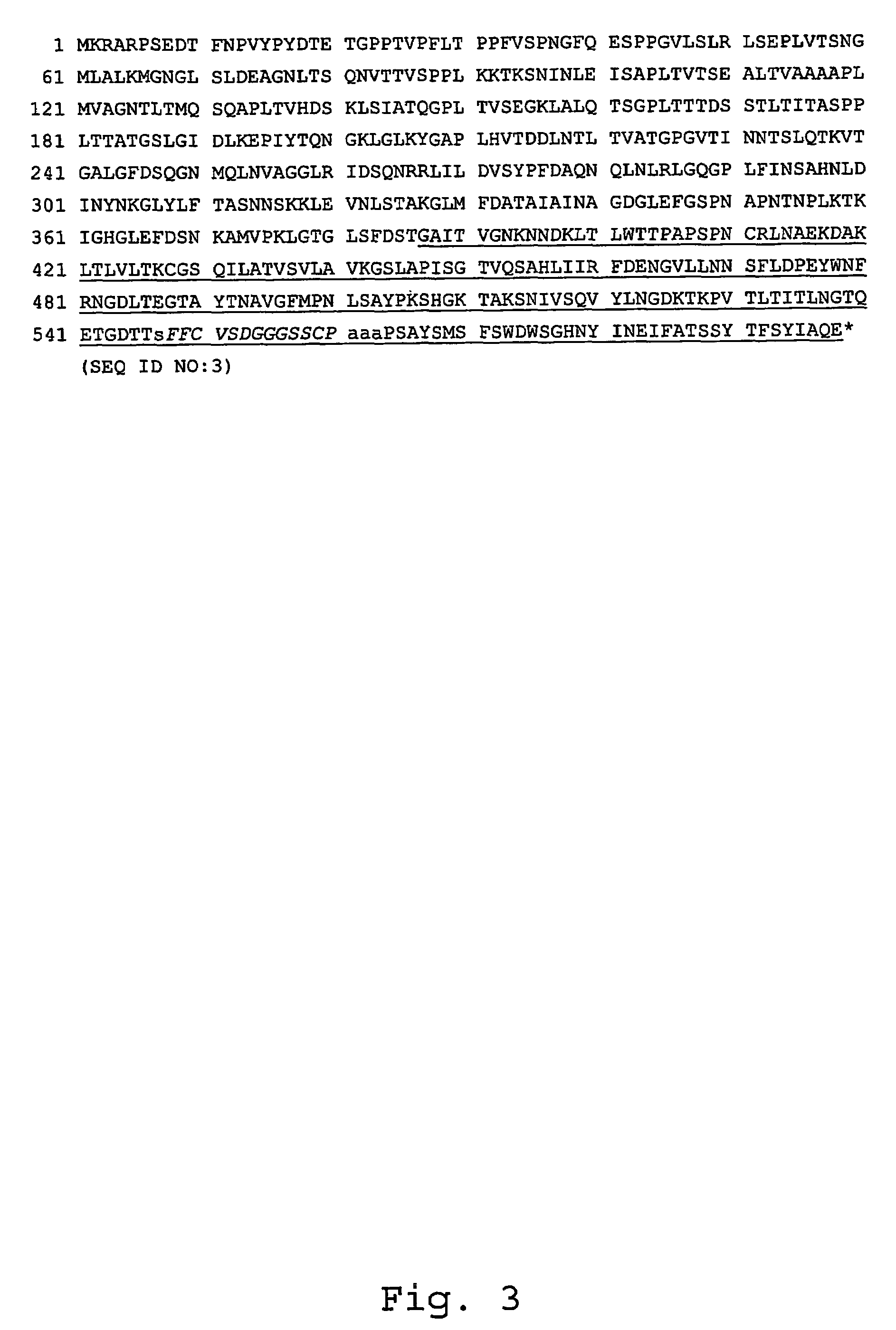
[0013]The present invention relates to a recombinant adenovirus having a modified fiber protein which comprises a peptide ligand for a cell surface binding site other than CAR. The peptide ligand comprises a core amino acid sequence of 14 residue positions such that residue positions one, two and thirteen are Xaa residues, preferably tyrosine (Tyr), tryptophan (Trp), or phenylalanine (Phe); residue positions three and twelve are cysteine (Cys) residues; residue positions four through eleven are Xaa residues, preferably glycine (Gly), serine (Ser), or valine (Val); and, residue position fourteen is an Xaa residue, preferably proline (Pro). The core amino acid sequence may be deleted by one or more residues at any residue position within the sequence, except at the fixed Cys residues at position three and position twelve. It is preferable that the region of the core amino acid sequence located between the fixed Cys residues comprises at least two amino acid residues. Additionally, up to 10 amino acid residues may be added between residue position three and residue position twelve. The amino acid sequence of the peptide ligand does not comprise a consecutive Arg-Gly-Asp (RGD) sequence between residue positions three and twelve, and the peptide ligand does not consist of the amino acid sequence as disclosed in SEQ ID NO:46. A recombinant adenovirus having a modified fiber protein with the ability to bind to a cell surface binding site other than CAR has an effect of broadening the wild-type tropism of the adenovirus, expanding the functionality of the adenovirus system for gene therapy.
[0053]The present invention also relates to methods of transducing a cell and / or tissue with a recombinant adenovirus described in this specification. By using a recombinant adenovirus as described herein to transduce cells / tissues, both the ability and efficiency of adenovirus infectivity increases. In one embodiment, these methods comprise transducing cells that have little or no expression of CAR and are refractory to Ad5 infection with a recombinant adenovirus of the present invention. Cells and / or tissues that may be transduced with a recombinant adenovirus of the present invention include, but are not limited to, synoviocytes, smooth muscle cells, endothelial cells, cancer cells, primary tumors, dendritic cells, skeletal muscle, melanocytes and murine melanoma cells. Another embodiment includes a method of transducing immature dendritic cells, including but not limited to those of mouse or human origin, as well as murine skeletal muscle and human primary melanocytes, with a recombinant adenovirus of the present invention.

Problems solved by technology

However, the flexibility of this “fiber swapping” approach is hampered by the number of serotypes available and their limited tropism.
In addition, impaired viability and reduced yield of the viral chimeras have limited the exploitation of this strategy.
The fact that ligands selected from phage libraries often do not retain their binding properties when grafted into a different protein location, i.e. the HI loop of the Ad5 fiber knob, has resulted in a paucity of successful reports using this strategy.
Furthermore, insertion of these peptides can affect fiber trimerization and virus assembly.

Method used


Image

  • Recombinant modified adenovirus fiber protein
  • Recombinant modified adenovirus fiber protein
  • Recombinant modified adenovirus fiber protein

Examples


example 1

Display of Ad5 Trimeric Knob Domain on the Capsid of Phage λ

[0168]Cells—E. coli strains BB4 and Y1090 were used for phage plating and amplification.

[0169]Construction of plasmid pDknob—Plasmid pNS3785 (Sternberg and Hoess, 1995, Proc. Natl. Acad. Sci. USA 92:1609-1613) was transformed into pDknob by modifying the 3′-end of the λD gene as indicated in FIG. 14.

[0170]An XbaI restriction site was introduced into plasmid pNS3785 to allow cloning of the entire plasmid into the unique XbaI site of lambda Dam15imm21nin5. Plasmid pNS3785 was amplified by inverse PCR using primers XbaI-NS.for (5′-TTTATCTAGACCCAGCCCTAGGAAGCTTCTCCTGAGTAGGACAAATCC-3′; SEQ ID NO:20) and XbaI-NS.rev (5′-GGGTCTAGATAAAACGAAAGGCCCAGTCTTTC-3′; SEQ ID NO:21) (XbaI site underlined). Reaction was performed using a mixture of Taq and Pfu DNA polymerases to increase fidelity of DNA synthesis (95° C.-30 sec, 55° C.-30 sec, 72° C.-20 min, for 25 amplification cycles). The PCR amplification product thus generated was digested...

example 2

λ-borne Ad5 Knob Specifically Binds Human CAR on 911 Cells

[0185]Cells—Human embryonic retinoblast 911 cells were obtained from Invitrogen (Rijswijk, The Netherlands). 911 cells were cultured in Dulbecco's modified Eagle's medium (DMEM) supplemented with 10% (v / v) fetal calf serum (FCS).

[0186]Viruses—The recombinant adenoviruses were propagated on Per.C6 cells and purified by centrifugation in CsCl gradients according to standard protocols (Fallaux et al, 1998, supra).

[0187]FACS Analysis—Binding of phage λknob-wt to human CAR displayed on the surface of 911 cells was monitored as follows. Phage particles (ranging from 0.4×10¹⁰ to 6×10¹⁰ pfu) were incubated with a suspension of 2×10⁶ cells / ml for 1 hour at room temperature (RT) in binding buffer composed of 3% bovine serum albumin (BSA), 10 mM MgCl2, 1 mM CaCl2 in PBS. Cells were washed with binding buffer and incubated for 45 min at 4° C. with rabbit anti-λ purified Ig in FACS buffer (2% FBS in PBS). Cells were then washed with FACS ...

example 3

Generating a Library of Modified Ad5 Fiber Knobs Displayed on λ

[0190]Viruses—The recombinant adenoviruses were propagated on Per.C6 cells and purified by centrifugation in CsCl gradients according to standard protocols (Fallaux et al, 1998, supra).

[0191]Construction of the λknobΔ-14aa.cys library—Plasmid pDknobΔ-L0 was generated by replacing the BglII / MscI fragment in pD-knobΔ with a PCR amplification product introducing unique SpeI and NotI sites in the HI loop. Two 326 bp and 93 bp DNA fragments were obtained by PCR-amplifying the plasmid pDknob using the primers knob.for2 (5′-AGGCAGTTTGGCTCCAATATCTG-3′; SEQ ID NO:35) and knobHI-Spe / Not.rev (5′-GCGGCCGCACCACTAGTTGTGTCTCCTGTTTCCTGTGTA-3′; SEQ ID NO:36), or knobHI-Spe / Not.for (5′-ACTAGTGGTGCGGCCGCTCCAAGTGCATACTCTATGTCATTT-3′; SEQ ID NO:37) and knob.rev3 (5′-GGATGTGGCAAATATTTCATTAAT-3′; SEQ ID NO:38). These DNA fragments were PCR-ligated. The resulting plasmid pDknobΔ-L0 was linearized by XbaI digestion and the entire plasmid was clo...


PUM


Abstract

Recombinant adenoviruses comprising modified fiber proteins which expand the tropism of the adenovirus in comparison to wild-type virus are disclosed. The modified fiber proteins described herein contain a peptide ligand for a cell surface binding site other than CAR comprising a 14 amino acid core sequence containing both fixed and variable amino acid residues. The invention includes isolated nucleic acid molecules encoding the modified adenovirus fiber proteins disclosed, as well as recombinant vectors and host cells containing said nucleic acid molecules. Methods of identifying peptide ligands that bind to cell binding sites other than CAR are included comprising screening a phage-display library of peptide ligands expressed within an adenovirus fiber knob context on CAR-negative cells. Recombinant adenoviruses of the present invention will increase the ability of an adenovirus to transduce important cell and tissue targets as part of a gene therapy / gene vaccination regime that have been shown to be refractory to adenoviral infection.

Description

CROSS-REFERENCE TO RELATED APPLICATIONS[0001]The present application is the §371 National Stage application of PCT International Application serial no. PCT / EP2004 / 004999, having an international filing date of May 10, 2004, which claims priority to U.S. Provisional Application Ser. No. 60 / 470,562, filed May 14, 2003, now expired.FIELD OF THE INVENTION[0002]The present invention relates generally to a recombinant adenovirus having a modified fiber protein that broadens the tropism of the adenovirus in comparison to wild-type virus. The modified fiber protein contains a peptide ligand for a cell surface binding site other than the coxsackievirus and adenovirus receptor (CAR) comprising a 14 amino acid core sequence containing both fixed and variable amino acid residues. The peptide ligand is inserted within the adenovirus fiber protein sequence such that it is accessible to the alternative cell surface binding site, and yet fiber knob trimerization is preserved. The present invention ...

Claims


Application Information

Patent Timeline
Patent Type & Authority Patents(United States)
IPC IPC(8): C07K14/75A61K38/00A61K48/00C12N15/861C07K14/005C07K14/075
CPCC07K14/005C12N15/86A61K48/00C07K2319/00C07K2319/735C12N2840/20C12N2710/10343C12N2710/10345C12N2810/40C12N2810/80C12N2710/10322
Inventor MONACI, PAOLOFONTANA, LAURA
Owner IST DI RICERCHE DI BIOLOGIA MOLECOLARE P ANGELETTI
Why Eureka
Eureka Blog
Learn More