Methods for treating rheumatoid arthritis using a CTLA-4 fusion protein
a fusion protein and rheumatoid arthritis technology, applied in the direction of peptide/protein ingredients, immunological disorders, drug compositions, etc., can solve the problem of limiting in vivo utility, achieve high cell or tissue specificity, minimize or avoid systemic side effects, and limit in vivo utility
- Summary
- Abstract
- Description
- Claims
- Application Information
AI Technical Summary
Benefits of technology
Problems solved by technology
Method used
Image
Examples
example 1
Production of Expression Vectors
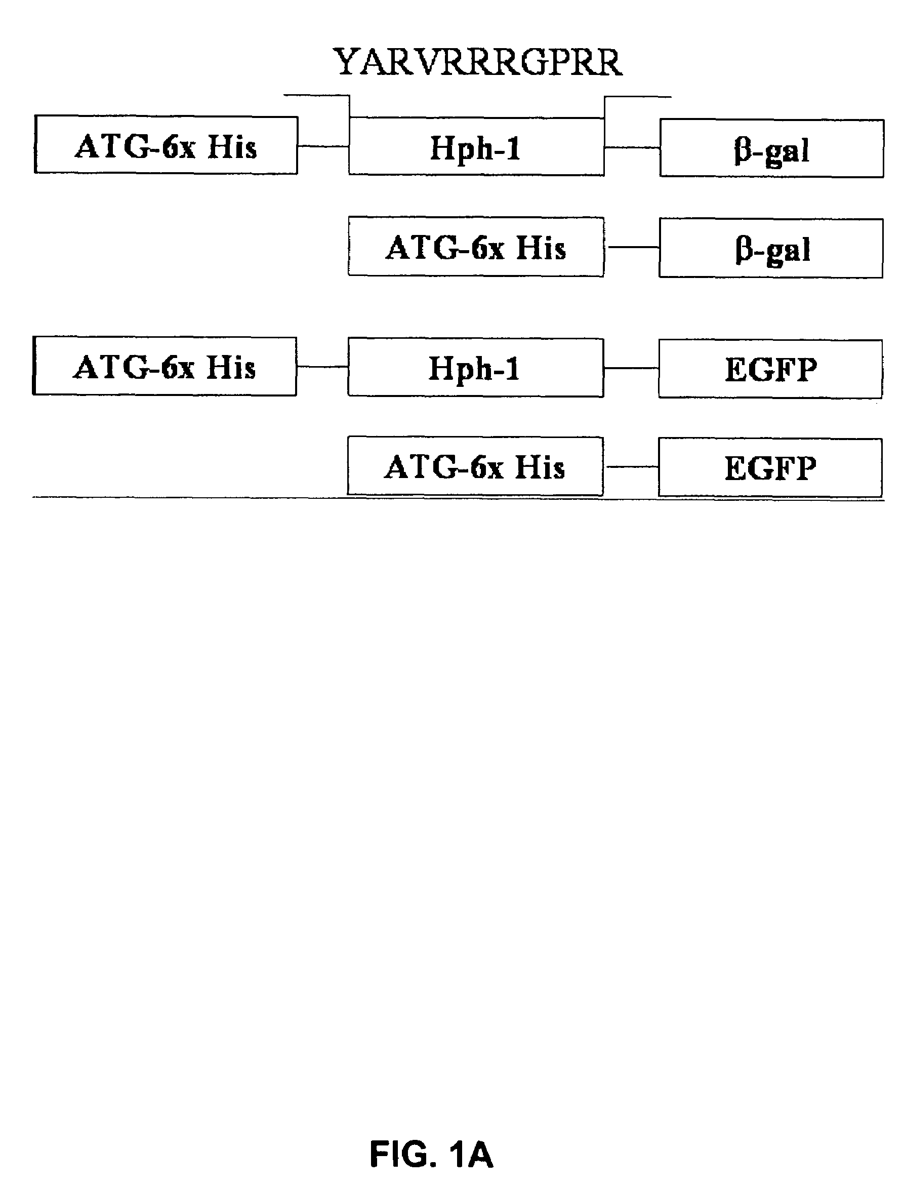
[0123]Based on common structural and functional features between TAT and uptake-targeting sequences which direct proteins from the cytosol to the intracellular organelles, an Hph-1 protein transduction domain (PTD) of 11 amino acids (YARVRRRGPRR) (SEQ ID NO:1) was identified from human transcriptional factor Hph-1. The terms “Hph-1-PTD” and “Hph-1” refer to this 11 amino acid sequence and are interchangeable.
[0124]Generation of Hph-1-PTD-ctCTLA-4, Hph-1-PTD-β-gal, Hph-1-PTD-ctCTLA-4-eGFP, and the corresponding genes not fused with Hph-1-PTD, was accomplished by generating PCR fragments and inserting each PCR fragment into the pRSET-B vector (Invitrogen) (FIG. 1A).
example 2
Preparation of the Recombinant Proteins
[0125]Site-directed mutagenesis was conducted to create the CTLA-4YF mutant. The fidelity of the reading frame was verified by sequencing. BL21 star (Novagen) transformed with each DNA construct was induced for 5 hours with 1 mM IPTG and sonicated in lysis buffer (6 M urea, 20 mM Tris-HCl, pH 8.0 and 500 mM NaCl). Lysates were clarified by centrifugation (20,000 r.p.m. for 30 min at 20° C.), adjusted to 10 mM imidazole (Tris-HCl, pH 8.0) and loaded on HisTrap chelating columns (Qiagen). Bound proteins were eluted with 50, 100, 250 mM or 3M imidazole in Tris-HCl buffer with >95% purity. The recombinant proteins contained in each fraction were desalted on PD-10 Sephadex G-25 (Amersham Pharmacia). The purified proteins were supplemented with 10% glycerol, aliquoted and flash-frozen at −80° C. (Becker-Hapak M., et al., Methods 24:247-256 (2001)).
example 3
Analysis of the In Vitro and In Vivo Transduction Efficiency of Hph-1-PTD
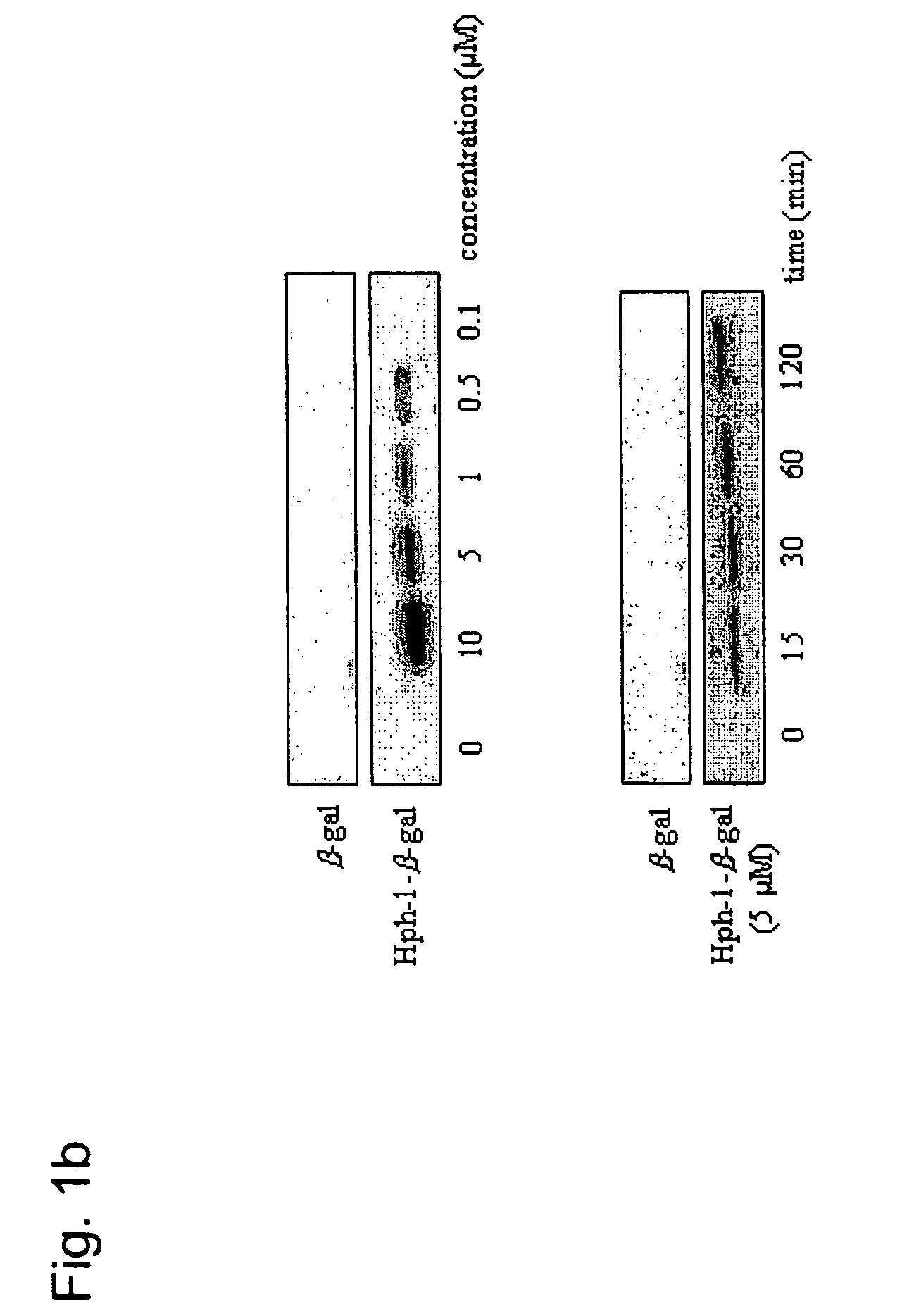
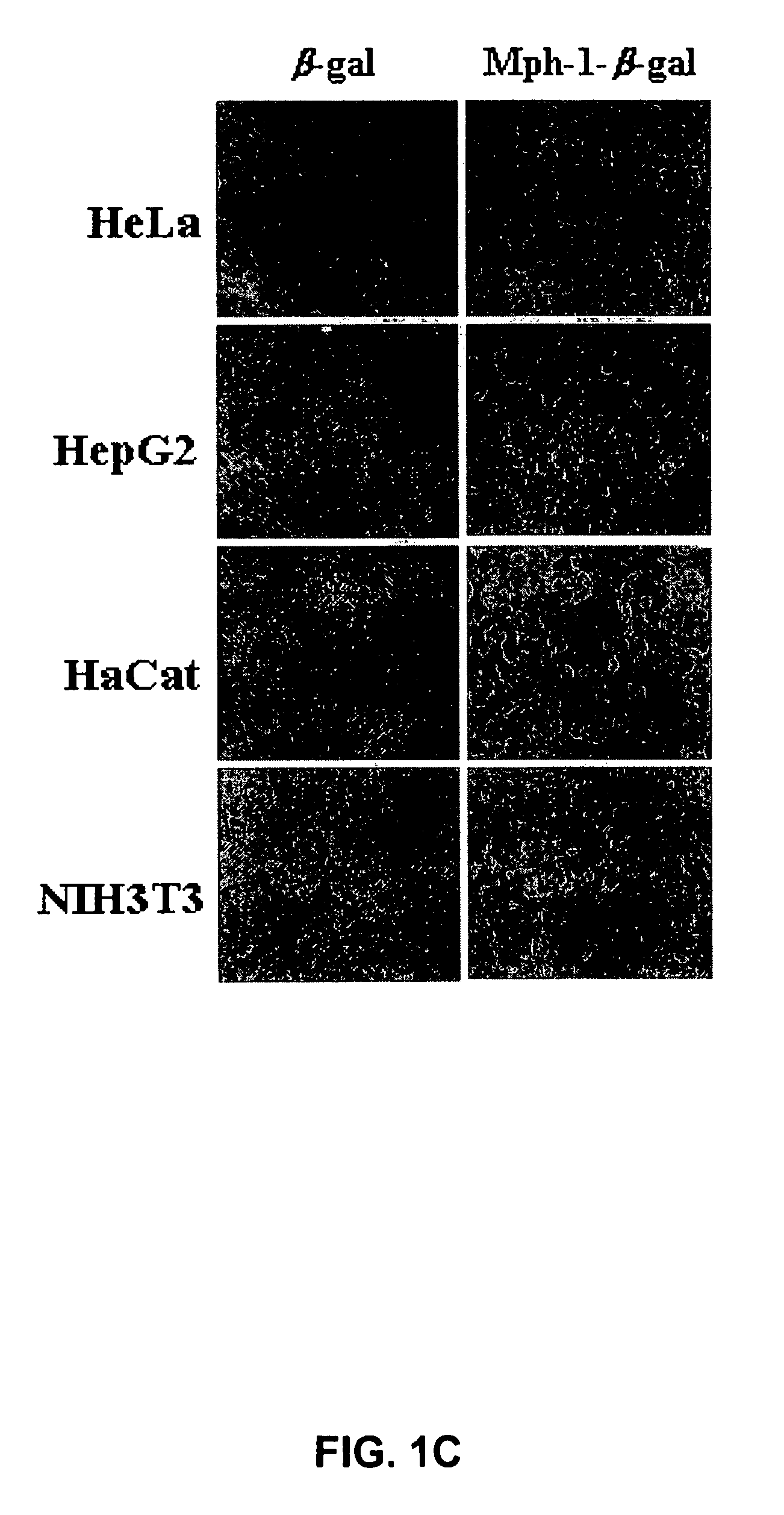
[0126]After the Hph-1-β-gal construct was transduced into each cell line for 1 hour, the cells were washed with PBS and fixed immediately. Then, the fixed cells were incubated with X-Gal solution for 1-3 hours at 37° C. The mice tissue samples from the liver, kidney, heart, lung, spleen and brain were isolated at 12 hours after i.p. injection of Hph-1-β-gal, sectioned (in 10- and 50-μm sections), and assayed using an X-Gal staining kit (Roche) (Schwarze S. R., et al., Science 285:1569-1572 (1999)). The image was captured by fluorescence microscopy (Nikon). Transduction capacity of Hph-1-β-gal was also analyzed by western blot analysis. Equal numbers of cells were solubilized in SDS-PAGE sample buffer and the proteins were resolved on a 12% gel, and blotted onto PVDF, which was probed with monoclonal antibody (1:1,000) to β-gal (Cell Signaling). Bound antibody was detected using horseradish peroxidase-conjugated...
PUM
| Property | Measurement | Unit |
|---|---|---|
| humidity | aaaaa | aaaaa |
| total volume | aaaaa | aaaaa |
| pH | aaaaa | aaaaa |
Abstract
Description
Claims
Application Information
 - R&D
- Intellectual Property
- Life Sciences
- Materials
- Tech Scout
- Unparalleled Data Quality
- Higher Quality Content
- 60% Fewer Hallucinations
Browse by: Latest US Patents, China's latest patents, Technical Efficacy Thesaurus, Application Domain, Technology Topic, Popular Technical Reports.
© 2025 PatSnap. All rights reserved.Legal|Privacy policy|Modern Slavery Act Transparency Statement|Sitemap|About US| Contact US: help@patsnap.com


