Antibody against uPA/uPAR

an antibody and upa technology, applied in the field of upa/upar antibodies, can solve the problems of destructive and invasive phenotype, high levels of released su-par, and poor prognosis

Inactive Publication Date: 2010-06-15
OSPEDALE SAN RAFFAELE SRL
View PDF6 Cites 0 Cited by
  • Summary
  • Abstract
  • Description
  • Claims
  • Application Information

AI Technical Summary

Problems solved by technology

Cell recruitment in itself, on the other hand, may cause destructive pathologies in which autologous cells attack and destroy cells and tissues, as in some auto-immune diseases.
Indeed, non-malignant stromal host cells do actively participate in establishing the malignant and hence the destructive and invasive phenotype.
In most cases high levels of released su-PAR can be related to poor prognosis (Stephens, 1999).
However, the presence of the chemotactic epitope in these fragments cannot be evaluated except by complex methods involving protein purification and sequencing.

Method used


Image

  • Antibody against uPA/uPAR
  • Antibody against uPA/uPAR
  • Antibody against uPA/uPAR

Examples


example 1

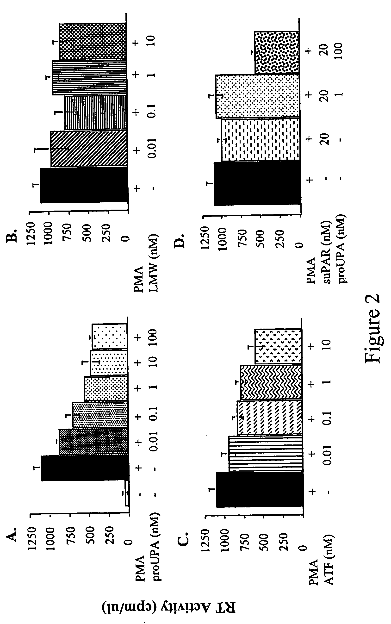
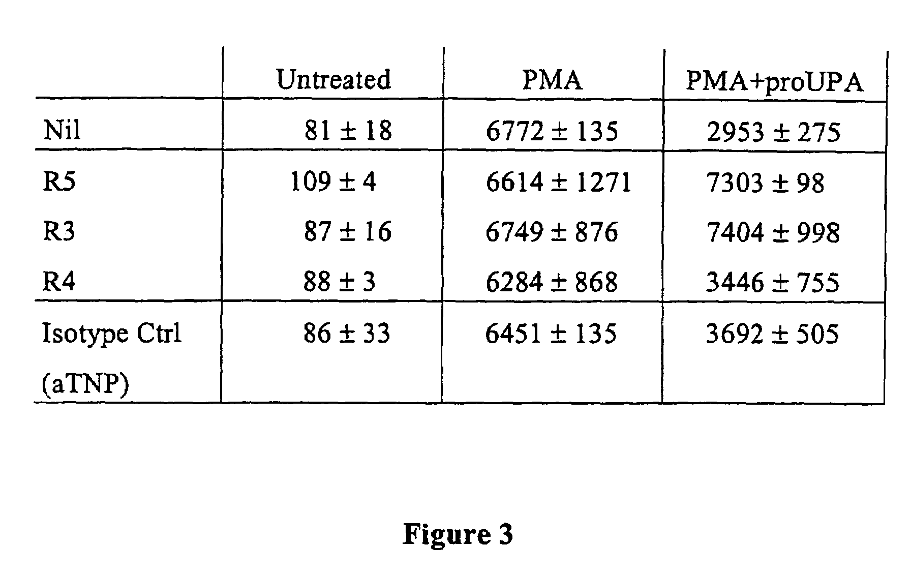
Pro-urokinase (pro-uPA) and its derivatives uPA and ATF, inhibit the replication of HIV-1 in latently infected monocytoid cell lines and in freshly infected human blood cells.

[0053]Latent HIV infected cells provide a cellular population resistant to anti-retroviral therapy and represent the mechanism for lifelong persistence of HIV (Finzi et al., 1999). The promonocytic U937 cell line is one of the most utilized models for studies on in vitro HIV infection. It displays both CD4 and CXCR4 and, therefore, can be infected by X4 viruses. From this cell line has been derived the chronically infected cell line named “U1”, containing two copies of integrated proviruses, and broadly used as a model of viral latency and reactivation (Folks et al., 1988). The U1 promonocytic cell line is one of the most thoroughly characterized models of post-integration latency. It was obtained from a population of U937 cells surviving the cytopathic effect of acute infection by HIV-1LAI / IIIB (X4), and conta...

example 2

Chemotaxis induced by uPA and “activated” uPAR fragments is mediated by the chemokine receptor FPRL1 / LXA4R.

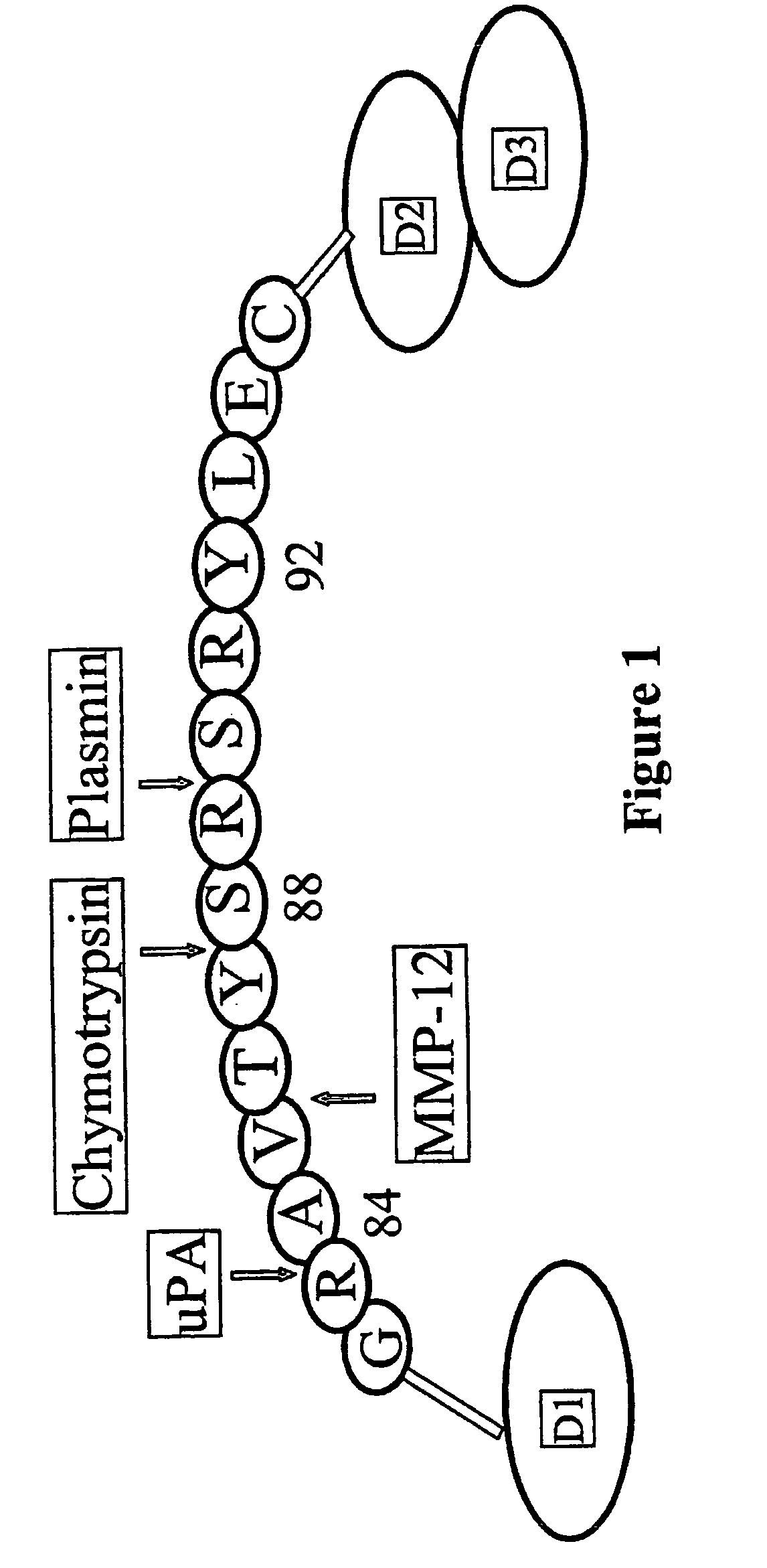
[0078]UPA and uPAR induce and are required for the chemotaxis of monocytes, THP-1 leukemic cells and several other cells. The derivative of uPA, ATF (amino terminal fragment), or the pro-enzyme pro-uPA have the same effect. In addition, the activated forms of uPAR (cleaved after residue 84) also activate chemotaxis. For all these stimulants, and in all cells tested, the mechanism remains the same (Degryse, 1999; Degryse, 2001; Fazioli, 1994; Resnati, 1996). In particular, these agents eventually rely on the activation of a trans-membrane mediator. After binding to uPAR, uPA cleaves uPAR or modifies its conformation (“activation”) to make it cleaved by between domain D1 and D2. The data show that the so-“activated” uPAR must interact with trans-membrane mediator that in turn transduces the migratory signals of pro-uPA / uPAR (Fazioli, 1994) (Blasi, 1997). This is due to the fact t...

example 3

Inhibition of uPA / ATF / uPAR chemotaxis by anti-FPLR1 / LXA4R reagents.

[0103]The specific response of FPRL1 / LXA4R containing cells to D2D384-278 (FIG. 10) strongly suggests that the G-protein-coupled FPRL1 / LXA4R receptor is the mediator of uPA and “activated” uPAR-induced chemotaxis. To substantiate this hypothesis, we have used cells that contain several chemokine receptors, including FPRL1 / LXA4R, and have tested whether specific antibodies to FPRL1 / LXA4R inhibit ATF or D2D384-278 induced chemotaxis.

Materials and Methods

[0104]Materials

[0105]The anti-LXA4R / FPRL1 antibodies (Fiore, 1995) were a generous gift of Dr. Mario Romano (Chieti, Italy) and Clarles Serhan (Boston, Mass., USA).

[0106]Cells

[0107]THP-1 cells have been described before (Resnati, 1996). Monocytes were obtained by two rounds of centrifugation over Ficoll (1.077 g / l) and Percoll (Pharmacia Biotech.) gradients from peripheral blood of healthy volunteers. Briefly, heparinized blood was diluted 1:1 with PBS and 3 ml of Ficol...


PUM

PropertyMeasurementUnit
concentrationsaaaaaaaaaa

Abstract

The invention herein disclosed relates to the anti-HIV action of uPAR-activators or activated uPAR molecules, the control of chemotaxis and cell migration by agents interfering with uPAR activation, a method for determining activated uPAR forms, the use of agents interfering with uPA/uPAR interaction for the diagnosis or therapy of diseases.

Description

CROSS REFERENCE TO RELATED APPLICATION[0001]This application is a 35 USC §371 National Phase Entry Application from PCT / EP02 / 00603, filed Jan. 22, 2002, and designating the U.S., which claims the benefit under 35 USC §119(e) of U.S. Provisional Application 60 / 263,198, filed Jan. 25, 2001.[0002]The present invention provides methods and agents useful for the modulation of uPA / uPAR function, which can be employed in the diagnosis or therapy of several pathologies, including HIV infection, inflammation and cancer.INTRODUCTIONCell Migration and Disease[0003]The human body responds to pathogenic stimuli by locally producing specific inflammatory molecules and recruiting specialized cells from blood and nearby tissues. This occurs in almost all diseases, and contributes in most cases to the general defenses of the organism. However, sometimes the inflammatory reaction, or the lack thereof, also contribute to the disease itself. Neutrophils, monocyte-macrophages, lymphocytes, endothelial c...

Claims


Application Information

Patent Timeline
Patent Type & Authority Patents(United States)
IPC IPC(8): C07K16/00A61K38/49A61K39/395A61P31/18C07K16/28G01N33/50G01N33/86
CPCA61K38/49A61K39/3955C07K16/28C07K16/2866C07K16/2896G01N33/86G01N33/505A61K2039/505C07K2317/34A61K2300/00A61P7/00A61P29/00A61P31/00A61P31/18A61P35/00
Inventor BLASI, FRANCESCOSIDENIUS, NICOLAIRESNATI, MASSIMOMONDINO, ANNAPOLI, GUIDOALFANO, MASSIMO
Owner OSPEDALE SAN RAFFAELE SRL
Why Eureka
Try Eureka