
Probes and methods for detection of Escherichia coli and antibiotic resistance

a technology applied in the field of probes and methods for detection of escherichia coli and antibiotic resistance, can solve the problems of life-threatening systemic infections, patient morbidity and health care expenditure, and the estimated annual cost of the united states health care system is approximately $1.6 billion, so as to accelerate the assay, detect the susceptibility to antimicrobial treatment, and rapid detection of antimicrobial susceptibility

Active Publication Date: 2010-07-27
U S GOVERNMENT REPRESENTED BY THE DEPT OF VETERANS AFFAIRS +1
View PDF4 Cites 11 Cited by
  • Summary
  • Abstract
  • Description
  • Claims
  • Application Information

AI Technical Summary

Benefits of technology

The invention provides methods and materials for quickly detecting and identifying bacteria that cause urological and other patient specimens. These methods involve using specific probes to detect specific bacteria and their resistance to antimicrobial treatments. The invention also provides a universal lysis buffer that can be used to quickly and easily prepare specimens for detection. The methods can be performed at room temperature and do not require a purification step. The invention also provides a method for detecting antibiotic resistance in a specimen by detecting the presence of a specific nucleic acid sequence. Overall, the invention provides a faster and more accurate way to detect and identify bacteria and their resistance to antimicrobial treatments.

Problems solved by technology

Urinary tract infection (UTI) is the most common urological disease in the United States and the second most common bacterial infection of any organ system, UTIs are a major cause of patient morbidity and health-care expenditure for all age groups.
The estimated annual cost to the United States health-care system is approximately $1.6 billion.
In patients who are at risk for complicated UTIs (e.g. obstructive uropathy, immunocompromised state, neurogenic bladder, congenital urinary tract anomalies, and indwelling foreign bodies), delay in diagnosis and initiation of appropriate medical intervention can lead to life threatening systemic infections or permanently reduced renal function.
The major drawback of the urine culture is the time lapse of approximately 1-2 days between specimen collection and pathogen identification.
Injudicious use of antimicrobial agents contributes to the incidence of adverse drug reactions and the emergence of antibiotic resistant pathogens.
More rapid realtime polymerase chain reaction (PCR) quantification involving amplification of target DNA or RNA currently requires technically demanding specimen processing procedures.
Despite the inherent advantages of molecular diagnostic approaches, this issue has thus far precluded widespread application of molecular techniques in clinical diagnostics.

Method used


Image

  • Probes and methods for detection of Escherichia coli and antibiotic resistance
  • Probes and methods for detection of Escherichia coli and antibiotic resistance
  • Probes and methods for detection of Escherichia coli and antibiotic resistance

Examples


example 1

Rapid Species-Specific Detection of Uropathogens Using an Electrochemical Sensor Array

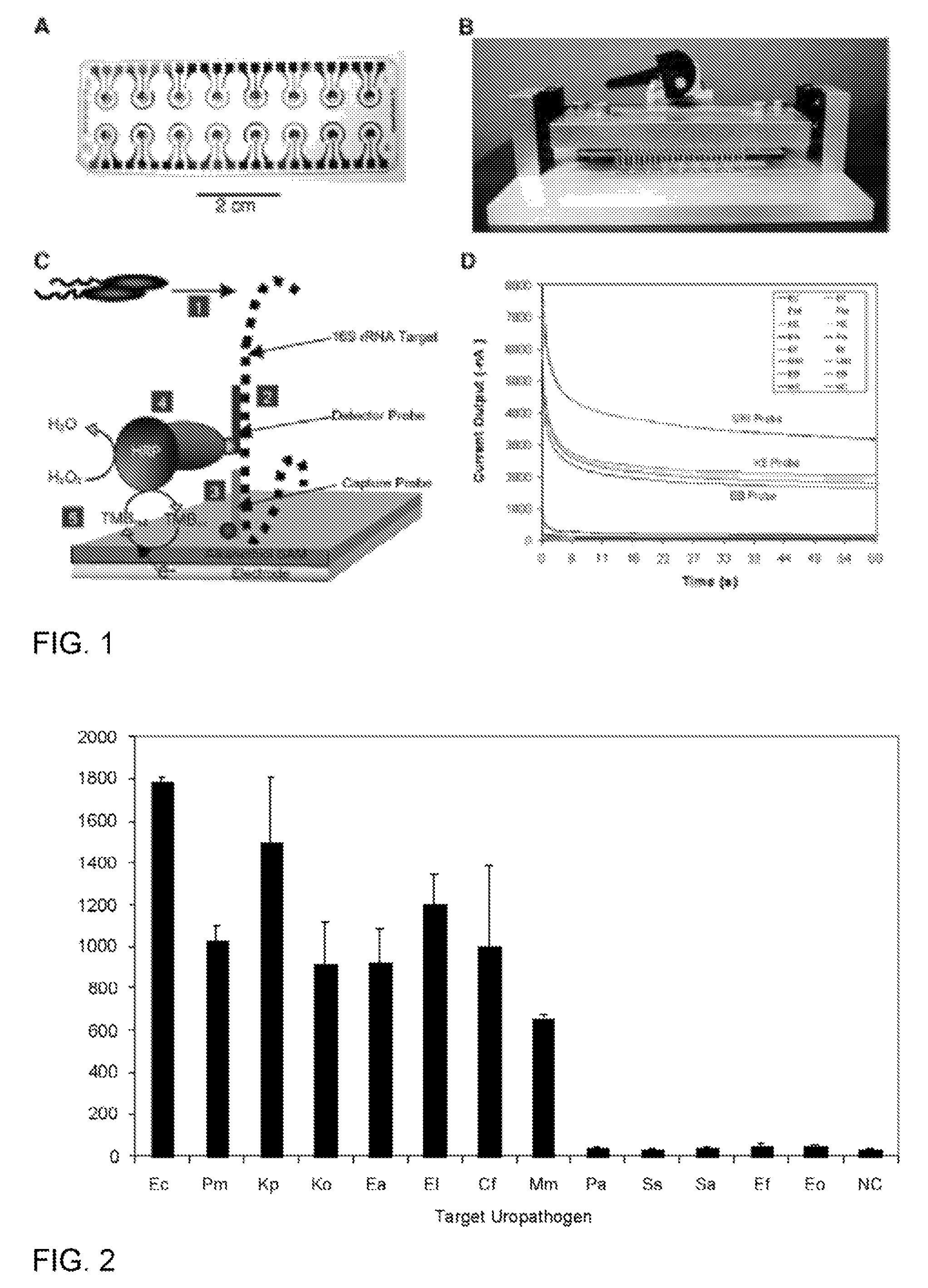
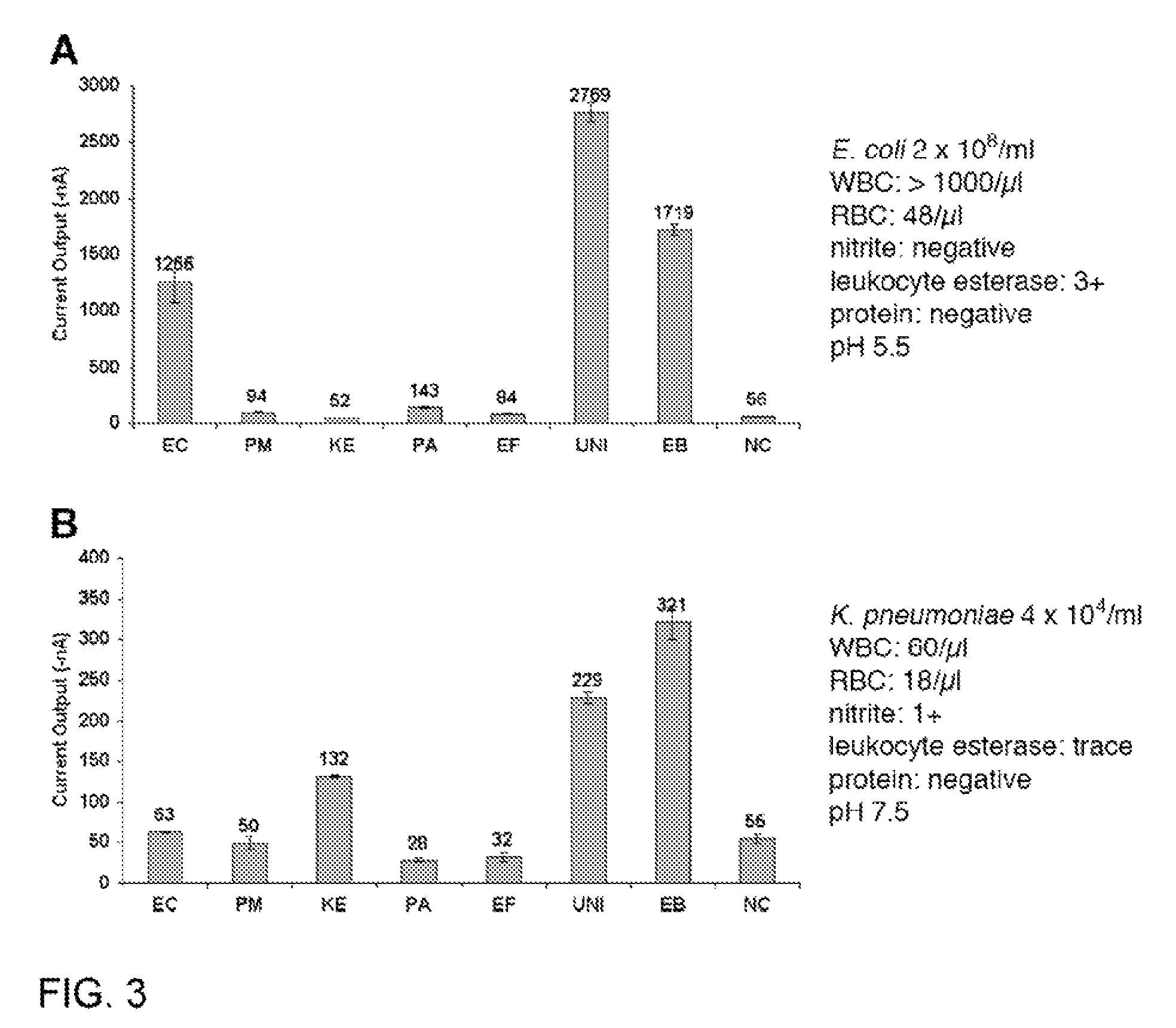
[0088]This example demonstrates species-specific detection of bacterial pathogens in clinical specimens using an electrochemical sensor. The sensor array can be an integral component of a point-of-care system for molecular detection of pathogens in body fluids.

[0089]Uropathogen isolates and clinical urine specimens. Uropathogen isolates and clinical urine specimens were obtained from the UCLA Clinical Microbiology Laboratory with approval from the UCLA Institutional Review Board and appropriate Health Insurance Portability and Accountability Act exemption. Isolates were received in vials containing Brucella broth with 15% glycerol (BBL, Maryland) and were stored at −70° C. Overnight bacteria cultures were freshly inoculated into Luria Broth (LB) grown to logarithmic phase as measured by OD600. Concentrations in the logarithmic phase specimens were determined by serial plating, typically yielding 10...

example 2

Determinants of Signal Intensity for Bacterial Pathogen Detection Using an Electrochemical DNA Biosensor Array

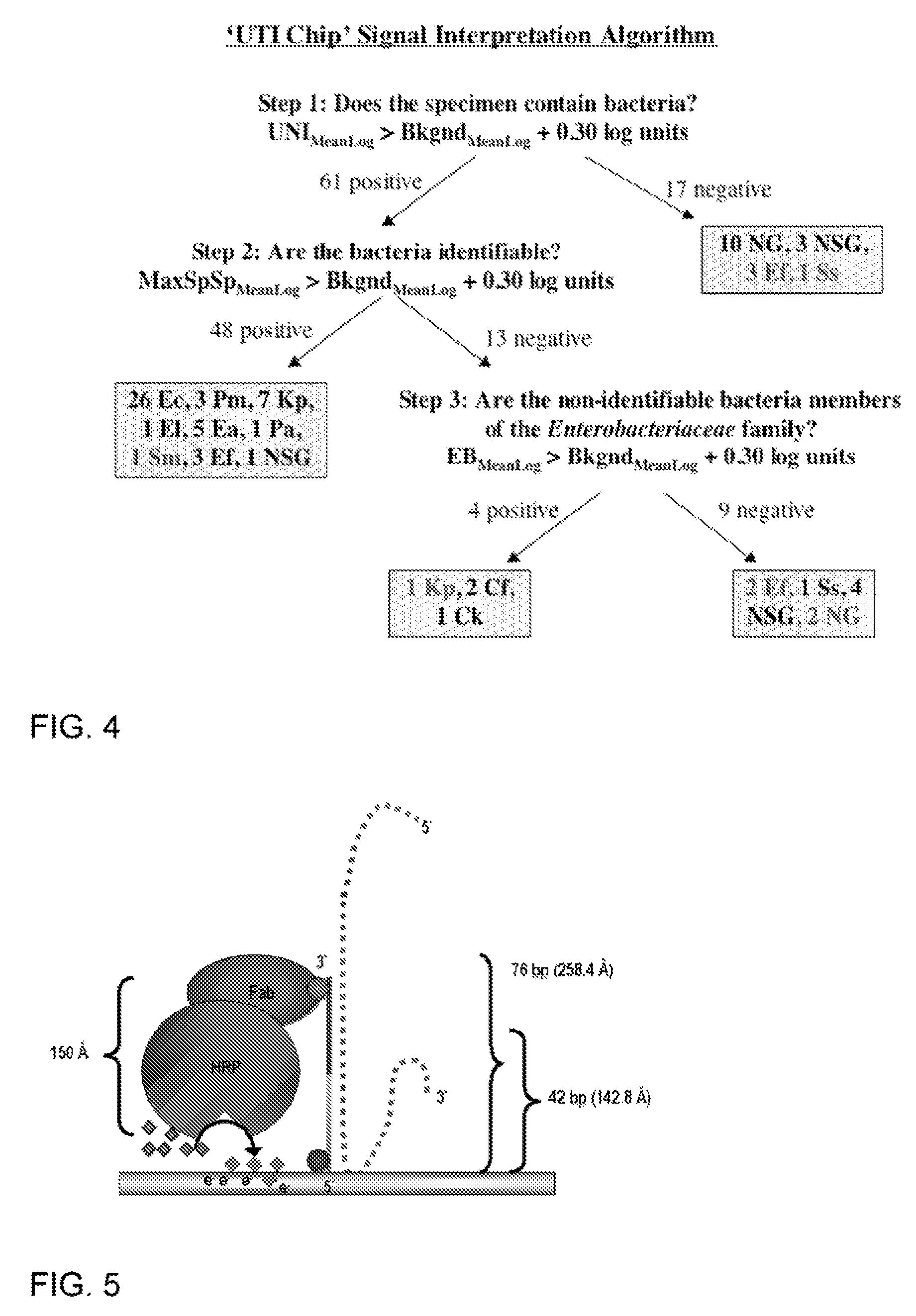
[0164]This example describes the determinants of electrochemical signal intensity using a sensor assay that involves hybridization of target rRNA to a fluorescein-modified detector probe and a biotin-modified capture probe anchored to streptavidin on the sensor surface. Signal is generated by an oxidation-reduction current produced by the action of horseradish peroxidase (HRP) conjugated to an anti-fluorescein monoclonal Fab bound to the detector probe. A 12-fold increase in electrochemical signal intensity for detection of Enterococcal 16S rRNA was achieved using a two-step approach involving initial treatment with Triton X-100 and lysozyme followed by alkaline lysis. This universal lysis system was shown to be effective for both Gram-positive and Gram-negative organisms. The location of fluorescein modification was found to be a determinant of signal intensity, indicating ...

example 3

Development of an Advanced Electrochemical DNA Biosensor

[0201]This example supplements Example 2 above with further data and additional probes for capture and detection.

[0202]The following flow chart shows the steps involved in the process of bacterial detection using the electrochemical sensor. Step 1: Bacterial lysis to release the 16S rRNA target. Step 2. Primary hybridization of the 16S rRNA target with the fluorescein-modified detector probe. Step 3. Secondary hybridization of the target-detector probe hybrid to the capture probe on the sensor surface. Step 4. Redox reaction generated by addition of anti-fluorescein horseradish peroxidase (αF-HRP), TMB substrate, and H2O2. The amount of time required for each step is shown. Washing occurs only before and after addition of αF-HRP. The entire assay was performed within 45 min.

[0203]

Bacterial Strains and Cultivation

[0204]The following American Type Culture Collection (ATCC) strains were obtained from the UCLA Clinical Microbiology...


PUM

PropertyMeasurementUnit
temperatureaaaaaaaaaa
timeaaaaaaaaaa

Abstract

Described are probes and methods for detecting pathogens and antibiotic resistance of a specimen. The method comprises contacting the specimen with a growth medium; and lysing the specimen to release nucleic acid molecules from the specimen. The lysate of the specimen is contacted with a capture probe immobilized on a substrate, wherein the capture probe comprises an oligonucleotide that specifically hybridizes with a first target nucleic acid sequence region of ribosomal RNA. The lysate is in contact with a detector probe that comprises a detectably labeled oligonucleotide that specifically hybridizes with a second target nucleic acid sequence region of ribosomal RNA. The presence or absence of labeled oligonucleotide complexed with the substrate is determined. Detection of labeled oligonucleotide complexed with the substrate is indicative of the presence of pathogen. Performing the method in the presence and absence of an antibiotic permits detection of antibiotic resistance.

Description

[0001]This application claims the benefit of U.S. provisional patent application No. 60 / 865,780, filed Nov. 14, 2006, and is a continuation-in-part of PCT application number US2005 / 39292, filed Nov. 1, 2005, which claims the benefit of U.S. provisional patent application No. 60 / 623,903, filed Nov. 1, 2004, the entire contents of which are incorporated herein by reference. Throughout this application various publications are referenced. Some of these references are indicated with numerals that refer to the list of references that can be found at the end of Example 1. Additional references noted in the other examples can be found in the list of literature cited at the end of the corresponding example. The disclosures of these publications in their entireties are hereby incorporated by reference into this application in order to describe more fully the state of the art to which this invention pertains.[0002]This invention was made with Government support via Grant No. EB00127, awarded ...

Claims


Application Information

Patent Timeline
Patent Type & Authority Patents(United States)
IPC IPC(8): C12Q1/68C07H21/02C07H21/04
CPCC12Q1/689
Inventor HAAKE, DAVID A.CHURCHILL, BERNARD M.LIAO, JOSEPH C.SUCHARD, MARC A.LI, YANGMASTALI, MITRA
Owner U S GOVERNMENT REPRESENTED BY THE DEPT OF VETERANS AFFAIRS
Learn More