
Implantable medical devices with antimicrobial and biodegradable matrices

a technology of biodegradable matrices and medical devices, applied in the field of implantable medical devices, can solve the problems of high mortality rate, hemodialysis requirement, poor glycemic control, etc., and achieve the effects of preventing the formation of initial bacterial biofilm, encouraging tissue growth, and preventing the formation of biofilm

Inactive Publication Date: 2010-09-14
MAQUET CARDIOVASCULAR LLC +1
View PDF70 Cites 13 Cited by
  • Summary
  • Abstract
  • Description
  • Claims
  • Application Information

AI Technical Summary

Benefits of technology

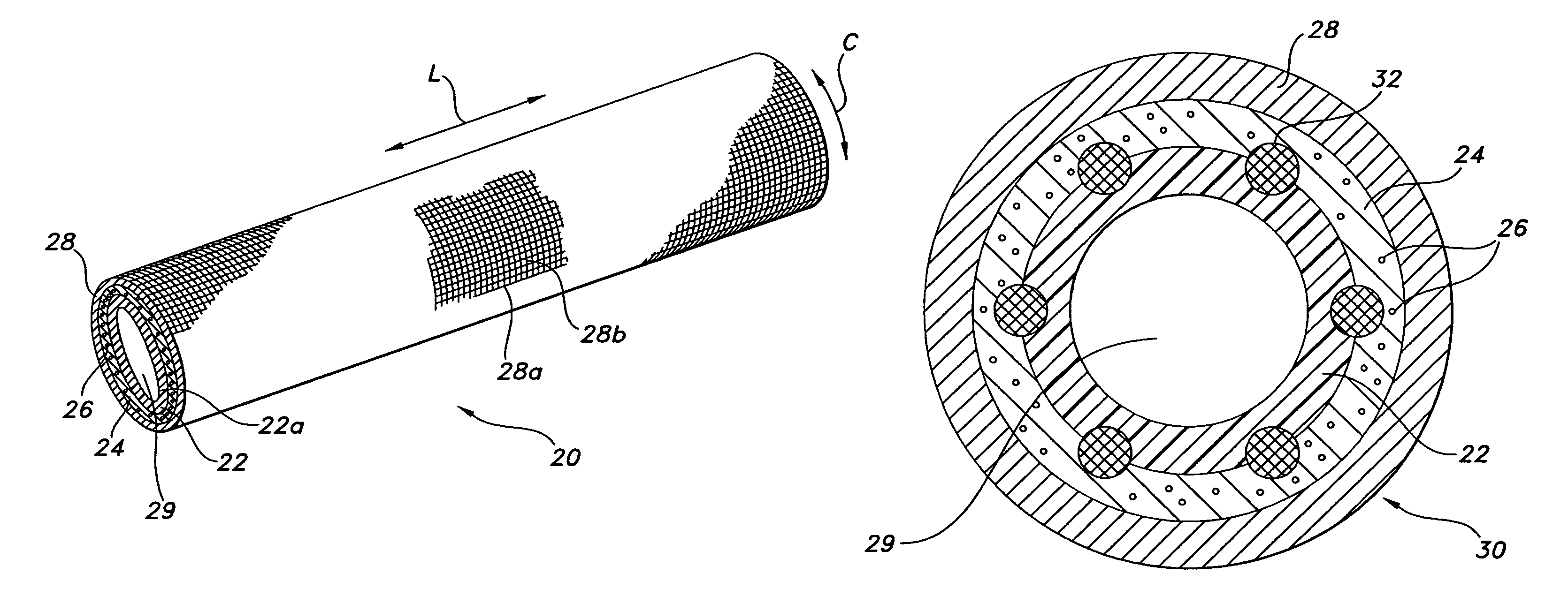
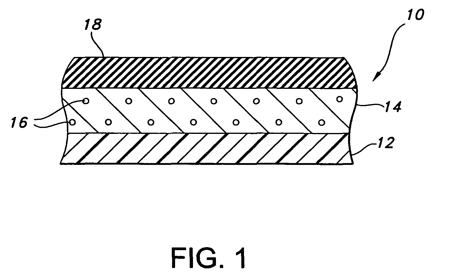
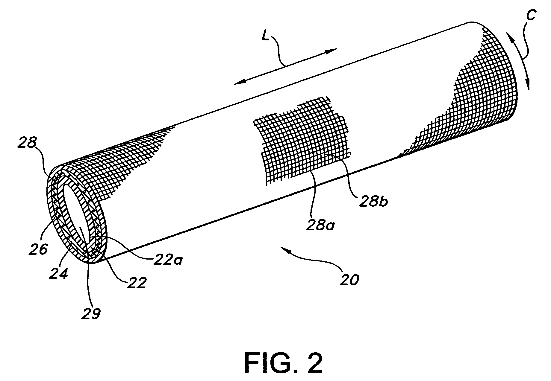
The present invention provides a vascular graft that can deliver bioactive agents to the region of a blood vessel in a controlled fashion. The graft includes a luminal layer of ePTFE, a biodegradable polymer layer that includes at least one bioactive agent, and a fabric layer that is porous enough to allow tissue ingrowth to replace the biodegradable polymer layer. The graft also has an internodal distance of up to about 90 microns, which encourages cellular communication between the blood and the perigraft tissue. The method of making the graft involves providing a luminal layer of ePTFE, positing a biodegradable polymer layer including a bioactive agent on the external side of the luminal layer, and positing a fabric layer on the external surface of the biodegradable layer. The graft can prevent infection and promote tissue ingrowth, which discourages biofilm formation around the graft.

Problems solved by technology

Poor glycemic control in diabetes and hypertension can lead to the requirement for hemodialysis.
It is known that 15-30% of all dialysis patients will have infection of their implant as a major cause of death.
It is this virulence that leads to septicemia and is one main factor in the high mortality rates.
There are severe complications as a result of vascular graft infections.
For example, anastonomic disruption due to proteolytic enzymes that the more virulent organisms produce can lead to a degeneration of the arterial wall adjacent to the anastomosis.
This can lead to a pseudoaneurism which can rupture and cause hemodynamic instability.
This increased blood platelet disruption leads to increased surface coverage of the implants with platelet cytoskeletal remains.
A disadvantage of this graft is that it is not formed of ePTFE, which is known to have natural antithrombogenic properties.
A further disadvantage is that the hydrophilic polyurethane matrix into which the silver salt is distributed does not itself control the release of silver into the surrounding body fluid and tissue at the implantation site of the graft.
A disadvantage of this device is that the porous polymeric outer layer needs to be applied without the use of solvents, catalysts, heat or other chemicals or techniques, which would otherwise degrade or damage the bioactive agent deposited on the surface of the stent.
Moreover, this graft is not designed to achieve fast tissue ingrowth within the tunnel to discourage initial bacterial biofilm formation.
A disadvantage of this device is that the biodegradable coating layer does not provide sufficient tensile strength for an outer graft layer.

Method used


Image

  • Implantable medical devices with antimicrobial and biodegradable matrices
  • Implantable medical devices with antimicrobial and biodegradable matrices
  • Implantable medical devices with antimicrobial and biodegradable matrices

Examples


example 1

[0070]The composite device of the present invention is formed by expanding a thin wall PTFE inner luminal tube at a degree of elongation on the order of 600-900%. The inner luminal tube is expanded over a cylindrical stainless steel mandrel at a temperature of 500° F. The luminal tube is fully sintered after expansion at a temperature of 660° F. for 15 minutes. The resulting luminal tube formed by this method exhibits an IND of greater than 40 microns, and in particular between 70 and 90 microns, and is spanned by a moderate number of fibrils.

[0071]The combination of the luminal ePTFE tube over the mandrel is then employed as a substrate over which the biodegradable layer is disposed. In particular, the biodegradable layer is formed of a synthetic hydrogel polymer. The hydrogel is a water-insoluble copolymer having a hydrophilic region, at least two functional groups that will allow for cross-linking of the polymer chains and a bioresorbable hydrophobic region. Silver iodate is blen...


PUM

PropertyMeasurementUnit
internodal distanceaaaaaaaaaa
temperatureaaaaaaaaaa
internodal distanceaaaaaaaaaa

Abstract

A composite vascular graft is provided, which incorporates bioactive agents that can be controllably delivered to the implantation site to deliver therapeutic materials and / or to reduce infection of the implant. The vascular graft of the present invention includes a luminal layer of ePTFE; and a biodegradable polymer layer including a bioactive agent, such as an antimicrobial agent. The biodegradable polymer layer is posited on the external surface of the luminal ePTFE layer. The graft also includes a fabric layer, which is posited on the external surface of the biodegradable layer. The graft is particularly useful as an arterial-venous graft for hemodialysis procedures.

Description

FIELD OF THE INVENTION[0001]The present invention relates to implantable medical devices, which inhibit or reduce bacterial growth during their use in a living body. More particularly, the present invention relates to composite vascular grafts which incorporate bioactive agents to deliver therapeutic materials and / or to inhibit or reduce bacterial growth during and following implantation, and which also desirably incorporate a luminal layer of ePTFE with an internodal distance of 70-90 microns to allow for intercellular communication.BACKGROUND OF THE INVENTION[0002]Poor glycemic control in diabetes and hypertension can lead to the requirement for hemodialysis. In order to facilitate treatment, a significant number of patients with these disorders will have a synthetic vascular graft surgically implanted between the venous and arterial systems to allow arterial-venous (A-V) access at the implantation type. The average time a synthetic graft will remain useful for A-V access is about...

Claims


Application Information

Patent Timeline
Patent Type & Authority Patents(United States)
IPC IPC(8): A61F2/06A61F2/00A61F2/02
CPCA61F2/06A61F2/07A61F2002/072A61F2210/0004A61F2250/0067
Inventor KING, TAMARAH L.
Owner MAQUET CARDIOVASCULAR LLC
Learn More