

Antibodies to osteoprotegerin binding proteins
a technology of osteoprotegerin and binding proteins, which is applied in the field of polypeptides, can solve the problems of reduced bone mass and strength, slow or incomplete repair of broken bones, and increased risk of fractures
- Summary
- Abstract
- Description
- Claims
- Application Information
AI Technical Summary
Benefits of technology
Problems solved by technology
Method used
Image

Examples
example 1
Identification of a Cell Line Source for an OPG Binding Protein
[0059]Osteoprotegerin (OPG) negatively regulates osteoclastogenesis in vitro and in vivo. Since OPG is a TNFR-related protein, it is likely to interact with a TNF-related family member while mediating its effects. With one exception, all known members of the TNF superfamily are type II transmembrane proteins expressed on the cell surface. To identify a source of an OPG binding protein, recombinant OPG-Fc fusion proteins were used as immunoprobes to screen for OPG binding proteins located on the surface of various cell lines and primary hematopoietic cells.
[0060]Cell lines that grew as adherent cultures in vitro were treated using the following methods: Cells were plated into 24 well tissue culture plates (Falcon), then allowed to grow to approximately 80% confluency. The growth media was then removed, and the adherent cultures were washed with phosphate buffered saline (PBS) (Gibco) containing 1% fetal calf serum (FCS). ...
example 2
Expression Cloning of a Murine OPG Binding Protein
[0063]A cDNA library was prepared from 32D mRNA, and ligated into the mammalian expression vector pcDNA3.1(+) (Invitrogen, San Diego, Calif.). Exponentially growing 32D cells maintained in the presence of recombinant interleukin-3 were harvested, and total cell RNA was purified by acid guanidinium thiocyanate-phenol-chloroform extraction (Chomczynski and Sacchi. Anal. Biochem. 162, 156-159, (1987)). The poly (A+) mRNA fraction was obtained from the total RNA preparation by adsorption to, and elution from, Dynabeads Oligo (dT)25 (Dynal Corp) using the manufacturer's recommended procedures. A directional, oligo-dT primed cDNA library was prepared using the Superscript Plasmid System (Gibco BRL, Gaithersburg, Md.) using the manufacturer's recommended procedures. The resulting cDNA was digested to completion with Sal I and Not I restriction endonuclease, then fractionated by size exclusion gel chromatography. The highest molecular weight...
example 3
OPG Binding Protein Sequence
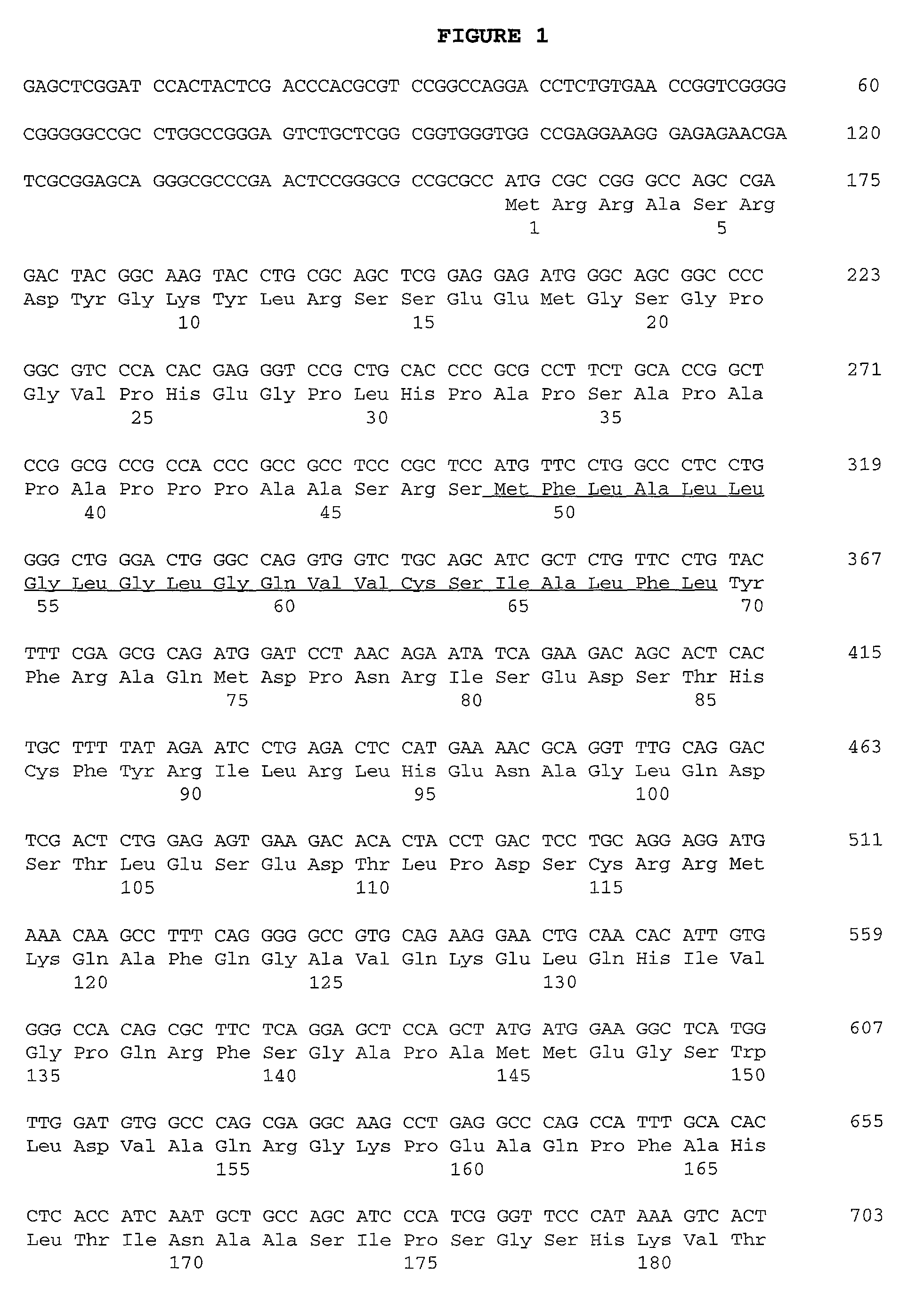
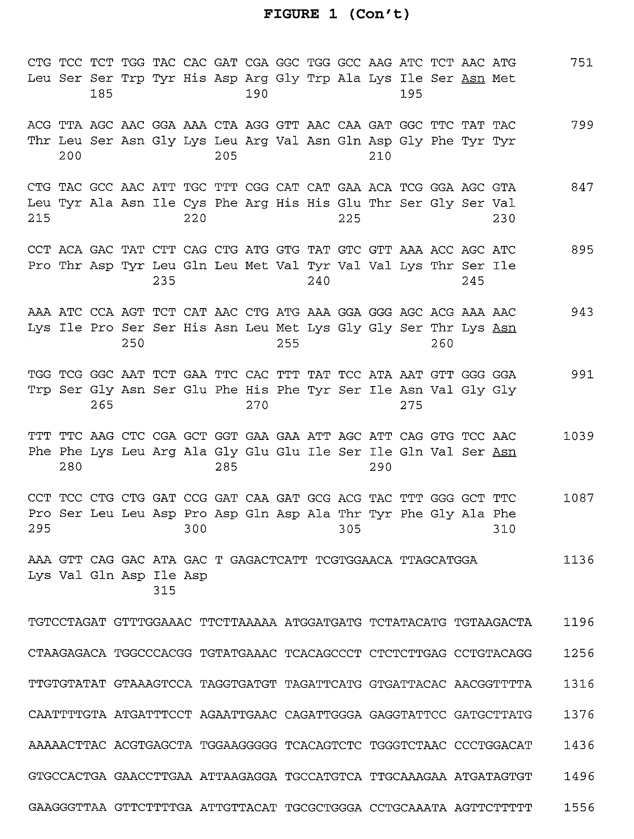
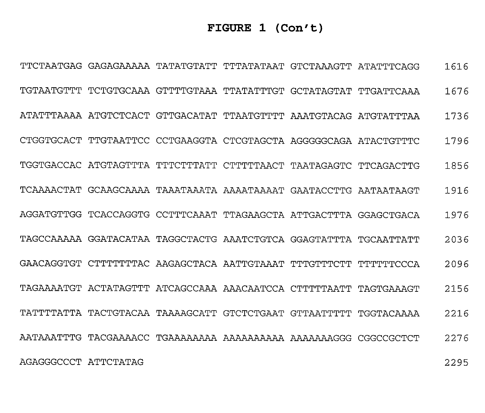
[0067]The 32D-F3 clone isolated above contained an approximately 2.3 kb cDNA insert (FIG. 1), which was sequenced in both directions on an Applied Biosystems 373A automated DNA sequencer using primer-driven Taq dye-terminator reactions (Applied Biosystems) following the manufacturer's recommended procedures. The resulting nucleotide sequence obtained was compared to the DNA sequence database using the FASTA program (GCG, University of Wisconsin), and analysed for the presence of long open reading frames (LORF's) using the “Six-way open reading frame” application (Frames) (GCG, University of Wisconsin). A LORF of 316 amino acid (aa) residues beginning at methionine was detected in the appropriate orientation, and was preceded by a 5′ untranslated region of about 150 bp. The 5′ untranslated region contained an in-frame stop codon upstream of the predicted start codon. This indicates that the structure of the 32D-F3 plasmid is consistent with its ability to ...
PUM
| Property | Measurement | Unit |
|---|---|---|
| concentrations | aaaaa | aaaaa |
| temperature | aaaaa | aaaaa |
| volume | aaaaa | aaaaa |
Abstract
Description
Claims
Application Information