Regulation of F1-ATPase beta subunit cellular location

a subunit and cellular location technology, applied in the field of f1-atpase beta subunit cellular location regulation, can solve the problems of limited enterostatin absorption across the intestine, inability to detect enterostatin selectively, and inability to analyze enterostatin in vivo, so as to improve the activity of plasma membrane atpase, improve the binding affinity of apoa-1, and affect the effect of cholesterol transpor

Inactive Publication Date: 2011-02-01
BOARD OF SUPERVISORS OF LOUISIANA STATE UNIV & AGRI & MECHANICAL COLLEGE
View PDF1 Cites 0 Cited by
  • Summary
  • Abstract
  • Description
  • Claims
  • Application Information

AI Technical Summary

Benefits of technology

[0020]We have discovered that enterostatin can cause the translocation of the F1-ATPase beta subunit to the plasma membrane. This should increase the level of plasma membrane ATPase activity and regulate membrane functions and intracellular metabolism through changes in ATP, ADP or AMP concentrations. The initial demonstration utilized Green Fluorescent Protein (GFP)-tagged protein transfected into HepG2 cells, and showed that enterostatin induced a movement of the protein to the perimeter of cells. Since the available literature has identified this protein as present in the plasma membranes of a number, but not all, cell types, the ability to induce translocation of this protein to the plasma membrane will have a variety of biological effects depending upon the cell type. Increasing its presence in the plasma membranes of hepatocytes would increase binding to ApoA-1, regulating endocytosis of HDL particles, and affecting cholesterol transport; of lymphocytes would increase angiostatin binding affecting angiogenesis and tumor growth; of endothelial cells would affect inflammatory responses or vascular integrity; and of pancreatic cells would modulate the inhibitory effect of enterostatin on insulin secretion. Thus, the ability to regulate the localization of enterostatin to the plasma membrane could regulate insulin secretion, cholesterol and lipid transport, angiogenesis and tumor growth.

Problems solved by technology

The in vivo concentration of enterostatin has not been established, due to problems in measuring enterostatin.
Antibodies that are selective to enterostatin that could be used to analyze tissue levels of enterostatin have been difficult to find.
Enterostatin absorption across the intestine was found to be limited and slow, occurring mainly into lymphatic system.
However, since enterostatin has been shown to be biologically active on food intake at extremely low doses compared to other peptides and to inhibit insulin secretion from isolated pancreatic islets at doses of 10−10 to 10−6 M, a proposed low affinity casomorphin binding site probably is not the biologically important enterostatin receptor that inhibits fat intake and insulin secretion.

Method used


Image

  • Regulation of F1-ATPase beta subunit cellular location
  • Regulation of F1-ATPase beta subunit cellular location
  • Regulation of F1-ATPase beta subunit cellular location

Examples


example 1

[0032]Materials and Methods

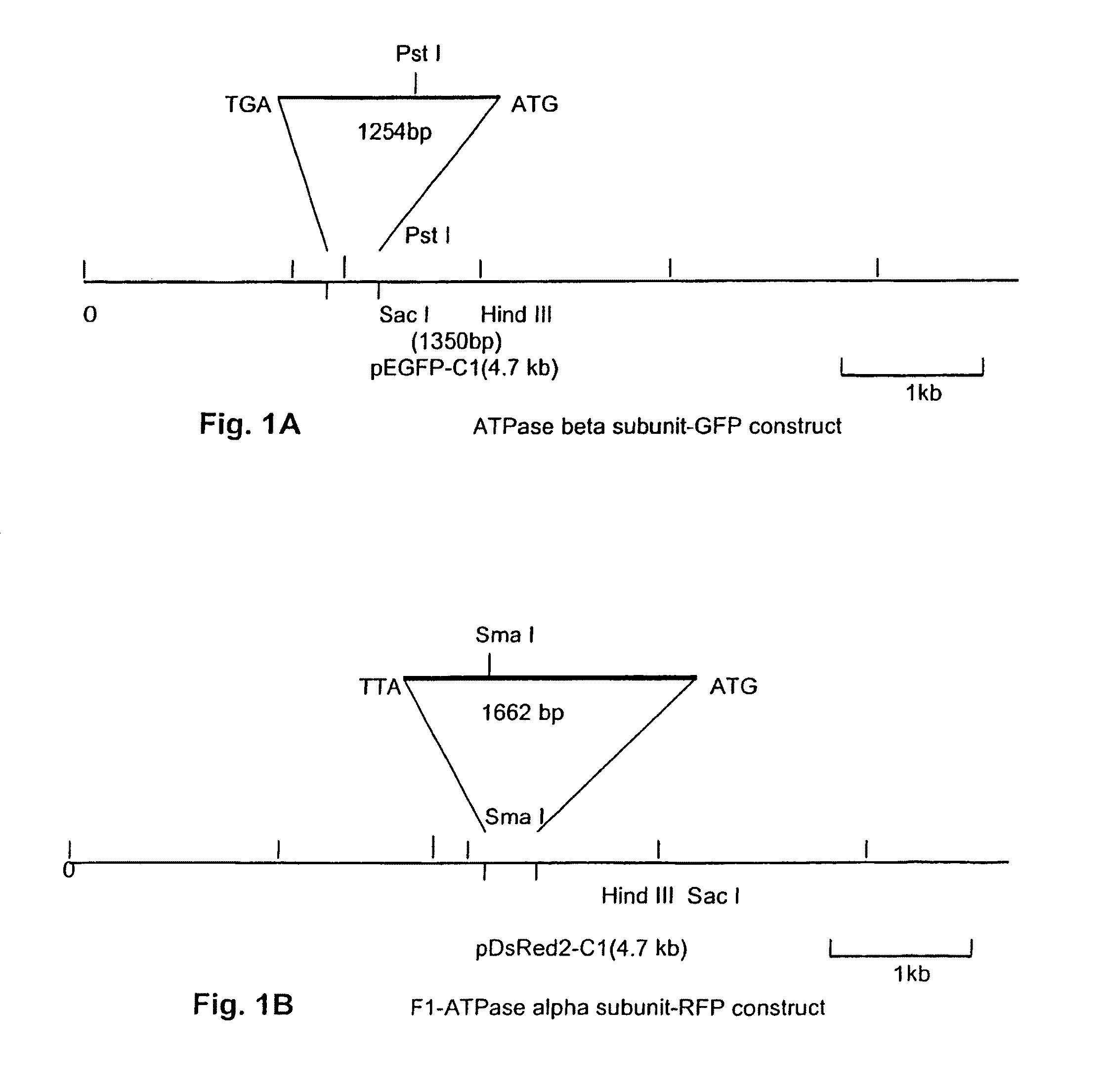
[0033]Generation of F1-ATPase beta subunit-GFP fusion protein. The F1-ATPase β-subunit clone (1,254 bp) was generated by PCR from an amygdala cDNA library using F1-ATPase β-subunit forward primer (5′GAGAGGAGCTCCACTATTGCTATGGATGGC3′ (SEQ ID NO. 1), Sac I recognition site underlined) and F1-ATPase β-subunit reverse primer (5′GAGAGAAGCTTCACGACCCATGCTC3′ (SEQ ID NO. 2), Hind III recognition site underlined), according to procedures described in Appendix B. The PCR product was cloned into the pEGFP-C1 vector (Clonetech, Palo Alto, Calif.) between the Sac I and Hind III sites as shown in FIG. 1A. The construct was then transformed into DH5α cells. Transformants were screened and positive clones containing plasmid with the correct orientation of the insert were cultured.

[0034]Generation of F1-ATPase alpha subunit-RFP fusion protein. The F1-ATPase α-subunit clone (1,662 bp) was generated by PCR from an amygdala cDNA library using F1-ATPase α-subunit forward primer...

example 2

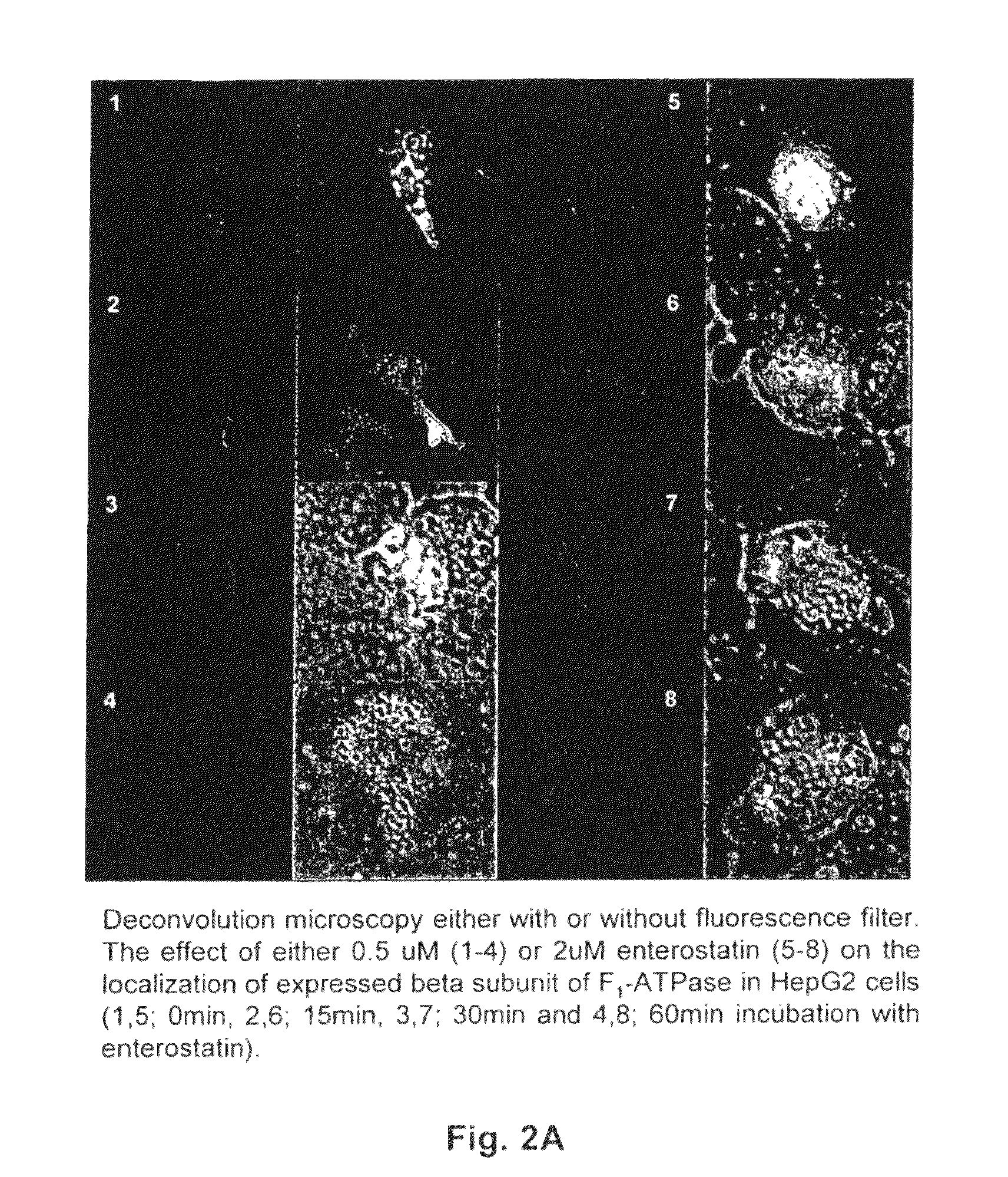
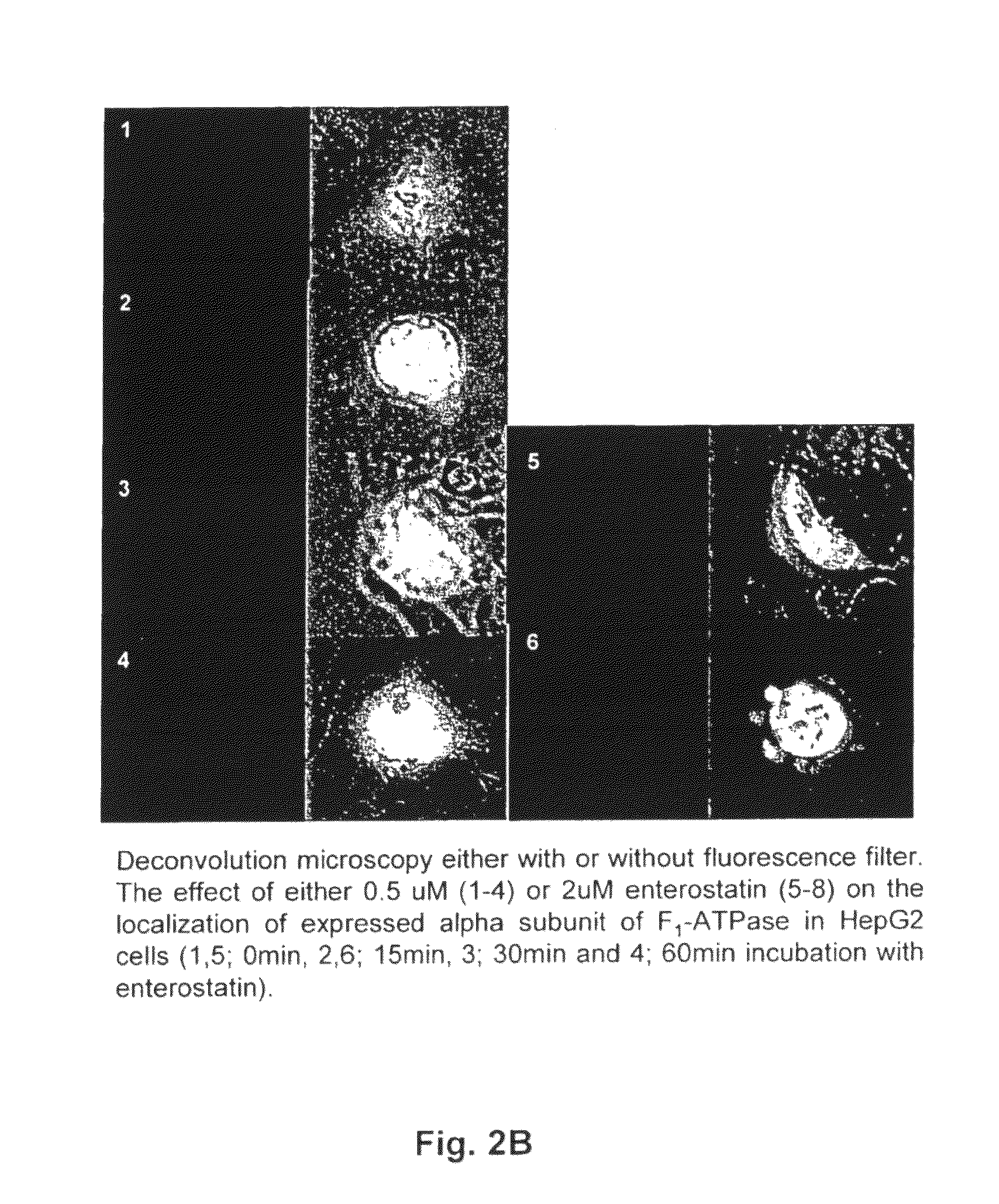
[0036]Enterostatin Alters F1-ATPase Beta Subunit Cell Location in HepG2 Cells

[0037]Either 0.5 or 2 μM enterostatin was added to the HepG2 cells after transfection with either alpha subunit-RFP or beta subunit-GFP for 16-18 hrs. After incubation with enterostatin for 15, 30, or 60 min, the cover slide was removed from the well and placed on a glass slide. Protein expression in HepG2 cells was then observed using deconvolution microscopy.

[0038]Enterostatin did not change the distribution of expressed F1-ATPase alpha subunit-RFP in HepG2 within the time of incubation with either concentration of enterostatin (FIG. 2B). However, after incubation with both 0.5 and 2 uM enterostatin, F1-ATPase beta subunit-GFP was clearly relocated toward the cell periphery (FIG. 2A). To study this event in real time, a confocal microscope with an inverted camera was used to observe protein localization in transfected HepG2 cells at the different incubation times with 2 μM enterostatin for both the beta s...

example 3

[0039]F1-ATPase Beta Subunit in Plasma Membrane of Liver, Pancreas and Amygdala Cells

[0040]Using Western blot analysis and immunohistochemistry, F1-ATPase β-Subunit was discovered present in the plasma membrane and mitochondrial membranes of liver, pancreas and amygdala cells from rats. The purified F1-ATPase β-Subunit (200 μg / ml) (produced as described in Appendix B) in Freunds complete adjuvant (Sigma Chemical Co., St. Louis, Mo.) was injected subcutaneously at multiple sites on the back of New Zealand white rabbits. Booster doses, diluted into Freunds incomplete adjuvant, were administered at weeks 4, 8 and 12. Blood was collected from injected animals on weeks 0, 4, 8, and 12. Rabbit antiserum was prepared by centrifugation (2000×g, 30 min, 4° C.) and stored at −20° C. in aliquots. The antibody was purified by passing over a protein A bead column (Sigma Chemical Co.). The antibody dilution necessary for antigen detection was assayed by an ELISA. Ninety six-well plates were coate...


PUM

PropertyMeasurementUnit
total volumeaaaaaaaaaa
pHaaaaaaaaaa
pHaaaaaaaaaa

Abstract

It has been discovered that the ability of analogues to affect binding of a labeled β-casomorphin (an enterostatin antagonist) to recombinant rat F1-ATPase β-subunit was closely correlated with their enterostatin-like biological activity. Using immunohistochemistry and western blots, the presence of the F1-ATPase β-subunit was demonstrated in plasma membranes of liver, pancreas and amygdala. The effects of enterostatin on the intracellular localization of the proteins were studied using deconvolution or confocal microscopy. Enterostatin did not alter the location of F1-ATPase a-subunit-RFP but induced movement of the F1-ATPase β-subunit-GFP to the periphery of cells. These studies, showing the plasma membrane localization of the F1-ATPase β-subunit, the influence of enterostatin on the cellular location of the protein, the appropriate Kd value for binding, together with the previous correlation of binding effects with biological activity for a number of analogues, indicate that this protein is the enterostatin receptor.

Description

[0001]This is the United States national stage of international application PCT / US05 / 37779, filed Oct. 19, 2005, which claims the benefit of the Nov. 12, 2004 filing date of U.S. provisional application Ser. No. 60 / 627,455 under 35 U.S.C. §119(e).[0002]The development of this invention was partially funded by the Government under a grant from the National Institute of Health (NIDDK45728). The Government has certain rights in this invention.TECHNICAL FIELD[0003]This invention pertains to a method to regulate the amount of a peptide receptor, the F1-ATPase β subunit, in the plasma membrane of mammalian cells, a method to screen patients for response to enterostatin treatment, and a method to screen for compounds that are agonists or antagonists for enterostatin.BACKGROUND ART[0004]Enterostatin[0005]Enterostatin is the aminoterminal pentapeptide of procolipase that is released by proteolytic activity when procolipase is converted into colipase (9). The procolipase gene is expressed in ...

Claims


Application Information

Patent Timeline
Patent Type & Authority Patents(United States)
IPC IPC(8): G01N33/573
CPCG01N33/5005G01N33/573G01N2333/914G01N2500/00
Inventor YORK, DAVID A.PARK, MIEJUNG
Owner BOARD OF SUPERVISORS OF LOUISIANA STATE UNIV & AGRI & MECHANICAL COLLEGE
Why Eureka
Try Eureka