Nucleic acid quantitation from tissue slides

a tissue slide and nucleic acid technology, applied in the field of nucleic acid extraction and quantitation from cells and tissues, can solve the problems of time-consuming, laborious, cumbersome, etc., and achieve the effects of improving the estimation of mrna copy counts, increasing sample analysis accuracy and sensitivity, and increasing the sensitivity to target fragmentation

Active Publication Date: 2011-06-28
AFFYMETRIX INC
View PDF12 Cites 15 Cited by
  • Summary
  • Abstract
  • Description
  • Claims
  • Application Information

AI Technical Summary

Benefits of technology

[0012]Methods of the invention are useful in addressing problems encountered in analysis of nucleic acid samples that are physically difficult to process or have experienced degradation. The methods can help obtain good representative test materials from samples that were previously processed for evaluation of histopathology. The methods can increase the accuracy and sensitivity of sample analyses by providing more representative standard materials. The condition of possibly degraded nucleic acids can be determined by an inventive offset bDNA assay configured for increased sensitivity to target fragmentation. The methods can improve estimates of mRNA copy counts for test materials derived from unknown numbers of normal and / or abnormal cells. The techniques can be used in combination to optimize nucleic acid analyses of, e.g., test materials derived from normal and / or aneuploid cells, formalin fixed cells, paraffin embedded cells, aged clinical samples, and the like.
[0013]Methods of the invention include combinations of inventive techniques working together to enhance the sensitivity and accuracy of a nucleic acid determination. For example, mRNA copy numbers can be estimated accurately by determining the number of cells in a test sample (e.g., by comparing a test sample rDNA value to a standard function of rDNA versus cell number), preparing a standard function for an RNA assay (e.g., assay output versus a degraded in vitro RNA standard assay input—degradation of sample or standard determined, e.g., by an offset bDNA assay), determining an amount of a test mRNA in the test sample using the RNA assay standard function, and determining the copy number of the mRNA in the test cells based on the number of cells and the determined amount of test mRNA. This procedure can effectively determine mRNA copy numbers in a variety of cell and tissue types, such as, e.g., tumor cells, cell lines, cells from a microscope slide, clinical samples more than a year old, fresh tissue, freshly fixed cells, freshly fixed paraffin embedded tissues, cells fixed with formalin, cells embedded in paraffin, normal and / or aneuploid cells, and the like e.g., originating from humans, plants, or animals.

Problems solved by technology

This procedure is time-consuming, cumbersome, and requires special handling, as xylene is a highly toxic chemical that emits noxious fumes.
The phase extraction dewaxing protocols are time consuming and laborious.
The repeated handling, aspirations and tube transfers can result in non-quantitative harvests of the nucleic acids.
The repeated vortexing and exposure to harsh solvents can cause sample degradation.
Additional problems exist in the quantitation of nucleic acids from preserved clinical specimens.
This can compromise, e.g., the ability to measure RNA in FFPE tissue blocks.
The nucleic acids ultimately extracted from embedded clinical samples are often highly degraded and fragmented.
Qualitative and quantitative assay errors often result when these extracts are evaluated by standard analytical techniques.
What's more, incomplete extractions can introduce error into calculations, such as mRNA copy number determinations.
One problem in measuring RNA from FFPE tissue blocks can be fragmentation of the RNA fragments, cross-linking, and base modifications induced by formalin-fixation procedures.
The exact causes for the fragmentation are not known, and thus it has been unclear how to solve this problem.
This problem is independent of whether the reverse transcription step uses oligo-dT or random priming.
However, this is often inadequate because adenines are more susceptible to alteration by formalin fixation and thus A / U rich sequences will be less accurately measured than G / C rich sequences (Masuda N, Ohnishi T, Kawamoto S, Monden M, Okubo K: Analysis of chemical modification of RNA from formalin-fixed samples and optimization of molecular biology applications for such samples, Nucleic Acids Res 1999, 27:4436-4443).
Consequently, there will be gene specific differences in the efficiency and reproducibility of measuring mRNA in formalin-fixed tissues.
Thus, there are severe limitations for PCR-based RNA measurements in formalin-fixed tissues.
A second problem that affects RNA quantification by QPCR is the required reverse transcription step to convert mRNA sequences of interest to cDNA.
Although, introduction of a high temperature heating step during PCR amplification steps may partially reverse some of the RNA base modifications, for many samples these modifications are irreversible.
With all these problems, QPCR has not been a satisfactory method of quantitating RNAs from FFPE samples.

Method used


Image

  • Nucleic acid quantitation from tissue slides
  • Nucleic acid quantitation from tissue slides
  • Nucleic acid quantitation from tissue slides

Examples


example 1

Comparison of Post-Homogenization Separation to Pre-Homogenization Extraction of FFPE Tissue Slide Hydrophobic Components

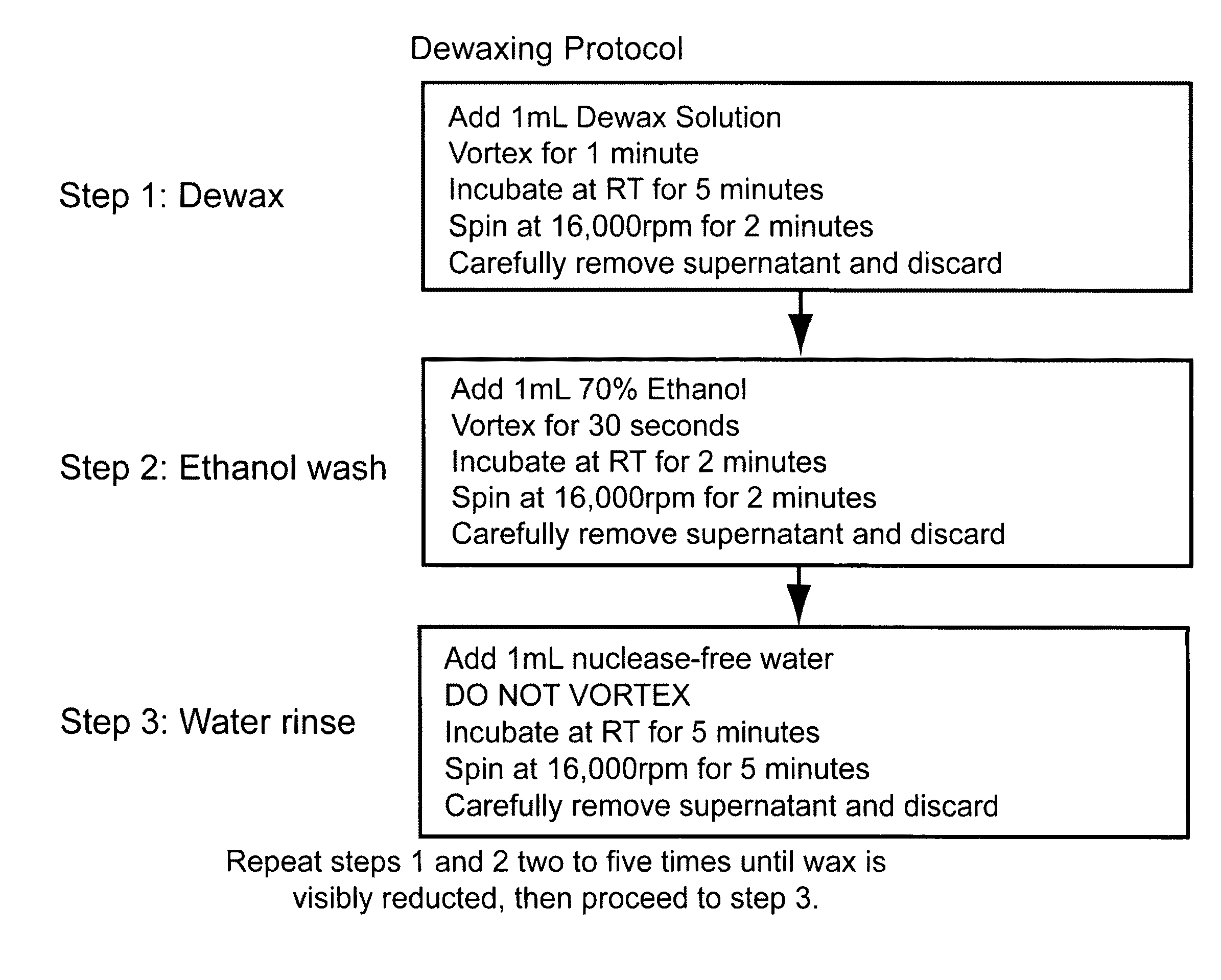
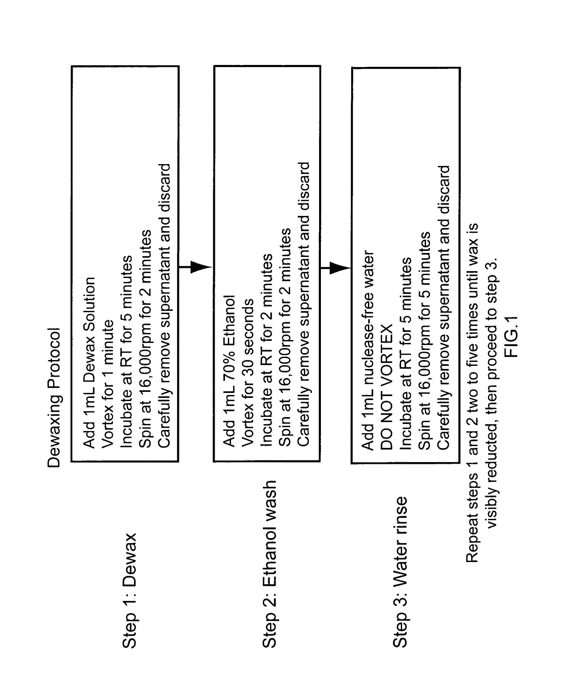
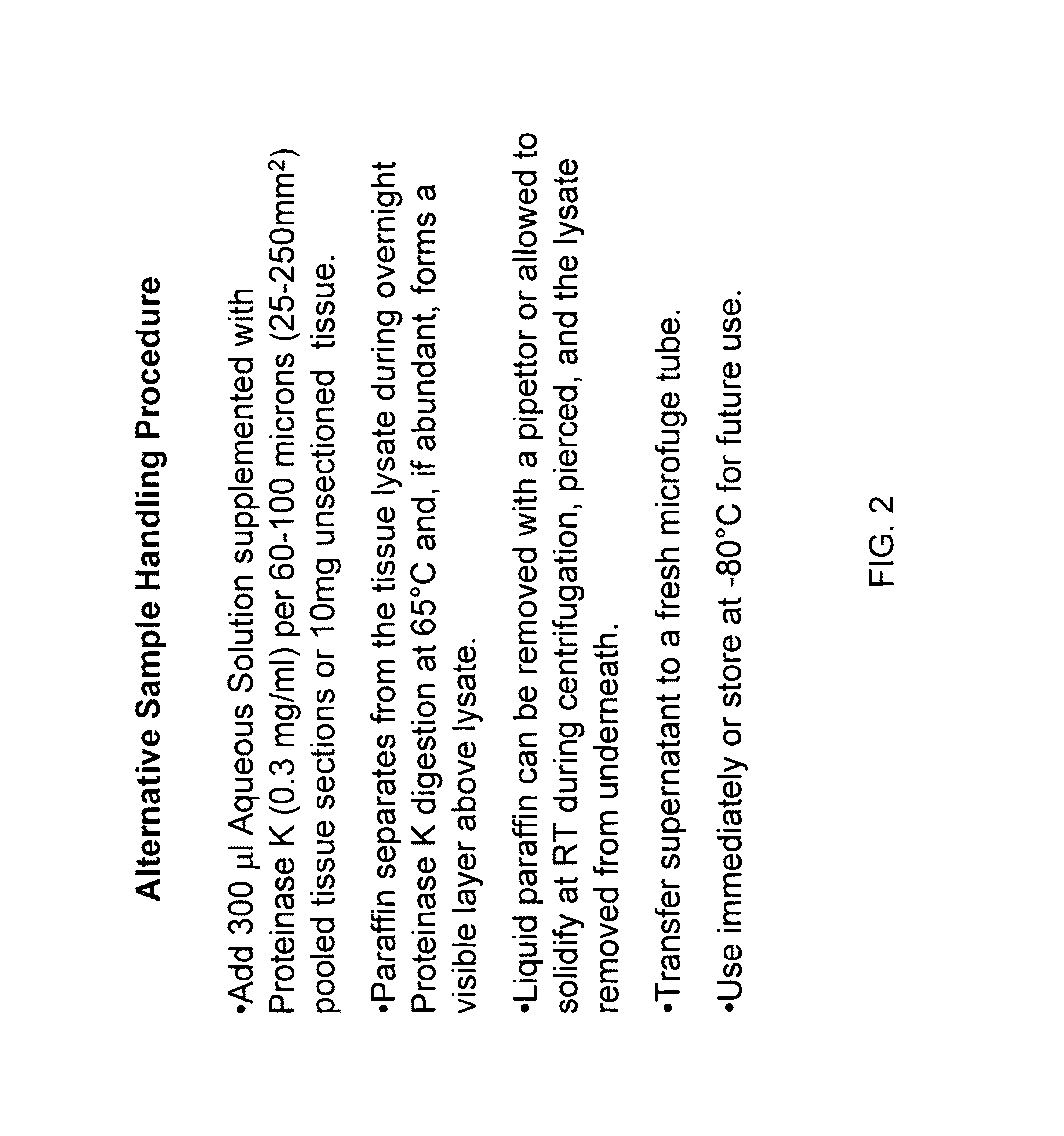
[0134]To demonstrate equivalence and improvements of post-homogenization separation over pre-homogenization organic extraction, FFPE sample mRNAs were compared using the branched DNA technology with and without pre-solubilization dewaxing. Samples from >10 yr-old matched human lung normal and tumor FFPE samples were pooled as three 10 micron sections. For one pair of samples (“0”), homogenates were prepared with wax separation after solubilization. For a second pair of samples (“1”), homogenates were prepared with 1× dewaxing organic extraction step before solubilization. For a third pair of samples (“2”), homogenates were prepared with 2× dewaxing organic extraction cycles. Extracts were tested using ribosomal protein S3 (RPS3, NM—001005 housekeeper or Reference control RNA) and Lactate dehydrogenase A (LDHA, NM—005566; 2-3× induction in tumor samples) as describ...

example 2

Equivalent Spike Recovery from FFPE Samples

[0136]An additional experiment was performed demonstrating that the recovery of RNA from FFPE sections is equivalent for post-solubilization separation and pre-solubilization extraction. Methods were as described above for the Lactate dehydrogenase A (LDHA, NM—005566) and ribosomal protein S3 (RPS3, NM—001005), except a known amount of in vitro transcribed (IVT) RNA from the bacterial gene dihydrodipicolinate reductase (dapB, L38424 not expressed in the human lung tissue) was added to a solublized FFPE sample. The data show yields were comparable between the 2× dewax extraction technique and physical wax separation technique. Using either procedure, the capture efficiency (spike recovery) of the spiked-in dapB IVT RNA ranged from 90-110%.

example 3

Use of Repetitive Ribosomal DNA to Determine Efficiency of FFPE Tissue Solubilization and Number of Cells in the FFPE Sample

[0137]An investigation was made to determine if accurate estimates of cell numbers can be made based on repetitive DNAs. In particular, cell counts for tumor and other aneuploid cells were determined using standard curves based on quantitation of ribosomal DNA genes.

[0138]Ten (10) cell lines were chosen with an average of about ten (10) ribosomal gene clusters per diploid genome. Four thousand (4000) cells of each cell line were lysed in QuantiGene Lysis Buffer (Panomics) and 10 cell line lysates were pooled; i.e. 10 cell lines in total or 40,000 cells are in a total volume of 110 ul.

[0139]Thirty microliters (30 ul) each were transferred to separate microfuge tubes, one to denature the DNA to measure the ribosomal DNAs (18S & 28S rDNA) and another undenatured control to measure background of the assay. Next, each tube was diluted to 300 ul total by adding 270 u...


PUM

PropertyMeasurementUnit
Tmaaaaaaaaaa
temperatureaaaaaaaaaa
melting pointaaaaaaaaaa

Abstract

This invention provides methods of quantitating nucleic acids from problematic samples, such as aged samples, formalin fixed samples, paraffin embedded samples, samples with aneuploid cells, and cells with fragmented nucleic acids. Methods include techniques to efficiently solublize the nucleic acids under non-denaturing conditions from preserved clinical samples without resort to organic extractions, to normalize cell counts regardless of aneuploidy, to access the fragmentation state of the nucleic acids, and to provide standard curves for degraded nucleic acid samples.

Description

CROSS-REFERENCE TO RELATED APPLICATIONS[0001]This application claims priority to and benefit of a prior U.S. Provisional Application No. 60 / 838,578, Nucleic Acid Quantitation from Tissue Slides, by Gary McMaster, et al., filed Aug. 17, 2006. The full disclosure of the prior application is incorporated herein by reference.FIELD OF THE INVENTION[0002]The present invention is in the field of nucleic acid extraction and quantitation from cells and tissues. Nucleic acids are extracted from embedded clinical samples without the use of hydrophobic solvents. Ribosomal DNA references and / or degraded in vitro RNAs are used to normalize standard curves and to establish nucleic acid copy numbers per cell.BACKGROUND OF THE INVENTION[0003]Formalin-fixed, paraffin-embedded (FFPE) tissue offers a vast source of biopsy specimens for which the clinical outcome is well documented and thus an optimal resource for retrospective studies (Lewis, F. et al. 2001; Yang et al. 2006). Access to and use of huma...

Claims


Application Information

Patent Timeline
Patent Type & Authority Patents(United States)
IPC IPC(8): C12Q1/68C12P19/34C12N1/08G01N33/00C07H21/04
CPCC12N1/06C12N15/1003C12Q1/6806C12Q1/6841Y10T436/143333C12Q2527/101C12Q2523/32C12Q2521/537C12Q2545/101
Inventor MCMASTER, GARYDAVIES, JOANMA, YUNQINGLUO, YULING
Owner AFFYMETRIX INC
Why Eureka
Try Eureka