Method for isolating and purifying oligodendrocytes and oligodendrocyte progenitor cells

a technology of oligodendrocytes and progenitor cells, applied in the field of separating cells of interest, can solve the problems of inability of damaged brain to function significantly structural self-repair, relative scarcity of adult brain tissue, and failure of mature brain to generate new neurons, etc., and achieve the effect of acceleration in the study

Inactive Publication Date: 2012-09-11
CORNELL RES FOUNDATION INC
View PDF21 Cites 20 Cited by
  • Summary
  • Abstract
  • Description
  • Claims
  • Application Information

AI Technical Summary

Benefits of technology

[0015]Extension of this approach to include fluorescent transgenes under the control of stage- and phenotype-specific promoters (both of which are intended to be covered by reference to “cell-specific” promoters herein) allows even more specific separations to be performed, for example, of oligodendrocytes over a range of developmental stages. More broadly, this strategy may be applied to any tissue of interest for which a cell-specific promoter is available, thereby allowing the identification, isolation and separation of progenitor cells and their products from any tissue for which constituent cells have been assigned phenotype-selective promoters. This strategy permits sufficient enrichment for in vivo implantation of the defined and separated progenitor pools, as well as for in vitro analyses of phenotypic specification and growth control.
[0016]By providing a means of identifying oligodendrocytic precursors while alive, even when present in small numbers in mixed populations, the use of fluorescent transgenes driven by cell type-selective promoters such as P/CNP2 will allow the specification of phenotype to be studied and perturbed on the single cell level, an approach that had previously only been feasible on larger populations. Indeed, when used in conjunction with post-transfection FACS, this strategy may permit the enrichment of any cell type for which stage- or phenotype-specific promoters are available. For instance, similar GFP constructs based upon early neuronal promoters, such as Tα1 tubulin (Wang et al., 1998), might similarly permit the enrichment of neuronal as well as oligodendrocytic precursors from adult brain tissue. As a result, spectrally d

Problems solved by technology

The damaged brain is largely incapable of functionally significant structural self-repair.
This is due in part to the apparent failure of the mature brain to generate new neurons (Korr, 1980; Sturrock, 1982).
However, the absence of neuronal production in the adult vertebrate forebrain appears to reflect not a lack of appropriate neuronal precursors, but rather their tonic inhibition and/or lack of post-mitotic trophic and migratory support.
A major impediment to both the analysis of the biology of adult neural precursors, and to their use in engraftment and transplantation studies, has been their relative scarcity in adult brain tissue, and their consequent low yield when harvested by enzymatic dissociation and purification techniques.
As a result, attempts a

Method used


Image

  • Method for isolating and purifying oligodendrocytes and oligodendrocyte progenitor cells
  • Method for isolating and purifying oligodendrocytes and oligodendrocyte progenitor cells
  • Method for isolating and purifying oligodendrocytes and oligodendrocyte progenitor cells

Examples


example 1

Materials and Methods

Plasmid Construction

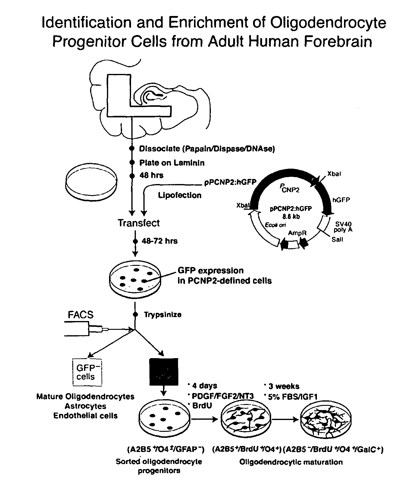
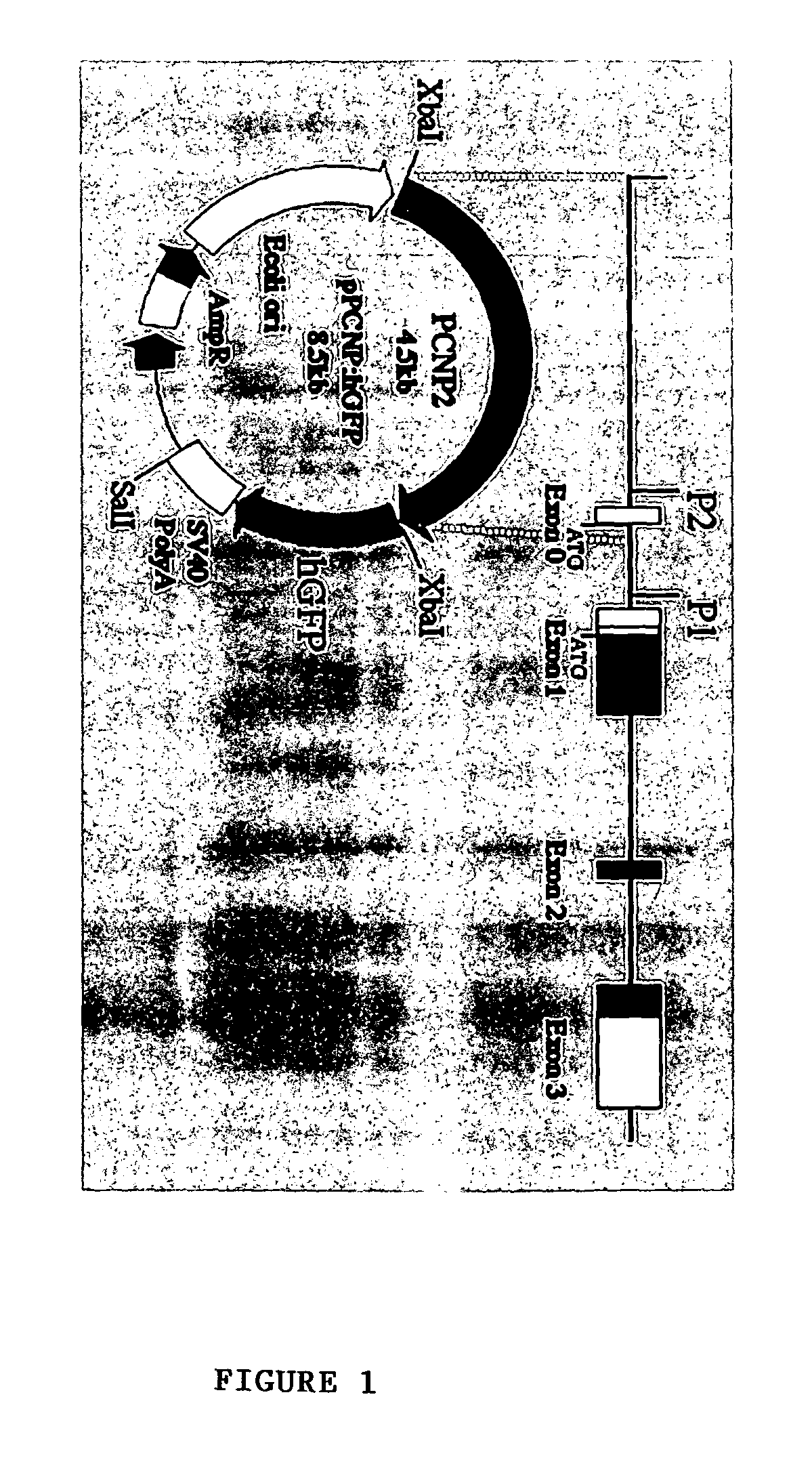
[0048]P / hCNP2:hGFP and P / hCNP2:lacZ Humanized GFP (hGFP), a mutant form of GFP optimized for expression in human cells (Levy, et al., 1996, which is hereby incorporated by reference), was placed under the control of the human CNP2 promoter (Douglas, et al., 1992; Monoh, et al, 1993; and Gravel, et al., 1996). The human CNP gene had been isolated previously (Gravel, et al., 1996), by screening a human fibroblast genomic library with a cDNA probe for rat CNP1. The human CNP gene was then subcloned into pBluescript, and the resultant plasmid was designated SK / hgCNP. This plasmid was digested with BgIII and XhoI to delete much of the gene downstream of the promoter region. The remaining BgIII and XhoI ends were then filled-in and blunt-end ligated, yielding plasmid SK / P1P2hCNP, in which both BgIII and XhoI were regenerated. A 1123 bp XhoI-XhoI fragment containing SV40 SD / SA-GFPh-SV40 poly(A) was then excised from pTα1:hGFP (Wang, et al., 1998), a...

example 2

Dissociates of Adult Human White Matter Harbored a Pool of Bipolar, A2B5+ Cells

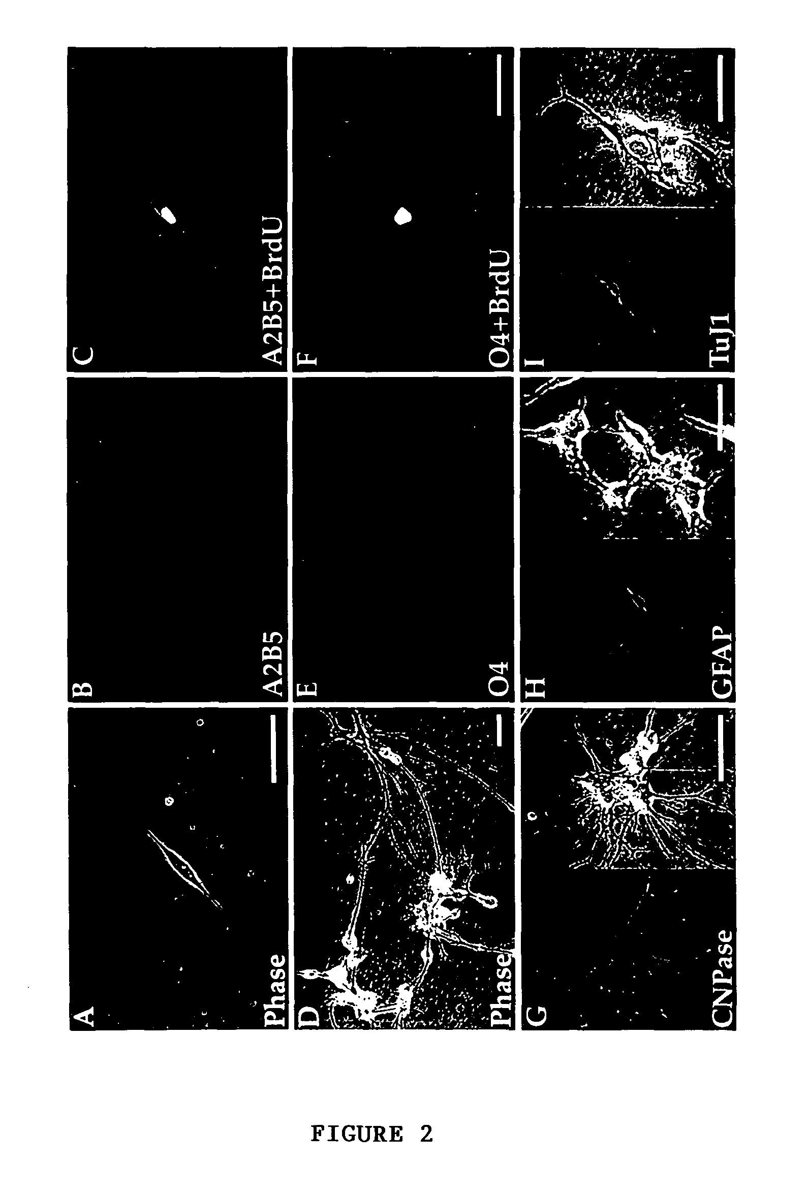
[0055]To fully characterize the cell phenotypes resident in adult human white matter, papain dissociates of surgically-resected frontal and temporal capsular white matter were obtained from 8 patients. These included 4 males and 4 females, who ranged from 24-65 years old. Three patients had temporal lobe resections for medication-refractory epilepsy; 2 were subjected to parenchymal excision during meningioma resection, 2 samples were taken during aneurysmal repair, and 1 was taken from the non-neoplastic approach to a histologically benign ganglioglioma. The monolayer cultures resulting from these white matter dissociations were stained after 5-7 days in vitro (DIV) for either of 2 oligodendrocytic markers, that included the epitopes recognized by the A2B5 and O4 antibodies. Additional, matched cultures were stained after 14 DIV, for either A2B5 or O4, for oligodendrocytic CNP protein, or for either neuro...

example 3

The CNP2 Promoter Targeted GFP Expression to a Bipolar, A2B5+ Phenotype

[0058]To identify either oligodendrocyte progenitor cells or their immature progeny, white matter dissociates were next transfected with plasmids encoding P / hCNP2:hGFP. Within 4 days after transfection with P / hCNP2:hGFP, a small proportion of GFP+ cells were noted. These were invariably small, bipolar cells, and constituted + / O4± / GFAP− / TuJ1−: 62.5±8.8% of P / hCNP2:hGFP+ cells expressed A2B5-IR, 21.1±7.5% were O4+, and another 7.3±3.2% expressed astrocytic GFAP. None were recognized by MAb TuJ1, which targets neuronal βIII-tubulin (Menezes, et al., 1994, which is hereby incorporated by reference). Thus, within the first 7-10 days in culture, P / hCNP2:hGFP selectively identified a population of bipolar, A2B5+ cells. When followed over the weeks thereafter, most of these P / hCNP2:hGFP+ cells developed into oligodendrocytes, that could be recognized by their small, multipolar, heavily branching profiles. Indeed, by 4 we...


PUM


Abstract

The present invention is directed to a method of separating oligodendrocyte cells or progenitor cells thereof from a mixed population of cells. It comprises selecting a promoter which functions only in the oligodendrocyte cells or progenitor cells thereof, introducing a nucleic acid molecule encoding a fluorescent protein under control of that promoter into the mixed population cells, allowing the oligodendrocyte cells or progenitor cells thereof to express the fluorescent protein, and separating the fluorescent cells from the mixed population cells, where the separated cells are the oligodendrocyte cells or progenitor cells thereof. The invention also relates to the isolated and enriched human oligodendrocyte cells or progenitor cells thereof.

Description

[0001]This application claims the benefit of U.S. Provisional Patent Application Ser. No. 60 / 104,809 filed Oct. 19, 1998.FIELD OF THE INVENTION[0002]The present invention relates generally to a method of separating cells of interest, in particular oligodendrocyte cells, from brain tissue.BACKGROUND OF THE INVENTION[0003]Throughout this application various publications are referenced, many in parenthesis. Full citations for these publications are provided at the end of the Detailed Description. The disclosures of these publications in their entireties are hereby incorporated by reference in this application.[0004]The damaged brain is largely incapable of functionally significant structural self-repair. This is due in part to the apparent failure of the mature brain to generate new neurons (Korr, 1980; Sturrock, 1982). However, the absence of neuronal production in the adult vertebrate forebrain appears to reflect not a lack of appropriate neuronal precursors, but rather their tonic i...

Claims


Application Information

Patent Timeline
IPC IPC(8): C12N5/02C12N5/071A61K35/12A61P25/00C12N5/079C12N5/0797
CPCC12N5/0622C12N5/0623A61K35/12C12N2510/00A61P25/00Y02A50/30
Inventor GOLDMAN, STEVEN A.WANG, SU
Owner CORNELL RES FOUNDATION INC
Why Eureka
Try Eureka