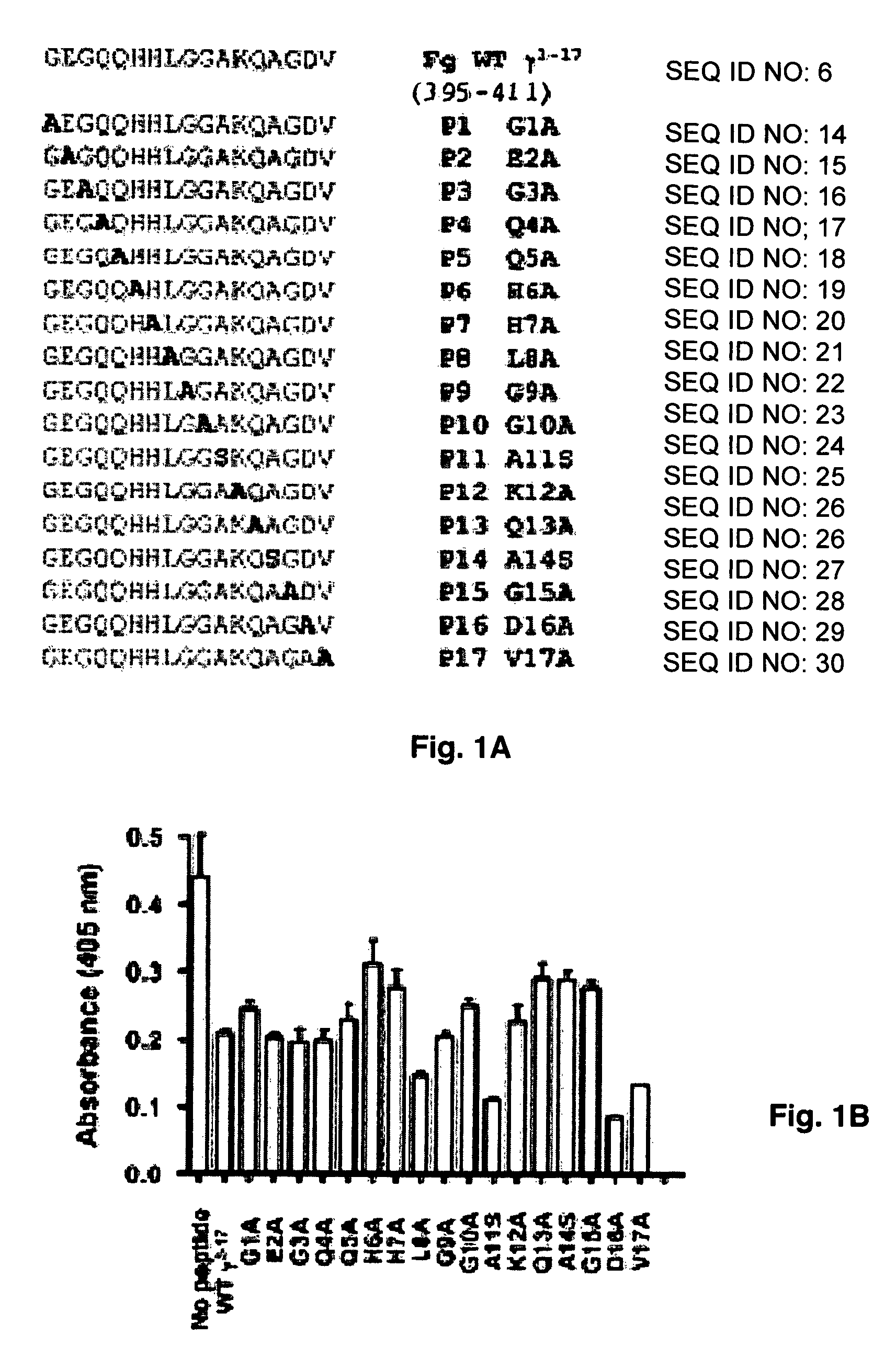
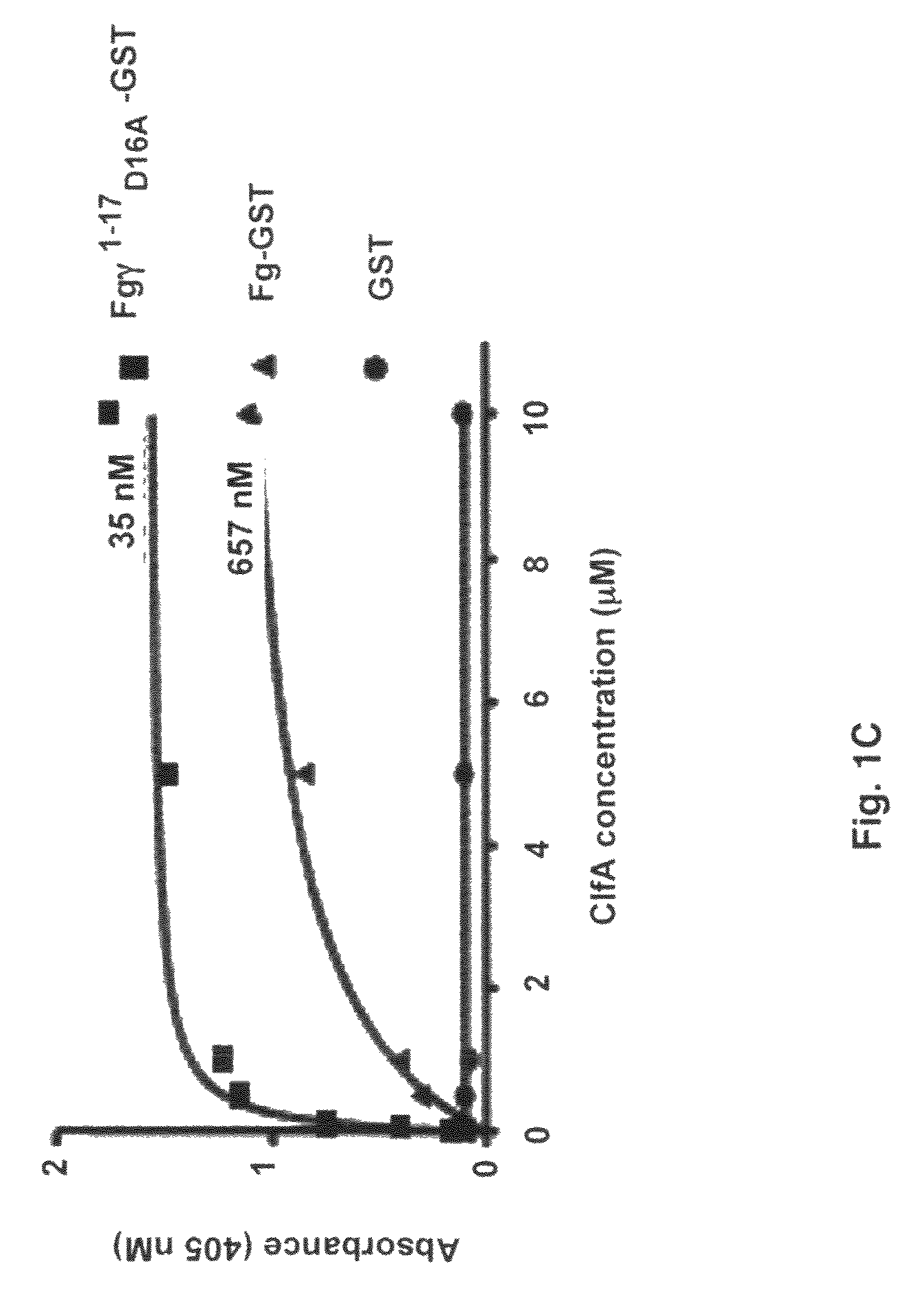
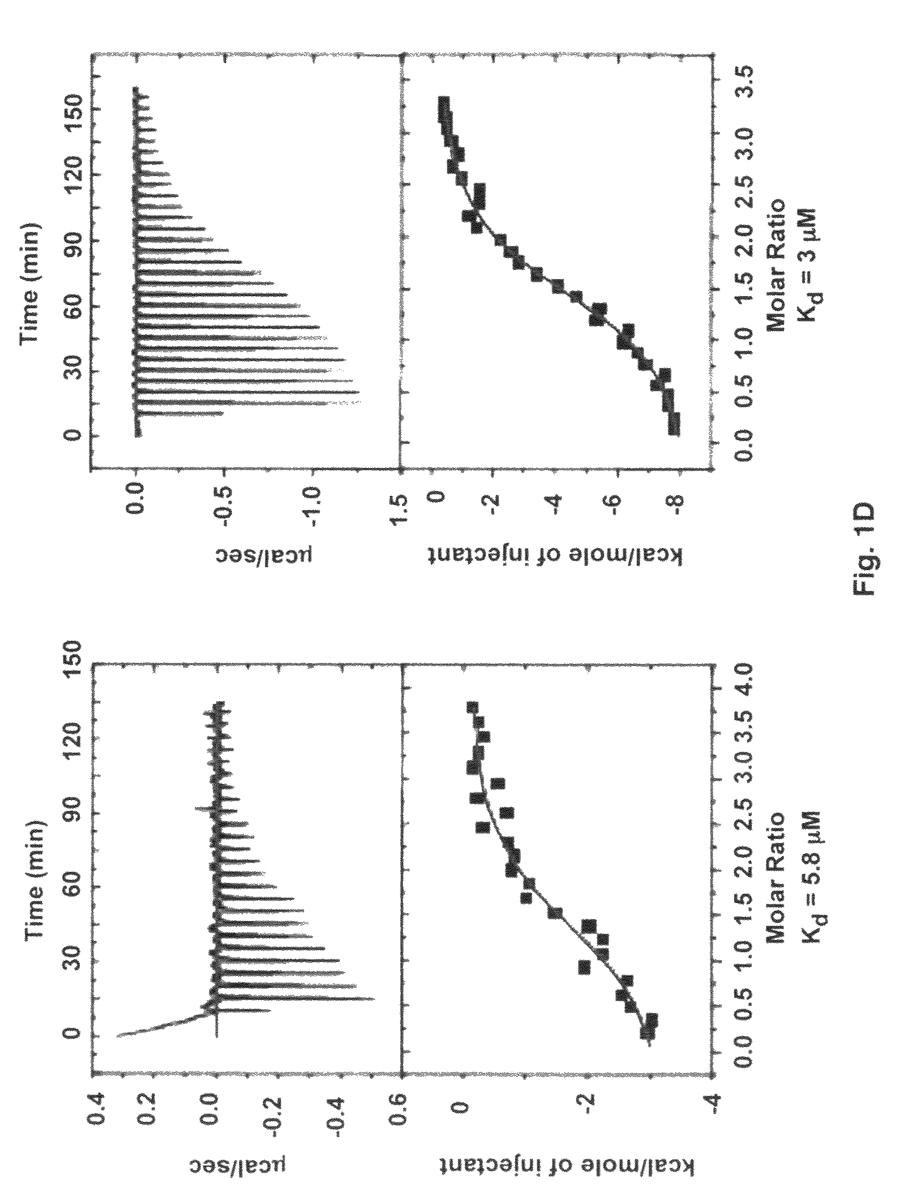
Crystal structure of Staphylococcus aureus clumping factor A in complex with fibrinogen derived peptide and uses thereof

a staphylococcus aureus and fibrinogen-derived peptide technology, applied in the field of crystal structure of staphylococcus aureus clumping factor a, can solve the problems of insufficient structural characterization of how clfa binds fg and its structure, and achieve the effect of little inhibitory effect and high affinity

Active Publication Date: 2012-10-02
TEXAS A&M UNIVERSITY
View PDF10 Cites 7 Cited by
  • Summary
  • Abstract
  • Description
  • Claims
  • Application Information

AI Technical Summary

Problems solved by technology

The continued emergence of antibiotic resistance among clinical strains has made the treatment of staphylococcal infections challenging, underscoring the need for new prevention and treatment strategies (1).
Thus, prior art is deficient in structural characterization of how ClfA binds Fg and its use in the design of vaccines and therapeutic compounds for the prevention and treatment of staphylococcal infections.

Method used


Image

  • Crystal structure of Staphylococcus aureus clumping factor A in complex with fibrinogen derived peptide and uses thereof
  • Crystal structure of Staphylococcus aureus clumping factor A in complex with fibrinogen derived peptide and uses thereof
  • Crystal structure of Staphylococcus aureus clumping factor A in complex with fibrinogen derived peptide and uses thereof

Examples


example 1

Bacterial Strains, Plasmids and Culture Conditions

[0062]Escherichia coli XL-1 Blue (Stratagene) was used as the host for plasmid cloning and protein expression. Chromosomal DNA from S. aureus strain Newman was used to amplify the ClfA DNA sequence. All E. coli strains containing plasmids were grown on LB media with ampicillin (100 μg / ml).

example 2

Manipulation of DNA

[0063]DNA restriction enzymes were used according to the manufacturer's protocols (New England Biolabs) and DNA manipulations were performed using standard procedures (30) (Sambrook and Gething, 1989). Plasmid DNA used for cloning and sequencing was purified using the Qiagen Miniprep kit (Qiagen). DNA was sequenced by the dideoxy chain termination method with an ABI 373A DNA Sequencer (Perkin Elmer, Applied Biosystems Division). DNA containing the N-terminal ClfA sequences were amplified by PCR (Applied Biosystems) using Newman strain chromosomal DNA as previously described (31). The synthetic oligonucleotides (IDT) used for amplifying clfA gene products and for cysteine mutations are listed in Table I.

[0064]

TABLE 1ClfA2295′-CCCGGATCCGGCACAGATATTACGAAT-3′(SEQ ID NO: 1)ClfA5455′-CCCGGTACCTCAAGGAACAACTGGTTTATC-3′(SEQ ID NO: 2)For disulfide mutant:rClfA3275′-TGCTTTTACATCACATTTAGTATTTAC-3′(SEQ ID NO: 3)fClfA3275′-GTAAATACTAAATGTGATGTAAAAGCA-3′(SEQ ID NO: 4)ClfA5415′-C...

example 3

Construction of Disulphide Mutants (Stable Form of ClfA)

[0065]Cysteine mutations were predicted by comparing ClfA221-559 to SdrG(273-597) disulfide mutant with stable closed conformations (32) and by computer modeling. A model of ClfA in closed conformation was built based on the closed conformation of the SdrG-peptide complex (25). The Cβ-Cβ distances were calculated for a few residues at the C-terminal end of the latch and strand E in the N2 domain. Residue pairs with Cβ-Cβ distance less than 3 Å were changed to cysteines to identify residues that could form optimum disulfide bond geometry. The D327C / K541C mutant was found to form a disulfide bond at the end of the latch. The cysteine mutations in ClfAD327C / K541C were generated by overlap PCR (33-34). The forward primer for PCR extension contained a BamHI restriction site and the reverse primer contained a KpnI restriction site. The mutagenesis primers contained complementary overlapping sequences. The final PCR product was digest...


PUM

PropertyMeasurementUnit
total volumeaaaaaaaaaa
pHaaaaaaaaaa

Abstract

The present invention provides a method for determining the structure of a microbial surface components recognizing adhesive matrix molecule in complex with fibrinogen, by providing a ClfA complexed with a fibrinogen gamma-peptide; determining a ClfA binding region of the fibrinogen gamma-peptide; determining one or more critical amino acid residues in the ClfA binding region of a native fibrinogen gamma-peptide that is critical for a ClfA:fibrinogen gamma-peptide interaction; determining one or more amino acid residues of the ClfA that binds to the ClfA binding region of the native fibrinogen gamma-peptide; modeling the structure of the ClfA binding region; determining the structure of the ClfA in complex with the :fibrinogen gamma-peptide interaction; and identifying one or more potential agent(s) that inhibit the ClfA:fibrinogen gamma-peptide interaction without affecting binding of other proteins to the fibrinogen gamma-peptide.

Description

CROSS-REFERENCE TO RELATED APPLICATIONS[0001]This non-provisional application claims benefit of priority under 35 U.S.C. §119(e) of provisional U.S. Ser. No. 61 / 133,537, filed Jun. 30, 2008, the entirety of which is hereby incorporated by reference.FEDERAL FUNDING LEGEND[0002]This invention was produced using funds obtained through a National Institutes of Health grant (AI20624). Consequently, the Federal government has certain rights in this invention.BACKGROUND OF THE INVENTION[0003]1. Field of the Invention[0004]The present invention relates generally to the fields of protein chemistry, immunology, therapeutic pharmaceuticals, and vaccine development. More specifically, the present invention discloses crystal structure of Staphylococcus aureus clumping factor A (ClfA) in complex with fibrinogen (Fg) derived peptide and its use in the design of ClfA targeted vaccines and therapeutic agents (including monoclonal antibodies).[0005]2. Description of the Related Art[0006]Staphylococcu...

Claims


Application Information

Patent Timeline
Patent Type & Authority Patents(United States)
IPC IPC(8): G06F19/00G01N31/00
CPCA61K38/363A61P31/04A61K38/00C07K14/31C07K2319/035C07K16/1271C07K16/18C07K14/75C07K16/36G01N33/68G01N2333/31G01N2333/75
Inventor HOOK, MAGNUSKO, YA-PINGSMEDS, EMANUELGANESH, VANNAKAMBADI K.
Owner TEXAS A&M UNIVERSITY
Why Eureka
Try Eureka