IGF-1R specific antibodies useful in the detection and diagnosis of cellular proliferative disorders

a specific antibody and cellular proliferative technology, applied in the field of biotechnology, can solve the problems of insufficient sensitive and specific detection of most types of early-stage cancer, slow and frustrating progress in understanding the progression of cancer and early detection, and achieve the effect of high affinity

Inactive Publication Date: 2013-01-01
MERCK SHARP & DOHME CORP +1
View PDF2 Cites 63 Cited by
  • Summary
  • Abstract
  • Description
  • Claims
  • Application Information

AI Technical Summary

Benefits of technology

[0023]Administration of the antibodies of the present invention in any of the conventional ways known to one skilled in the art (e.g., topical, parenteral, intramuscular, etc.), will provide an extremely useful method of detecting dysplastic cells in a sample as well as allowing a clinician to monitor the therapeutic regiment of a patient undergoing treatment for a hyperproliferative disorder associated with or mediated by expression of IGF-1R.
[0052]In accordance therewith, an embodiment of the invention is drawn to a method of diagnosis, preferably in vitro, of illnesses connected with an overexpression or an under expression, preferably overexpression of the IGF-IR receptor. Samples are taken from the patient and subject to any suitable immunoassay with IGF-1R specific antibodies to detect the presence of IGF-1R. Preferably, the biological sample is formed by a biological fluid, such as serum, whole blood, cells, a tissue sample or biopsies of human origin. The sample, may for example include, biopsied tissue, which can be conveniently assayed for the presence of a pathological hyperproliferative oncogenic disorder associated with expression of IGF-1R.
[0053]Once a determination is made of the amount of IGF-1R present in the test sample, the results can be compared with those of control samples, which are obtained in a manner similar to the test samples but from individuals that do not have or present with a hyperproliferative oncogenic disorder associated with expression of IGF-1R, e.g., ovarian cancer. If the level of the IGF-1R polypeptide is significantly elevated in the test sample, it may be concluded that there is an increased likelihood of the subject from which it was derived has or will develop said disorder, e.g., ovarian cancer.
[0062]In certain embodiments, detection of dysplastic cells or neoplastic tissue associated with expression of IGF-1R is made possible by exposing cells expressing IGF-1R to a conventional IGF-1R antagonist moiety (treated cells) and then contacting the treated cells to the antibodies disclosed herein to assess the ability of the conventional IGF-1R antagonist moiety in down-regulating cell surface expression of IGF-1R. The step of contacting the treated cells to the anti-IGF-1R antibodies of the invention may be carried out simultaneously with the antibodies disclosed herein or at a subsequent time point. Alternatively, IGF-1R expression in treated cells may be assayed over several time points post treatment to ascertain whether the patient is responding to treatment. This entails the same steps as above except over time. Thus, a down regulation of IGF-1R expression as indicated by low binding of the treated cells with the 12B1 antibody may indicate that the patient is responding favorably to treatment with the conventional IGF-1R specific treatment, while no change in expression levels or an increase in expression levels of IGF-1R as determined by the binding of the antibodies of the invention to IGF-1R on treated cells may indicate that the patient is not responding favorably to treatment with the conventional IGF-1R antagonist moiety. A consequence of the above assay is that it will provide the treating physician valuable information in determining whether intensive or invasive protocols such as colonoscopy, surgery or chemotherapy would be needed for effective diagnosis or treatment. Such detection would be helpful not only for patients not previously diagnosed with a hyperproliferative oncogenic disorder such as cancer but also in those cases where a patient has previously received or is currently receiving therapy for a pathological effect of any oncogenic disorder associated with expression of IGF-1R.

Problems solved by technology

The progress in the understanding of cancer progression and early detection has been slow and frustrating due to the complex multifactorial nature and heterogeneity of the cancer syndrome.
One of the challenges in drug development is to show in pre-clinical development, in clinical trials and with an approved agent that the anti-IGF-1R therapeutic is effective.
Currently employed diagnostic techniques such as medical imaging, tissue biopsy and bioanalytical assay of body fluids by enzyme linked immunosorbent assay (ELISA) are insufficiently sensitive and specific to detect most types of early-stage cancers.
Moreover, these assays are labour intensive, time consuming, expensive and don't have multiplexing capability.
Indeed, the paucity of reliable biomarkers that show efficacy in detecting IGF-1R has hampered industry efforts in evaluating the efficacy of numerous anti-IGF-1R therapeutic protocols.

Method used


Image

  • IGF-1R specific antibodies useful in the detection and diagnosis of cellular proliferative disorders
  • IGF-1R specific antibodies useful in the detection and diagnosis of cellular proliferative disorders
  • IGF-1R specific antibodies useful in the detection and diagnosis of cellular proliferative disorders

Examples


example-1

Generation and Selection of the Murine Monoclonal Antibody (MAb)

[0378]With the aim of generating antibodies, particularly monoclonal antibodies specifically directed against IGF-IR that do not cross-react with IR, a protocol comprising 4 screening steps was performed.

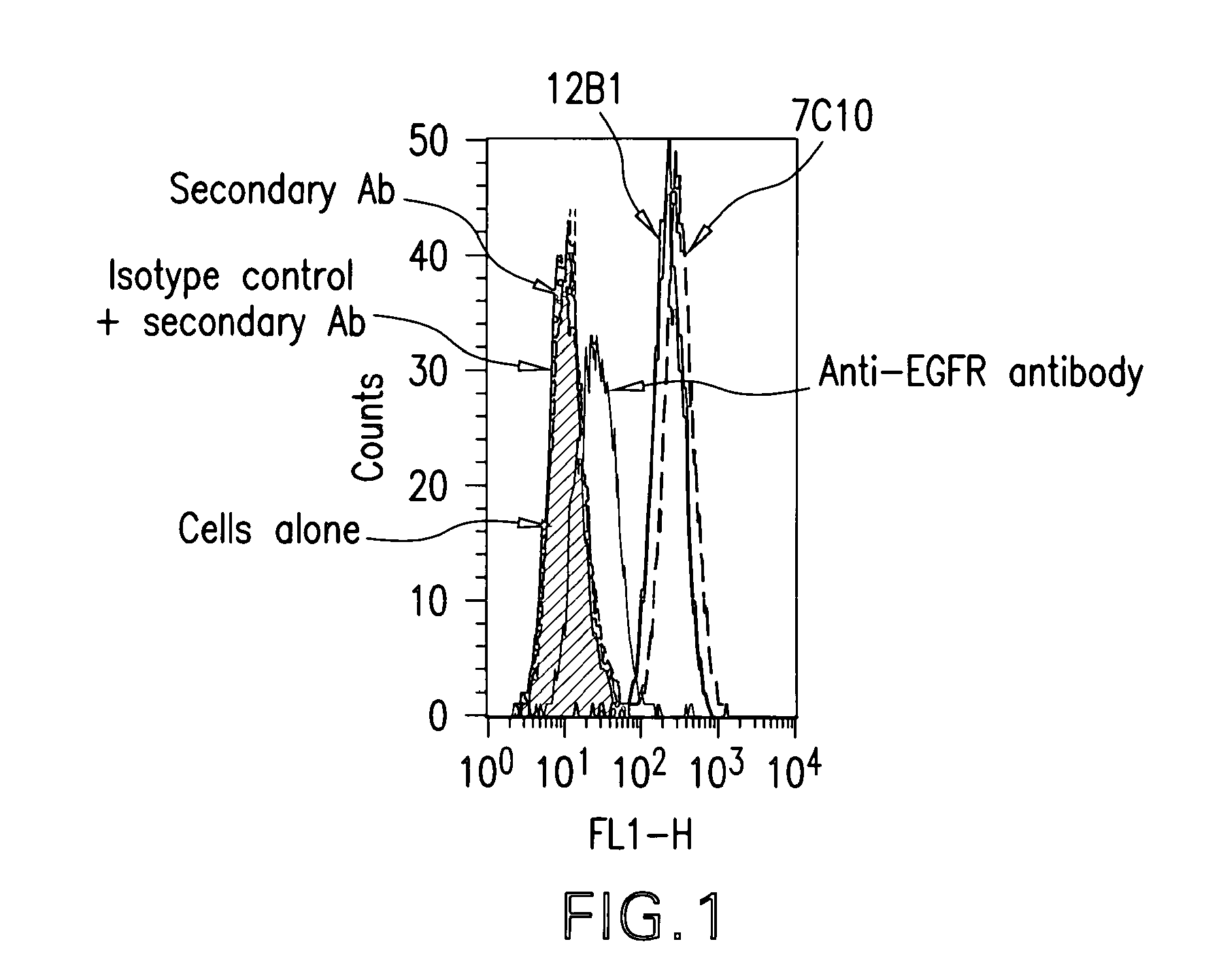
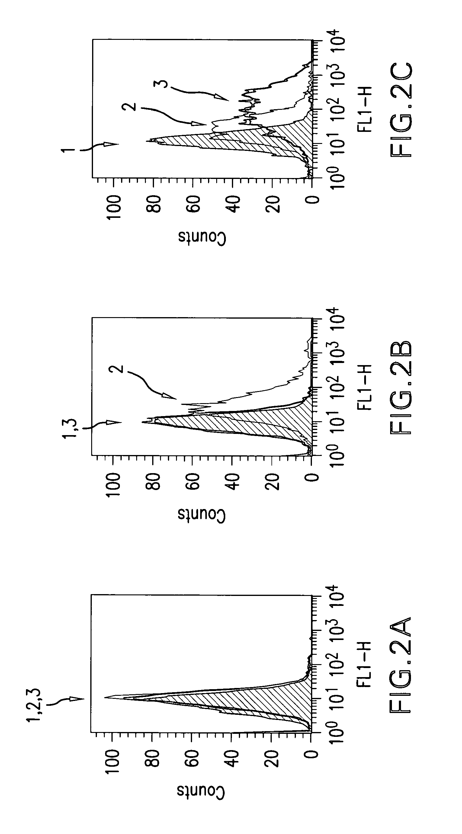
[0379]The protocol comprised:[0380]immunizing mice with the human recombinant IGF-IR, in order to generate hybridomas,[0381]screening the cell culture supernatants by ELISA on the human recombinant protein used for immunization,[0382]testing all the positive supernatants of hybridomas resulting of this first ELISA on the native receptor overexpressed on MCF-7 tumor cells,[0383]evaluating the supernatants of hybridomas positive in the two first screenings in terms of differential recognition of IGF-IR versus IR on insect cells infected with baculoviruses respectively expressing either IGF-IR or IR.

[0384]The various steps outlined above are detailed herebelow.

[0385]For the immunization stage, mice were injected subcutaneo...

example 2

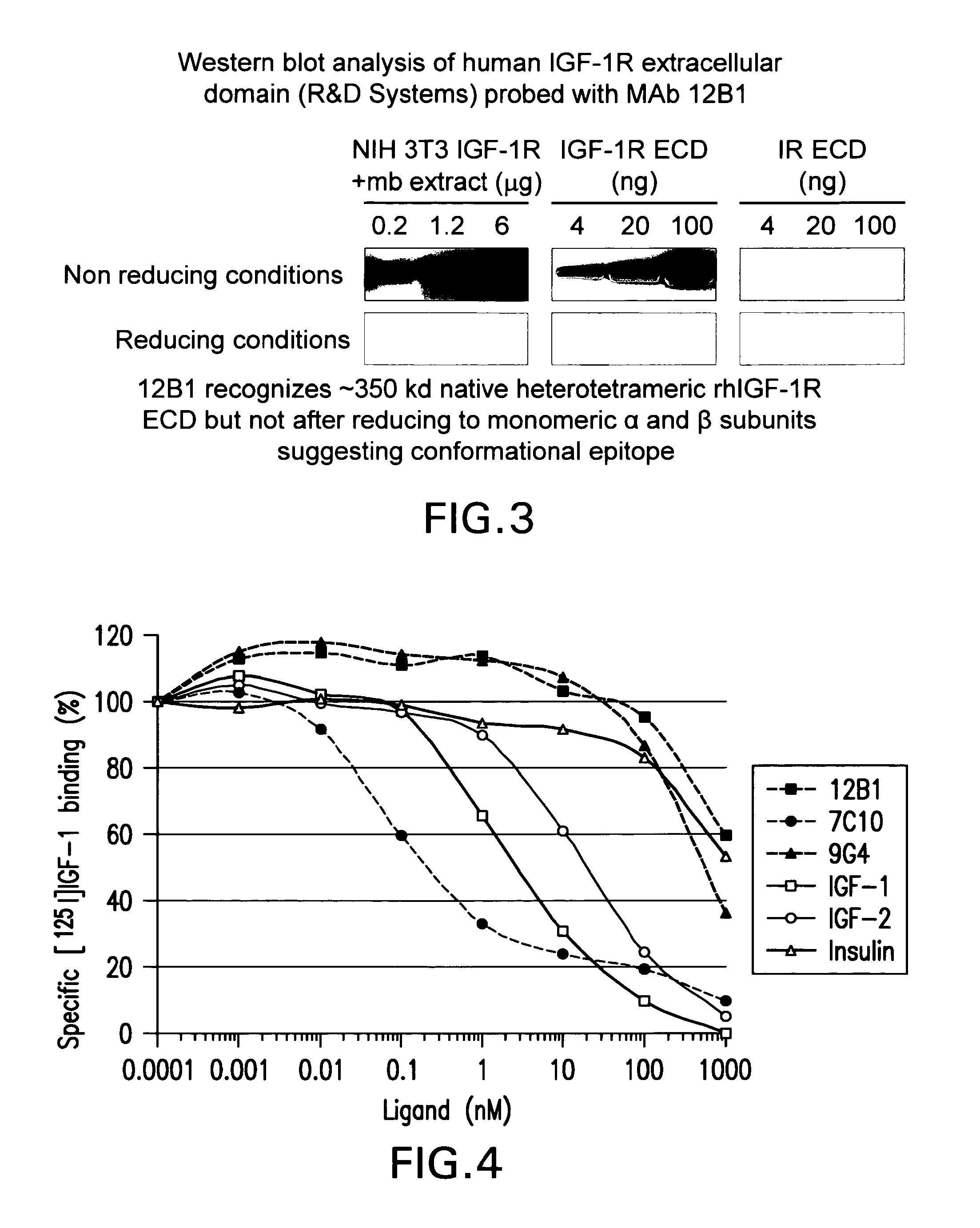
Western Blot Experiments

Material and Methods

Proteins and Membrane Extract

[0390]Recombinant human insulin receptor (IR) and insulin-like growth factor 1 receptor (IGF-1R) extracellular domains (ECD) were purchased from R&D Systems (Lille, France). Membrane extracts of NIH 3T3 cells overexpressing IGF-1R were obtained as detailed here below. Briefly, after cell lysis in 10 mM Tris-HCl pH 7.5 buffer, whole cell membranes were collected by centrifugation at 105,000 g for 1 h at 4° C. The pellet was re-suspended in 50 mM Tris-HCl pH 7.5 buffer containing 150 mM NaCl, 0.5% IGEPAL, 0.5% Triton X-100, 0.25% sodium deoxycholate and protease inhibitors, and stirred overnight at +4° C. Insoluble material was separated from the soluble extract containing hIGF-1R by centrifugation at 10,000 g for 10 mM at +4° C. Soluble membrane extracts were analyzed for protein concentration by the bicinchoninic assay.

Electrophoresis and Western Blot

[0391]Proteins were analyzed by SDS-PAGE electrophoresis on C...

example 3

Cloning Strategy of Genes Coding for the Variable Regions of the Heavy and Light Chains of the Monoclonal Antibody (mAb) 12B1

[0394]Total RNA was extracted from 107 cells of hybridomas secreting the antibody 12B10 by using the TRI REAGENT™ (according to the instructions given by the supplier, SIGMA, T9424). The first cDNA strand was synthesized with the aid of the ‘First strand cDNA synthesis’ kit of Amersham-Pharmacia (#27-9621-01, according to the instructions given by the supplier). For the two chains, the reaction was primed with the oligonucleotide Not I-d(T)18, comprised in the Kit.

[0395]The cDNA:mRNA hybrid thus obtained was used for the amplification by PCR of the genes coding for the heavy and light chains of the 12B1 mAb. The PCR were carried out by using a combination of oligonucleotides specific for the heavy and light (Kappa) chains of mouse immunoglobulins. The primers corresponding to the 5′ ends hybridize in the region corresponding to the signal peptides (Table 2 for...


PUM

PropertyMeasurementUnit
flow rateaaaaaaaaaa
W/Waaaaaaaaaa
W/Waaaaaaaaaa

Abstract

The present invention relates to mammalian antibodies, designated 12B1 and antigen-binding portions thereof that specifically bind to insulin-like growth factor I receptor (IGF-IR), preferably human IGF-IR. Also included are chimeric, bispecific, derivatized, single chain antibodies derived from the antibodies disclosed herein. Nucleic acid molecules encoding the mammalian antibodies as well as methods of use thereof are also disclosed. Also included are pharmaceutical compositions comprising these antibodies and methods of using the antibodies and compositions thereof for treatment and diagnosis of pathological hyperproliferative oncogenic disorders associated with expression of IGF-1R.

Description

CROSS REFERENCE TO RELATED APPLICATIONS[0001]This application is a U.S. National Phase application under 35 U.S.C. §371 of PCT Application No. PCT / US2008 / 009065 filed Jul. 25, 2008, which claims priority from U.S. Provisional Application Ser. No. 60 / 962,688, filed Jul. 31, 2007.FIELD OF THE INVENTION[0002]This invention is related to the field of the biotechnology and in particular with new recombinant monoclonal antibodies, which recognize epitopes expressed on the insulin-like growth factor 1 receptor 1(IGF-1R), preferably human IGF-1R.BACKGROUND OF THE INVENTION[0003]The present invention relates to novel antibodies that are selective for the IGF-1R cell surface receptor. Also included is derivation of recombinant antibodies, e.g., chimeric, humanized or veneered versions including single chain Fv fragments (scFv) from the mammalian antibodies detailed herein and designated “12B1”. The invention likewise comprises utilization of the murine or recombinant antibodies derived theref...

Claims


Application Information

Patent Timeline
Patent Type & Authority Patents(United States)
IPC IPC(8): C07K16/00A61K39/395
CPCC07K16/2863C07K2317/56C07K2317/565G01N33/574G01N2333/71C07K2317/76A61P35/00
Inventor GOETSCH, LILIANEBROOKS, DAVIDCHASTAIN, MICHAELZHANG, ZHI-QIANGCORVAIA, NATHALIE
Owner MERCK SHARP & DOHME CORP
Why Eureka
Try Eureka