Digital light processing hyperspectral imaging apparatus

a hyperspectral imaging and digital light technology, applied in the field ofspectroscopic imaging, can solve the problems of increasing infection and pain, irritating surrounding structures, and inflammation often spreading to the outer covering, and achieve the effect of maintaining body hea

Active Publication Date: 2013-03-26
BOARD OF RGT THE UNIV OF TEXAS SYST
View PDF23 Cites 280 Cited by
  • Summary
  • Abstract
  • Description
  • Claims
  • Application Information

AI Technical Summary

Benefits of technology

[0030]Another embodiment of the present invention is a method for hyperspectral surgery of a human subject by capturing image data of one or more fluorescent targets generating a wide spectrum electromagnetic radiation comprising: covering the human subject with NIR transparent material to provide privacy, accessibility and maintain body heat; separating the electromagnetic radiation into at least one spectral band or the band spectrum using a spatial light modulator; illuminating the fluorescent target with the at least one spectral band or the band spectrum; collecting the reflected at least one spectral band or the band spectrum resulting from the one or more fluorescent targets; and, collecting at least one spectral band or the band spectrum to one or more detector to form the image data, wherein the hyperspectral surgery is used in surgical procedures selected from a cholecystectomy, amputation, burn, skin flap evaluation, visualizing areas of angiogenesis, probes that bind antigens and absorbs near-infrared during pathological evaluations and in vivo, quality control of pharmaceuticals, monitoring vascular changes and drug discovery in response to pharmaceuticals, monitoring diabetic retinopathy, cancer, diabetes, sickle cell, anemia, bilirubin, raynauds, ulcers, burns, skin flaps, surgery, gallbladder, brain, monitoring wound healing, measuring oxygenation of retina, measuring optic nerve oxygenation, measuring macular pigments, measuring pigments in retinal photoreceptors and retinal pigment epithelium, diagnosis of autoimmune retinitis, diagnosis of infectious retinitis, diagnosis of infiltrative neoplastic conditions, evaluating disease biomarkers, diagnosis of ocular trauma injuries, measuring oxygenation of a kidney and early detection of wound infections.
[0045]In another aspect, the present invention includes a method for hyperspectral surgery by capturing image data of one or more object generating a wide spectrum electromagnetic radiation including, by not limited to the following steps: separating the electromagnetic radiation into one or more single wavelength bands or a spectrum band using a spatial light modulator; illuminating the object with the one or more single wavelength bands or the spectrum band; collecting the reflected one or more single wavelength bands or the spectrum band resulting from the one or more object; and, directing the collected or more single wavelength bands or spectrum band to one or more detector to form the image data, wherein the hyperspectral surgery is used in a surgical procedure selected from the group consisting of: a cholecystectomy, amputation, burn, skin flap evaluation, visualizing areas of angiogenesis, probes that bind antigens and absorbs near-infrared during pathological evaluations and in vivo, quality control of pharmaceuticals, monitoring vascular changes and drug discovery in response to pharmaceuticals, monitoring diabetic retinopathy, diseases such as cancer, diabetes, sickle cell, anemia, bilirubin, raynauds, ulcers, burns, skin flaps, surgery, gallbladder, brain, monitoring wound healing, measuring oxygenation of retina, measuring optic nerve oxygenation, measuring macular pigments, measuring pigments in retinal photoreceptors and retinal pigment epithelium, diagnosis of autoimmune retinitis, diagnosis of infectious retinitis, diagnosis of infiltrative neoplastic conditions, evaluating disease biomarkers, diagnosis of ocular trauma injuries, measuring oxygenation of a kidney and early detection of wound infections. The method can further include the step of covering the human subject with NIR transparent material to provide privacy, accessibility and maintain body heat.

Problems solved by technology

Extreme cases may result in necrosis and rupture, and could cause further infection and pain resulting from such inflammation.
Inflammation often spreads to its outer covering, irritating surrounding structures such as the diaphragm and bowel.
Nausea and vomiting may also occur.
Complaints of gas, nausea, and abdominal discomfort after meals are the most common, but they may be vague and indistinguishable from similar complaints in people without gallbladder disease.
However, any surgery involves risk factor associated with it, which can cause complications.
The most common associated risk with Cholecsytectomy is the injury to the common bile duct (CBD) which is hidden, as it lies below a layer of fat.
Under normal conditions, patients recover within a day or two; however, complications may still occur.
During a cholecystectomy, one of the important problems faced by the surgeon is visibility of structures below the fat layer.
While this process is current the industry standard, the procedure is time consuming.
It requires removal of tissue from the patient, processing time, and has inherent sampling error.
In addition, the major limitation of this process is the delay in providing the surgeon with clinically relevant information at the time of surgery.
The aforementioned hyperspectral imaging systems are slow for routine clinical practice.
In addition, there are no methods for directly imaging the in vivo level of the biomolecules in live humans or animals during clinical visits or during surgical (open, endoscopic or laparoscopic) operations.

Method used


Image

  • Digital light processing hyperspectral imaging apparatus
  • Digital light processing hyperspectral imaging apparatus
  • Digital light processing hyperspectral imaging apparatus

Examples


Embodiment Construction

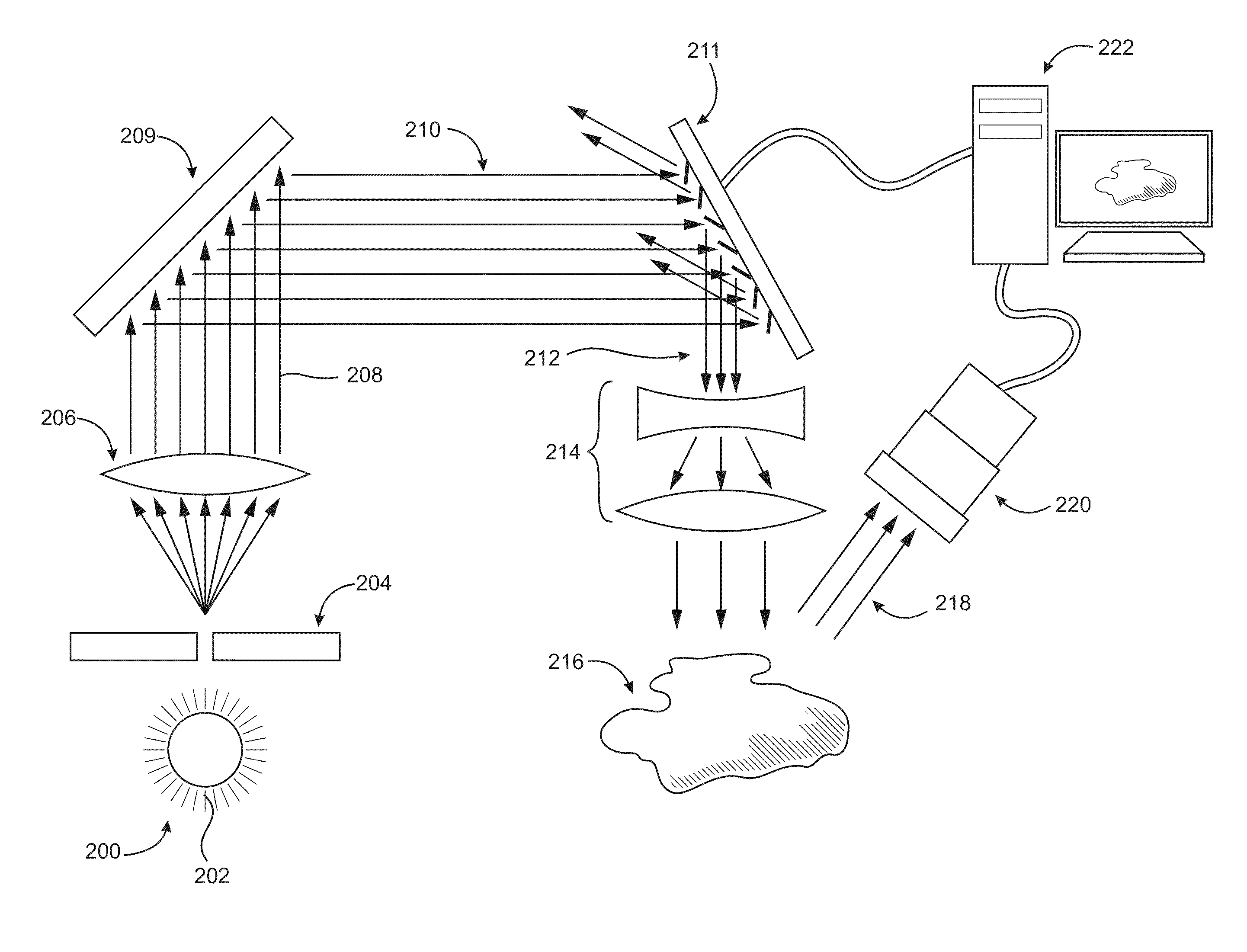
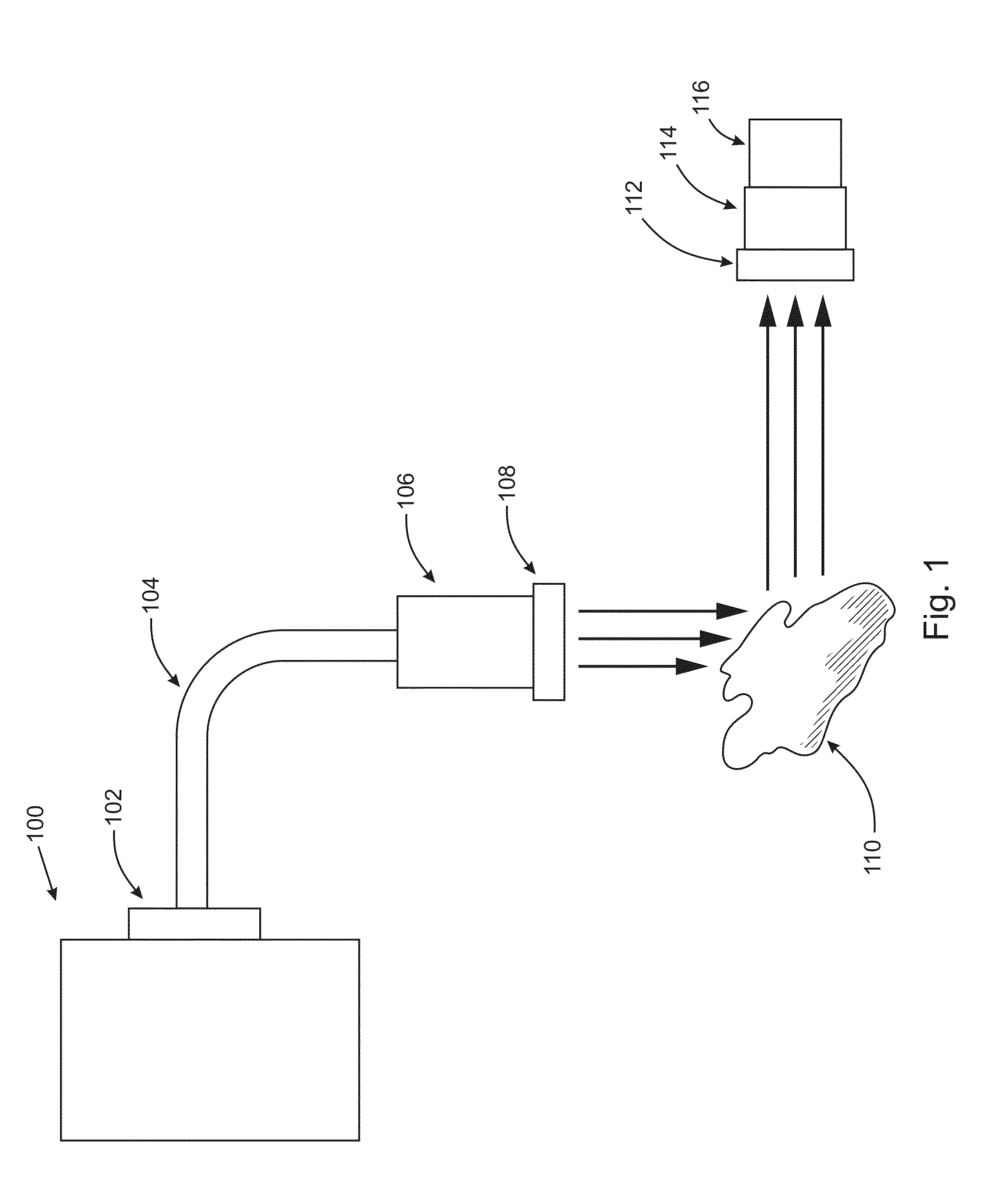
[0159]At the outset, it should be appreciated that like drawing numbers on different drawing views identify identical, or functionally similar, structural elements of the invention. While the present invention is described with respect to what is presently considered to be the preferred aspects, it is to be understood that the invention as claimed is not limited to the disclosed aspects.

[0160]Furthermore, it is understood that this invention is not limited to the particular methodology, materials and modifications described and as such may, of course, vary. It is also understood that the terminology used herein is for the purpose of describing particular aspects only, and is not intended to limit the scope of the present invention, which is limited only by the appended claims.

[0161]Unless defined otherwise, all technical and scientific terms used herein have the same meaning as commonly understood to one of ordinary skill in the art to which this invention belongs. As used herein, D...


PUM


Abstract

A hyperspectral imaging system having an optical path. The system including an illumination source adapted to output a light beam, the light beam illuminating a target, a dispersing element arranged in the optical path and adapted to separate the light beam into a plurality of wavelengths, a digital micromirror array adapted to tune the plurality of wavelengths into a spectrum, an optical device having a detector and adapted to collect the spectrum reflected from the target and arranged in the optical path and a processor operatively connected to and adapted to control at least one of: the illumination source; the dispersing element; the digital micromirror array; the optical device; and, the detector, the processor further adapted to output a hyperspectral image of the target. The dispersing element is arranged between the illumination source and the digital micromirror array, the digital micromirror array is arranged to transmit the spectrum to the target and the optical device is arranged in the optical path after the target.

Description

CROSS-REFERENCE TO RELATED APPLICATIONS[0001]This patent application claims priority to U.S. Provisional Patent Application No. 61 / 087,714, filed Aug. 10, 2008, U.S. Provisional Patent Application No. 61 / 087,715, filed Aug. 10, 2008, and U.S. Provisional Patent Application No. 61 / 168,347, filed Apr. 10, 2009, which applications are incorporated herein by reference.STATEMENT REGARDING FEDERALLY SPONSORED RESEARCH OR DEVELOPMENT[0002]This invention was made with government support under Grant Number 5 U24 DK076169-02 awarded by the National Institutes of Health. The government has certain rights in the invention.FIELD OF THE INVENTION[0003]The present invention relates generally to the field of spectroscopic imaging that results in collecting / measuring spectroscopic image data and the associated algorithms that result in chemically encoded images of tissue, including devices, methods and systems for fluorescence imaging of tissues in real-time. Additionally, the present invention rela...

Claims


Application Information

Patent Timeline
Patent Type & Authority Patents(United States)
IPC IPC(8): A61B6/00
CPCA61B5/0071A61B5/0075A61B5/0084A61B5/0086A61B5/14551G01J3/02G01J3/0208G01J3/021G01J3/0229G01J3/0264G01J3/2823G01N21/31G01N21/39G01J3/10A61B5/0059G01J3/2803A61B5/411A61B5/413G01J2003/1213G01J2003/1282A61B5/4842A61B5/4064A61B3/12G06T2207/10036
Inventor ZUZAK, KARELCADEDDU, JEFFREY A.UFRET-VINCENTY, RAFAELFRANCIS, ROBERT P.LIVINGSTON, EDWARD
Owner BOARD OF RGT THE UNIV OF TEXAS SYST
Why Eureka
Try Eureka