
Surgical applications for BMP binding protein

a technology of bmp and binding protein, which is applied in the field of surgical applications of bmp binding protein, can solve the problems of limiting clinical use, limiting the usefulness of bmp-7 (also known as op-1), and the potential systemic adverse effects of high-dose bmps when used for longer fusions, etc., and achieves the effects of increasing the rate and degree, and reducing the risk of op-1

Active Publication Date: 2013-04-09
RGT UNIV OF CALIFORNIA +1
View PDF28 Cites 12 Cited by
  • Summary
  • Abstract
  • Description
  • Claims
  • Application Information

AI Technical Summary

Benefits of technology

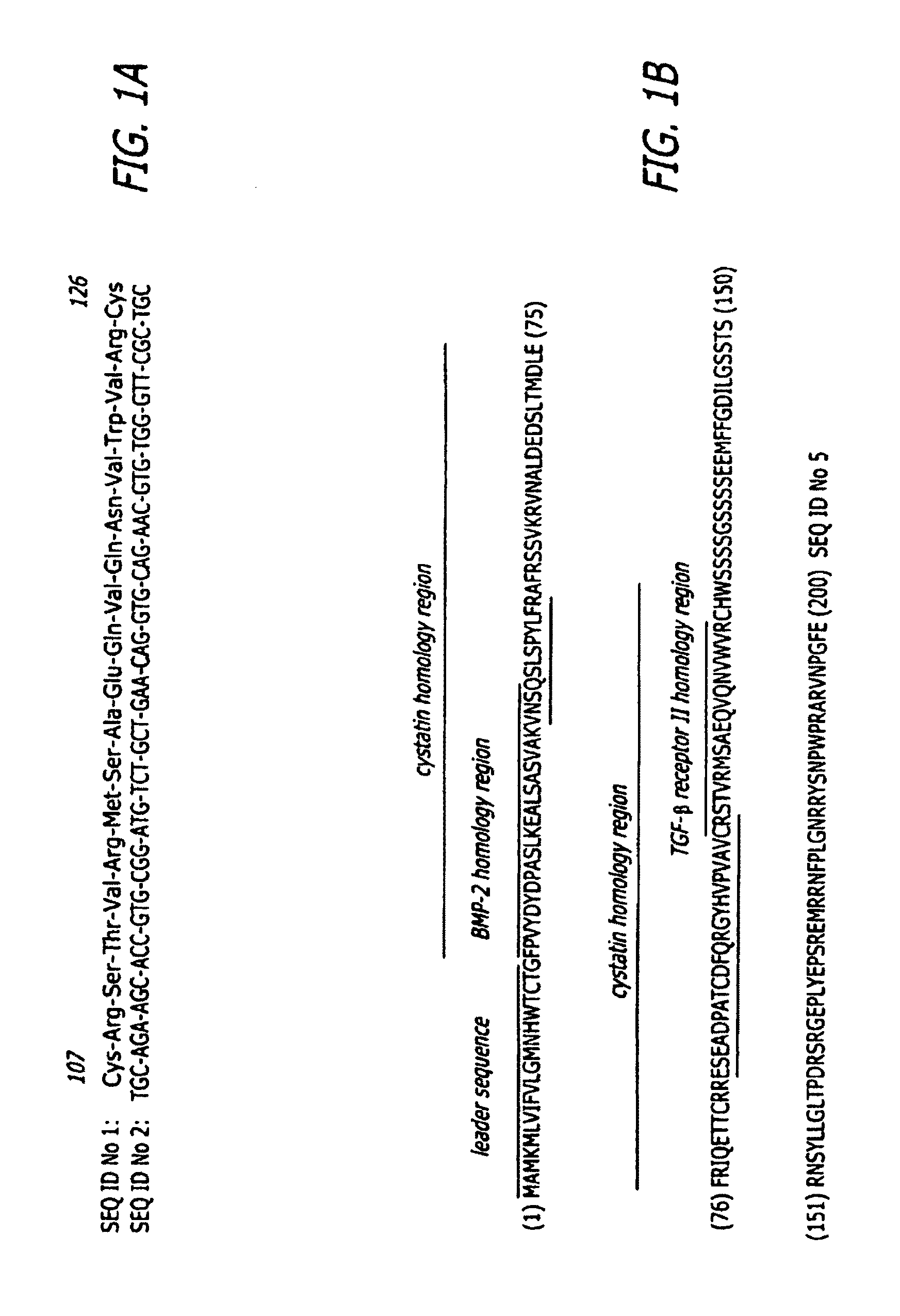
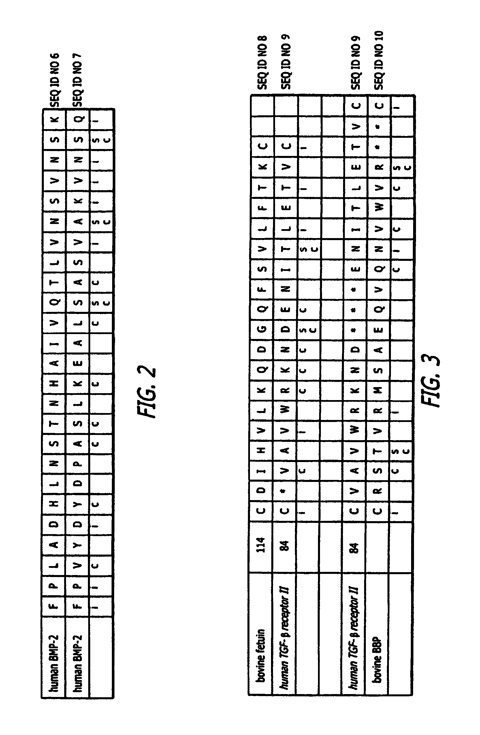
[0010]The inventions are related to a cyclic peptide designated BMP Binding Peptide (BBP) that avidly binds growth factors, such as rhBMP-2. BBP increases the rate and degree to which rhBMP-2 induces bone formation. BBP may accomplish this by increasing the residency rates of other bone growth factors. However, BBP alone induces calcification of chondrogenic, osteogenic and osteoblastic cells. Compositions and substrates including BBP, antibodies to BBP and methods of using BBP are useful in applications relating to bone.
[0012]In one embodiment, the invention includes methods and devices for increasing the residency times of bone inducing substances, such as BMP. In one embodiment, the invention includes methods and devices for increasing the rate and overall osteogenic activity of bone inducing substances, such as BMP. Further, in one embodiment, the invention includes methods and devices for reducing time required for the effect of osteogenesis and calcification of bone inducing substances.
[0015]This invention is advantageous at least in that BBP alone or in combination with other growth factors enhances calcification of chondrogenic or osteogenic precursor cells. Further, this invention is advantageous at least in that BBP enhances osteogenesis to occur faster to a greater extent, which may improve the clinical rate and effectiveness of treatment with BMP, and reduce doses and therefore the costs and side effects of BMP treatment alone.

Problems solved by technology

However, the cost of using minimally effective dosages of BMP-7 (also known as OP-1), for example has been a limiting factor in clinical use.
However, their usefulness is limited by expense and by local adverse effects such as unwanted ectopic bone formation and inflammatory responses associated with doses currently used for spinal fusion procedures.
Furthermore, potential systemic adverse effects of high dose BMPs when to be used for longer fusions has not been fully determined.
However, its high expense as well as the reported local side effects such as unwanted bone formation and dangerous swelling at the neck after the anterior cervical fusion procedure has prevented its extensive use for spinal procedures.
The main limitation is the need for better delivery systems that provide a sustained, biologically appropriate concentration of BMP-2 at the side of fusion bed.
Delivery needs to be sustained, because BMPs have exceedingly short biological half-lives, usually the order of minutes or hours, rather than the days or weeks needed to stimulate a complete osteogenic response.

Method used


Image

  • Surgical applications for BMP binding protein
  • Surgical applications for BMP binding protein
  • Surgical applications for BMP binding protein

Examples


example 1

Extraction and Separation of Non-Collagenous Bone Proteins (NPCs)

[0085]Methods: NCPs were extracted from defatted, demineralized human cortical bone powder with 4 M GuHCl, 0.5 M CaC12, 2 mM N-ethylmalemide, 0.1 mM benzamidine HCl, and 2 mM NaN3 for 18 hr at 6° C. Residual collagen and citrate-soluble NCPs were extracted by dialysis against 250 mM citrate, pH 3.1 for 24 hours at 6° C. The residue was pelleted by centrifugation (10,000×g at 6° C. for 30 min), defatted with 1:1 (v / v) chloroform: methanol for 24 hr at 23° C., collected by filtration and dried at 22° C. The material was resuspended in 4 M GuHCl, dialyzed against 4 M GuHCl, 0.2% (v / v) Triton X-100, 100 mM Tris-HCl, pH 7.2 for 24 hr at 6° C., then dialyzed against water, and centrifuged at 10,000×g for 30 min at 6° C. The pellet was lyophilized and subsequently separated by hydroxyapatite chromatorgraphy.

[0086]Chromatography was conducted using a BioLogic chromatography workstation with a CHT-10 ceramic hydroxyapatite colu...

example 2

In Vivo Activity of BBP

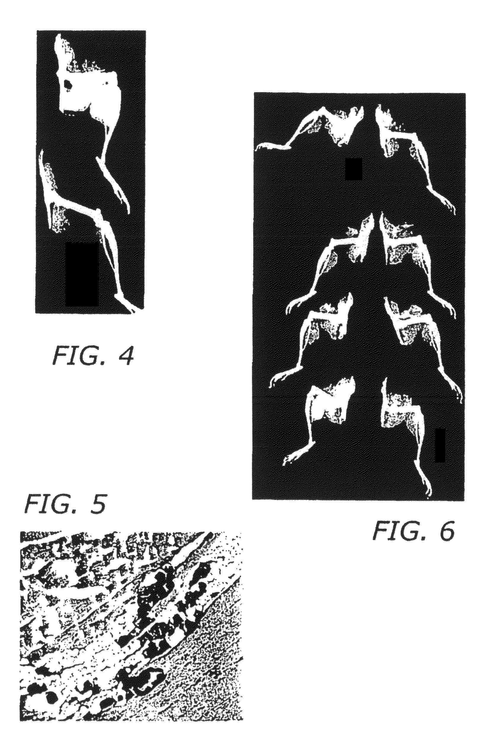
[0093]Methods: The osteogenic activity of material was tested using male Swiss-Weber mice aged 8 to 10 weeks were used (Taconic Farms, Germantown, N.Y.). Prior to the assay, the BBP was solublized and lyophilized into 2 mg of atelocollagen. The dried material was placed in a #5 gelatin capsule and sterilized by exposure to chloroform vapor. To conduct the assay, mice were anesthetized using 1% isoflurane delivered in oxygen at 2 l / min through a small animal anesthesia machine (VetEquip, Pleasanton, Calif.). Animals were affixed to a surgery board and the fur over the hindquarters shaved. The skin was cleaned with 70% ethanol and a midline incision made over the spine adjacent to the hindquarters. Blunt dissection with scissors was used to expose the quadriceps muscle on one side. A small pouch was made in the muscle using the point of scissors and the #5 capsule containing the test material was inserted into the pouch. The skin was then closed with three 11 mm...

example 3

Surface Plasmon Resonance to Determine the Interaction of BMP-2 and the Synthetic Peptide

[0105]Methods: The binding interaction between rhBMP-2 and BBP was characterized using surface plasmon resonance employing a Biacom X instrument (Biacore, Piscataway, N.J.). Buffers and chips for the procedure were obtained from Biacore. RhBMP-2 was dialyzed into 10 mM sodium acetate, pH 5.5 at a concentration of 1 mg / ml. This material was then attached to a CM-5 sensor chip using reagents and procedures supplied by the manufacturer. Running buffer was 10 mM HEPES, pH 7.4, 150 mM NaCl, 3 mM EDTA, 0.005% Surfactant P20. The peptide was dissolved in running buffer at concentrations ranging from 1×10⁻⁵ to 1×10⁻⁴ M. Flow rates from 5 to 50 μl / min and injection volumes of 20 to 100 μl were employed. The regeneration solution was 10 μM glycine-HCl, pH 2.0.

[0106]Results: Results of the surface plasmon resonance studies to determine the interaction between rhBMP-2 and BBP are shown in FIG. 9.

[0107]FIG. ...


PUM

PropertyMeasurementUnit
pHaaaaaaaaaa
pHaaaaaaaaaa
concentrationaaaaaaaaaa

Abstract

The present invention relates to the clinical application of BBP, alone or in combination with other growth factors, for use in bone healing applications, such as spinal surgery. Additional applications include use in orthopedic implantable prostheses and implantation into other surgical sites (e.g., surgical reconstruction, regional osteopenia, etc.) where bone is desired.

Description

PRIORITY CLAIM[0001]This application is a national stage application of PCT International Patent Application Number PCT / US2007 / 026315, filed Dec. 26, 2007, which claims priority to U.S. Provisional Patent Application No. 60 / 876,821, filed Dec. 22, 2006, and is a continuation-in-part of U.S. patent application Ser. No. 11 / 985,745, filed Nov. 16, 2007 now U.S. Pat. No. 8,193,312, which is a continuation-in-part of U.S. patent application Ser. No. 10 / 587,313, which has a 371 date of Apr. 28, 2008 now U.S. Pat. No. 8,188,219 and which is a national stage application of PCT / US05 / 02722, filed Jan. 28, 2005, which claims priority to U.S. Provisional Patent Application No. 60 / 539,903, filed Jan. 28, 2004.[0002]The instant application contains a Sequence Listing which has been submitted in ASCII format via EFS-Web and is hereby incorporated by reference in its entirety. Said ASCII copy, created on Oct. 30, 2012, is named 38586375.txt and is 10,900 bytes in size.BACKGROUND OF THE INVENTION[00...

Claims


Application Information

Patent Timeline
Patent Type & Authority Patents(United States)
IPC IPC(8): A61K38/10C07K14/495C07K14/51C07K5/12A61K38/18A61K38/12C07K4/12
CPCA61K38/1841A61K38/1875C07K14/51A61K38/10A61K2300/00C07K14/47A61P19/00A61K35/12A61K35/32A61K38/12C07K7/64
Inventor MURRAY, SAMUEL S.BROCHMANN-MURRAY, ELSA J.BEHNAM, KEYVANWANG, JEFFREY C.
Owner RGT UNIV OF CALIFORNIA
Learn More