Method for simultaneous detection and discrimination of bacterial, fungal, parasitic and viral infections of eye and central nervous system

a technology which is applied in the field of simultaneous detection and discrimination of eye and central nervous system bacterial, fungal, parasitic and viral infections, can solve the problems of inability to generally use diagnostic purposes, inability to detect and treat diseases, and inability to accurately detect diseases and diseases, etc., to achieve high specificity of identification and high sensitivity of pcr assay

Active Publication Date: 2013-06-18
COUNCIL OF SCI & IND RES
View PDF6 Cites 14 Cited by
  • Summary
  • Abstract
  • Description
  • Claims
  • Application Information

AI Technical Summary

Benefits of technology

[0083]The present invention combines the high sensitivity of PCR assay and the high specificity of identification by hybridization on to a macro-array with the detection of hybridization by color detection methods, the end result of which can be monitored by naked eye. However fluorescent labels such as Quantum Dots, Cy3, Cy5, FITC can also be used and the product could be visualized by fluorescence microscopy.

Problems solved by technology

The culture techniques referred to here are mostly useful in diagnosis of bacterial infections and fungal infections and they are not generally employed for diagnostic purposes in case of viral infections especially since the frequency of the isolation of viruses by culturing from a clinical sample is less than 15%.
The appropriate sample for the diagnosis of infections such as eye infections and infections of nervous system is an additional critical issue in success of diagnosis in detection of the aetiological agent of the underlying syndrome such as kerato conjunctivitis, endophthalmitis, uveitis, retinitis, meningitis, and encephalitis.
Simple aspirate of vitreous quite often is inadequate to diagnose fungal and bacterial endophthalmitis by smear examination and culture.
It is possible to introduce errors that sometimes lead to ambiguous identification of pathogens, and therefore result in a wrong diagnosis / treatment while performing classical methods.
Some difficulties, however, are associated with PCR-based methods, such as false-positive results due to contaminating nucleic acids and inhibition of the PCR reaction due to complex samples.
However the nested PCR has the attendant problem of introduction of amplicon contamination within the lab as the PCR product of the first round of amplification has to be transferred to a second PCR tube containing a second set of primers amplifying a smaller region of the gene amplified in the first round of PCR.
In all these samples there is a limitation of the amount of sample that can be obtained from a patient in a single sitting.
Up to 5 ml of CSF sample can be drawn but the volume of CSF sample required for PCR will be less than 0.5 ml.Consequent to the limitation of the volume of the sample is the limited number of bacteria / viruses / parasites present in the sample.
Below a critical mass, the infectious agents bacteria and fungi fail to grow in culture and are thus difficult to diagnose.
There are no easy detection systems for parasites such as Toxoplasma gondii and the number of parasites are also too few for detection.
The tests such as IgM or IgG detection for HSV, CMV, VZV, Adenoviruses, Chlamydia and toxoplasma are very non-specific and are not of diagnostic significance.Another major difficulty in diagnosis is all the afflictions of the eye described above are of acute nature and require an immediate and accurate diagnosis for institution of appropriate therapy.
The methods such as mass spectrometry are not practicable even in advanced tertiary medical care centers and microarrays detection based on expensive scanners cannot be afforded in clinical settings.
A line probe assay is prone for amplicon contamination.

Method used


Image

  • Method for simultaneous detection and discrimination of bacterial, fungal, parasitic and viral infections of eye and central nervous system
  • Method for simultaneous detection and discrimination of bacterial, fungal, parasitic and viral infections of eye and central nervous system
  • Method for simultaneous detection and discrimination of bacterial, fungal, parasitic and viral infections of eye and central nervous system

Examples


example 1

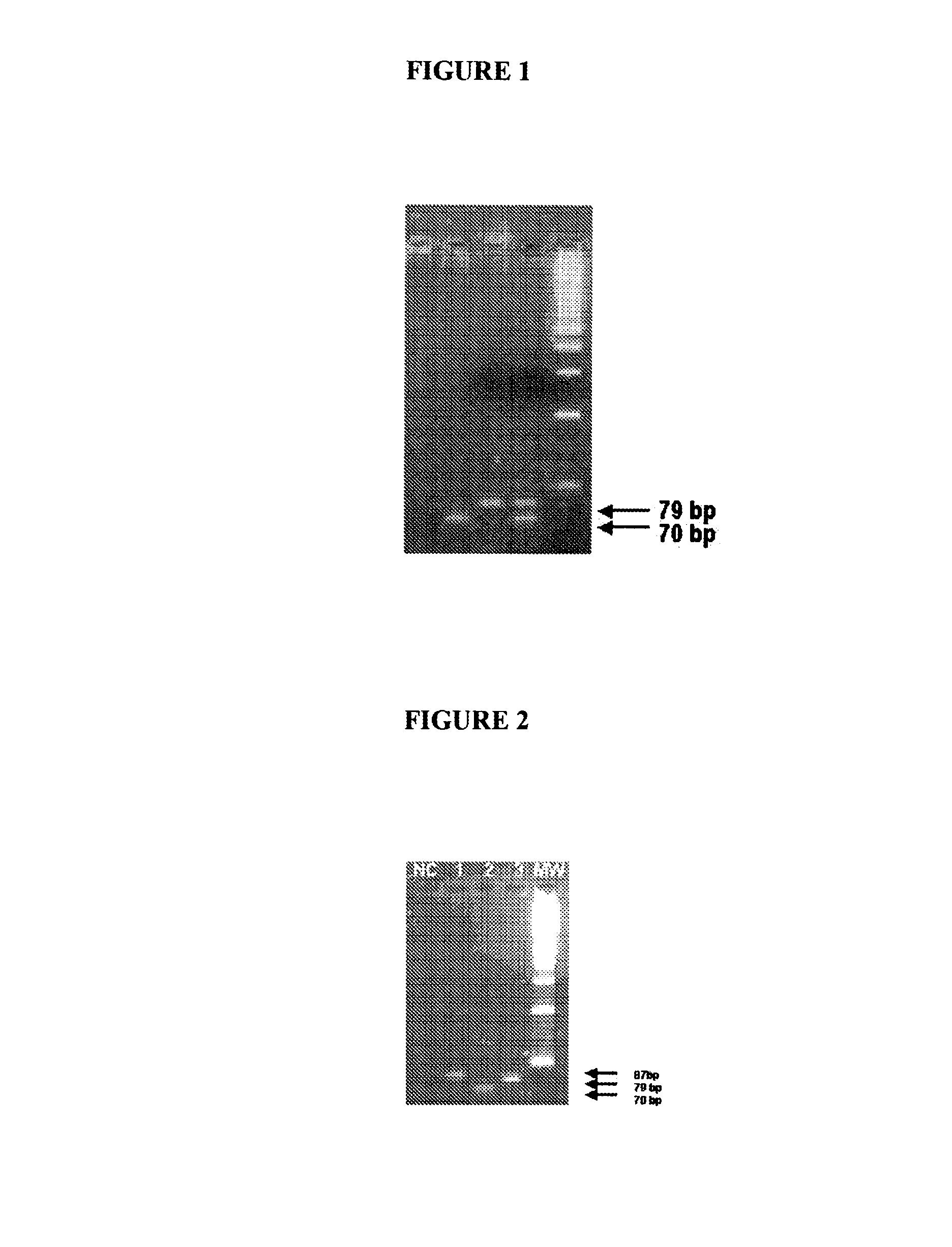
[0239]A multiplex PCR was carried out with primer sets 9 and 18, which can amplify the hexon gene of adenoviruses and polymorphic protein II gene of Chlamydia trachomatis respectively. The PCR mix contained 10 to 20 pmoles each of the forward and reverse primers, 200 μM of each d-ATP, d-UTP, d-CTP and d-GTP, 2 units of Taq polymerase in 10 mM Tris-HCl pH 9.0, 1.5 mM MgCl2, 5 mM KCl, 0.01% gelatin, 1 mM EDTA and 1 unit of UDP glycosylase to prevent amplicon contamination. The cycling conditions are being incubation at 37° C. for 30 minutes for complete digestion of any amplicon contaminants, 2 minutes at 50° C., a denaturing step of 94° C. for 5 minutes, followed by 40 cycles of 45 seconds at 60° C., 45 seconds at 72° C. and 45 seconds at 94° C. followed by 10 minutes extension of the reaction at 72° C. The product was analysed by 6% agarose gel. As can be seen in FIG. 1 both the genes got amplified. Standard DNA of 1 pg of adenovirus and 10 fg of Chlamydial DNA was used for amplific...

example 2

[0241]A multiplex PCR was carried out with primer sets 1, 2 and 3, which can amplify the Glycoprotein D, UL 44 and DNA Polymerase genes respectively. The PCR mix contained 10 pmoles each of the forward and reverse primers, 200 μM of each d-ATP, d-UTP, d-CTP and d-GTP, 2 units of Taq polymerase in 10 mM Tris-HCl pH 7.5, 1.5 mM MgCl2, 5 mM KCl, 0.01% gelatin, 1 mM EDTA and 1 unit of UDP glycosylase to prevent amplicon contamination. The cycling conditions are being incubation at 37° C. for 30 minutes for complete digestion of any amplicon contaminants, 2 minutes at 50° C. a denaturing step of 94° C. for 5 minutes, followed by 40 cycles of 45 seconds at 60° C., 45 seconds at 72° C. and 45 seconds at 94° C. followed by 10 minutes extension of the reaction at 72° C. The product was analysed by 6% agarose gel. As can be seen in FIG. 2 all the three genes got amplified

[0242]FIG. 2 shows 4% Agarose gel electrophoretogram showing the amplified products of glycoprotein D, DNA polymerase, UL-4...

example 3

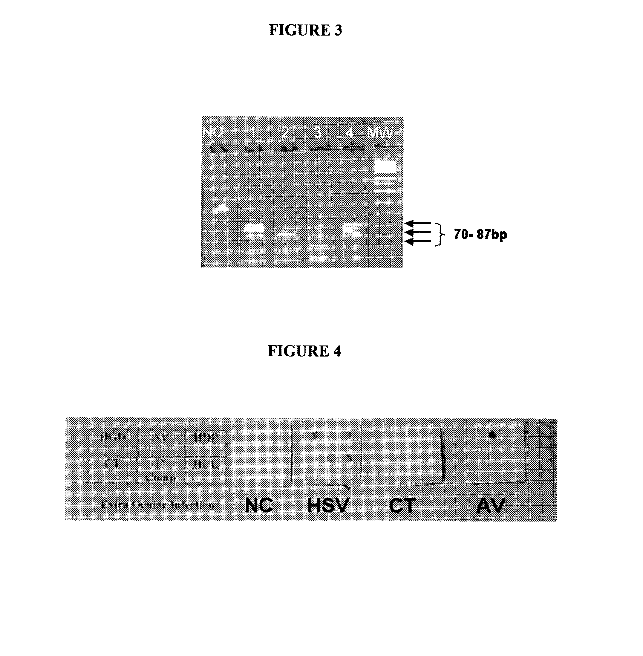
[0243]A multiplex PCR was carried out with primer sets 1, 2, 3, 9 and 17 which can amplify the Glycoprotein D gene, UL 44 gene and DNA Polymerase genes of HSV, hexon gene of adenoviruses and polymorphic protein II gene of Chlamydia trachomatis respectively. The PCR mix contained 10 pmoles each of the forward and reverse primers, 200 μM of each d-ATP, d-UTP, d-CTP and d-GTP, 2 units of Taq polymerase in 10 mM Tris-HCl pH 9.0, 1.5 mM MgCl2, 5 mM KCl, 0.01% gelatin, 1 mM EDTA and 1 unit of UDP glycosylase to prevent amplicon contamination. The cycling conditions are being incubation at 37° C. for 30 minutes for complete digestion of any amplicon contaminants, 2 mins at 50° C. and a denaturing step of 94° C. for 5 minutes, followed by 40 cycles of 45 seconds at 60° C., 45 seconds at 72° C. and 45 seconds at 94° C. followed by 10 minutes extension of the reaction at 72° C. Five tubes of the PCR mix mentioned above were incubated with the following DNA preparations where in the tube NC di...


PUM

PropertyMeasurementUnit
volumeaaaaaaaaaa
volumeaaaaaaaaaa
temperatureaaaaaaaaaa

Abstract

The present invention relates to the diagnostic methods for identification of the single causative agent or more than one causative agent of ocular and nervous system infections among many probable pathogens, which can cause the infection. All the pathogens affecting a discrete area of eye or nervous system generally cause same clinical manifestations or syndromes. The present invention relates to detection and discrimination of the pathogen among the set of probable pathogens in a single test without resorting to a battery of tests each being directed at detection of one pathogen. The current invention aims at the syndrome based diagnostic replacing the diagnostics based on detection of individual pathogens.

Description

FIELD OF INVENTION[0001]The present invention relates to the diagnostic methods for identification of the single causative agent or more than one causative agent of ocular and nervous system infections among many probable pathogens, which can cause the infection. All the pathogens affecting a discrete area of eye or nervous system generally cause same clinical manifestations or syndromes. The present invention relates to detection and discrimination of the pathogen among the set of probable pathogens in a single test without resorting to a battery of tests each being directed at detection of one pathogen. The current invention aims at the syndrome based diagnostic replacing the diagnostics based on detection of individual pathogens.BACKGROUND OF THE INVENTION[0002]Infections of the eye can be clinically classified according to the anatomical compartment harboring and consequently affected by the infection. There are many organisms which can cause ocular infections and are in detail ...

Claims


Application Information

Patent Timeline
Patent Type & Authority Patents(United States)
IPC IPC(8): C12P19/34C07H21/04
CPCC12Q1/689C12Q1/701C12Q1/705C12Q1/6895C12Q2600/16C12Q2527/101C12Q2561/113C12Q2537/143
Inventor RAO, CHINTALAGIRI MOHANRAO, IKUNCHALA SRIDHARRAMCHANDER, PUPPALA VENKATMADHAVAN, HAJIB NARAHARIRAOSHARMA, SAVITRISATPATHY, GITARAVI KUMAR, VENKATA BANDA
Owner COUNCIL OF SCI & IND RES
Why Eureka
Try Eureka