Method of measuring electrical resistance value of corneal trans-epithelium

a technology of electrical resistance value and transepithelial, which is applied in the field of measuring the electrical resistance value of the corneal transepithelium, can solve the problems of difficult to quantitate the disorder of the corneal epithelium, serious corneal ulcers, and inability to cur

a technology of electrical resistance value and transepithelial, which is applied in the field of measuring the electrical resistance value of the corneal transepithelium, can solve the problems of difficult to quantitate the disorder of the corneal epithelium, serious corneal ulcers, and inability to cur

US8478395B2Inactive Publication Date: 2013-07-02NAGASAKI UNIVERSITY +1

Image

  • Method of measuring electrical resistance value of corneal trans-epithelium
  • Method of measuring electrical resistance value of corneal trans-epithelium
  • Method of measuring electrical resistance value of corneal trans-epithelium

Examples


example 1

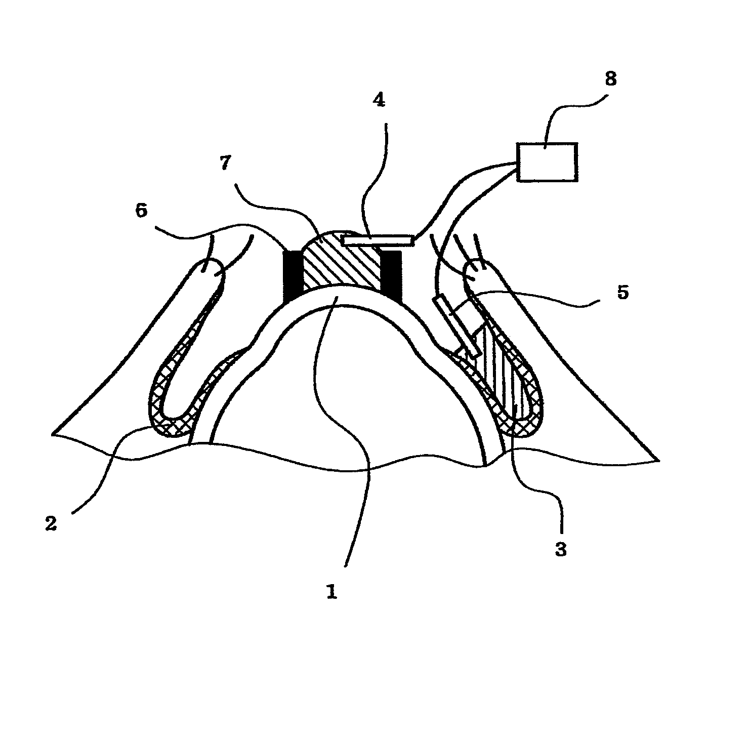
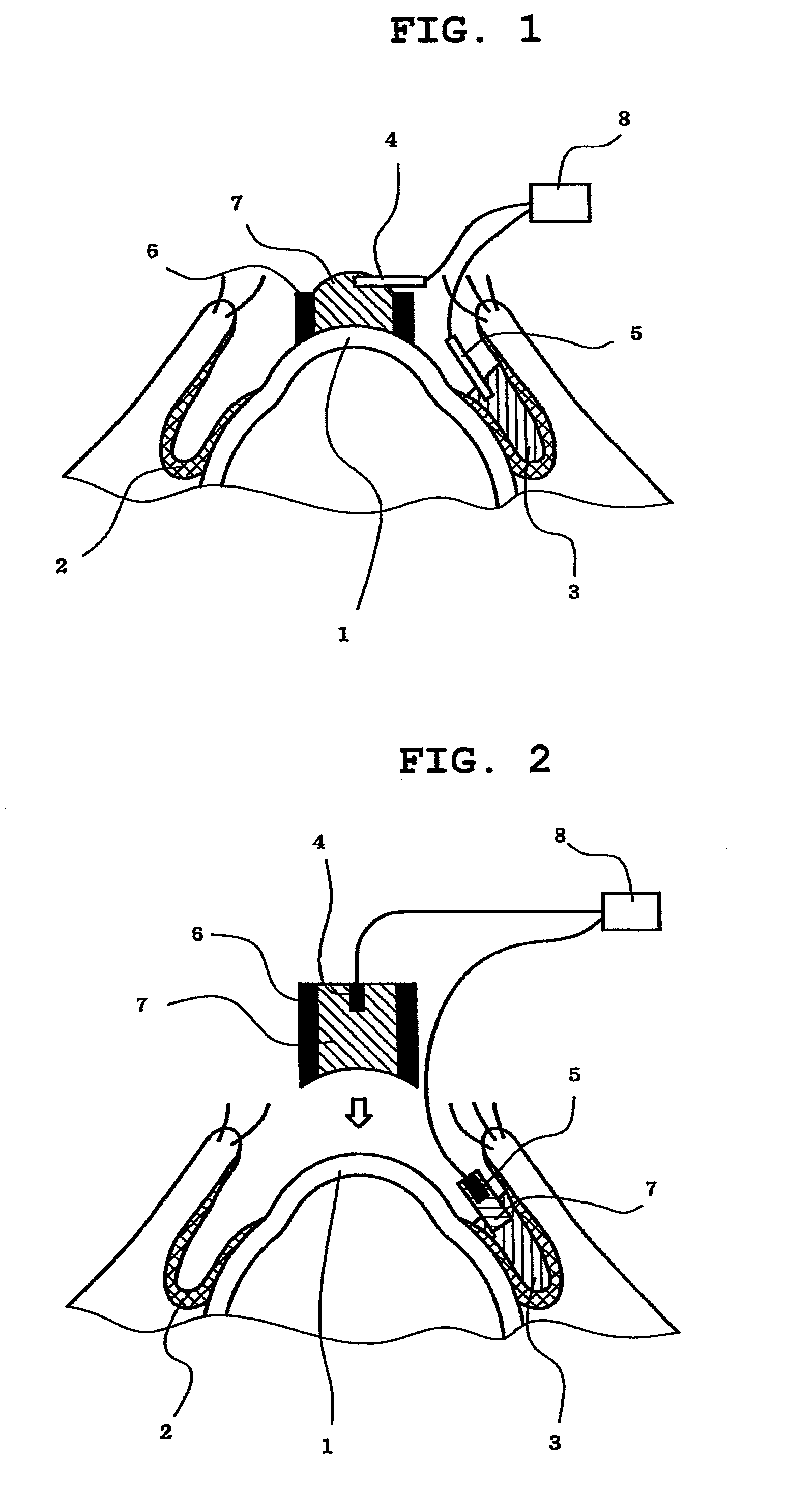
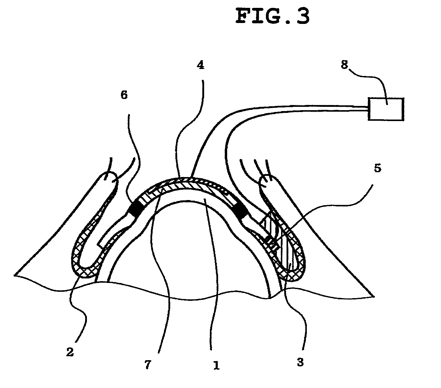
Evaluation of Corneal Disorder Due to BAC by Living Corneal TER Measurement Requiring No Anterior Chamber Centesis (Referred to as Conjunctival Sac Method) (FIG. 1)

[0099]Silver-silver chloride electrode having a rod shape was placed on a cornea and into a conjunctival sac of a living rabbit, and a 0.02% BAC was administered thereto. A change in the corneal TER due to exposure to BAC was measured by a voltage-electric resistance value measuring device. A rubber ring (6 mm in the inner diameter) was fixed onto the cornea around the periphery of the electrode by using a bioadhesive (Aron alpha A from Sankyo Co., Ltd.) for insulation, so that an electric current is surely passed through the epithelium of the cornea. An AC current of ±20 μA was flowed, and a change curve of TER, a reduction of TER and a TER value after a lapse of 60 seconds from the exposure to 0.02% BAC, were compared with those from the conventional measuring method in which the anterior chamber centesis was carried ou...

example 2

Preparation of Living Corneal TER Measuring Device (FIG. 6)

Corneal Electrode Portion

[0102]A silver electrode and a silver-silver chloride electrode having a diameter of 1 mm were placed on a round cover glass having a diameter of 12 mm. PDMS having a circular cylinder shape with 10 mm in diameter and 6 mm in inner diameter and having a curvature radius of the corneal contact surface of 7.9 mm was secured onto the cover glass by using an adhesive. An Eppendorf tube was attached to a rear side (on the side opposite to the corneal contact surface (cover glass side)) for mount onto a slit-lamp. The inner space of the PDMS was filled with an atelocollagen gel and its curvature radius was adjusted to 7.9 mm.

Conjunctival Electrode Portion

[0103]A silver electrode and a silver chloride electrode having a diameter of 1 mm were placed on a PET sheet of 0.005×0.5×1 cm, at position of 1 mm from the distal end of the sheet on the eyeball side. The PET sheet, the silver electrode and the silver ch...


PUM


Abstract

The invention provides a method for evaluating a corneal disorder quantitatively and is applicable to living eyes. In particular, the invention provides a method for measuring a corneal transepithelial electric resistance, which method comprises: (1) a step of placing a first electrode on the cornea and a second electrode on the conjunctiva; and (2) a step of flowing an electric current between the first electrode and the second electrode to measure the electric resistance. The invention also provides a device for measuring a corneal transepithelial electric resistance value.

Description

CROSS-REFERENCE TO RELATED APPLICATIONS(S)[0001]This application is the U.S. national phase of International Patent Application No. PCT / JP2009 / 053968, filed on Mar. 3, 2009, which claims priority to Japanese Patent Application No. 052567 / 2008, filed Mar. 3, 2008.TECHNICAL FIELD[0002]The present invention relates to a method for measuring a corneal transepithelial electric resistance value, which is less invasive to respective tissues of eyes including a corneal epithelium.BACKGROUND ART[0003]A cornea is a transparent membrane with a thickness of about 0.5 mm, and has a five-layer structure which consists of corneal epithelial cells, a Bowman membrane, corneal stroma, a Descemet's membrane and corneal endothelial cells, in this order from the surface of body. Among these, although the corneal epithelial cells have high regenerating capability, a serious corneal ulcer might occur, or a nonreversible corneal opacity might be left, in the case where a disorder of the epithelial cells is...

Claims


Application Information

Patent Timeline
02 Jul 2013
Publication
US8478395B2
IPC
A61B5/053; A61B5/296
CPC
A61B5/6821; A61B5/053; A61B2562/0215
Inventors
UEMATSU, MASAFUMI; KITAOKA, TAKASHI