Immunoglobulin cleavage fragments as disease indicators and compositions for detecting and binding such

Active Publication Date: 2013-08-06
JANSSEN BIOTECH INC
View PDF5 Cites 6 Cited by
  • Summary
  • Abstract
  • Description
  • Claims
  • Application Information

AI Technical Summary

Problems solved by technology

The detection of the actual cleavage products have been reported (Fick et al., 1985; Goldberg and Whitehouse, 1970; Waller, 1974) but a robust assay which would allow these fragments to serve as biomarkers has not been developed possibly due to the low concentrations in serum resulting from rapid clearance of the various fragments or to technical problems in detecting the fragments amidst the large amount of intact immunoglobulin in blood and tissues.
Further, the ability to correct loss of IgG functions in vivo due to cleavage and the ability to compensate the process of IgG cleavage in the body of subjects suffering from protease specific disease activity has not been heretofore envisioned as a therapeutic strategy.

Method used


Image

  • Immunoglobulin cleavage fragments as disease indicators and compositions for detecting and binding such
  • Immunoglobulin cleavage fragments as disease indicators and compositions for detecting and binding such
  • Immunoglobulin cleavage fragments as disease indicators and compositions for detecting and binding such

Examples


example 1

Cleavage Analysis of Human IGG Heavy Chain

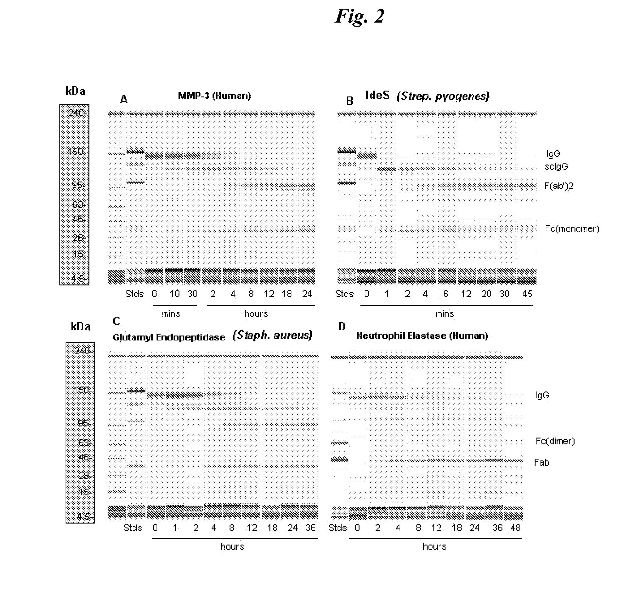
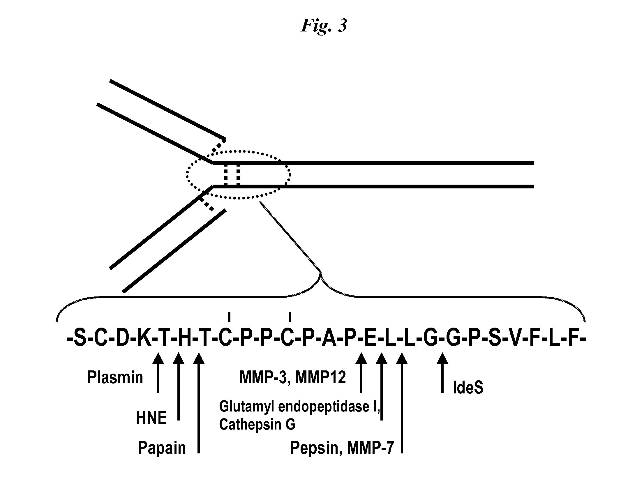
[0082]Proteolysis of human IgG heavy chain by matrix metalloproteases, cathepsins, human neutrophil elastase (HNE), and selected pathogen enzymes such as staphylococcal glutamyl endopeptidase (V8 protease), and immunoglobulin degrading enzyme of streptococcus (IdeS) was studied.

[0083]A purified monoclonal antibody comprising a human IgG heavy chain was contacted with the proteases described and sampling was conducted over various durations of contact. Fragmentation in the samples was evaluated using the Agilent microfluidics “lab-on-a-chip” technology for in vitro biosizing (Goetz H et al. Biochemical and Biophysical Methods 60; 281-293, 2004).

[0084]Antibody Substrates.

[0085]Monoclonal antibodies were either fully human, recombinant humanized murine antibodies or human / murine chimeric antibodies possessing human constant domains and hinge regions of the IgG1kappa class / subclass and species: Mab1 is a human IgG1 which binds a pathogen, Mab2 (...

example 2

Cleavage of IGG in an Inflammatory Exudate

[0107]Inflammatory exudates and other such fluids are expected to possess proteolytic enzymes associated with the inflammation and wound healing. For this purpose, samples of wound fluid were obtained from Ethicon Inc.

[0108]First, an antibody substrate, which comprises human heavy chain constant domains was randomly biotinylated. Ten microL of the biotinylated substrate antibody was added to 190 microL of the wound fluid and incubated at 37° C. for 8-24 hours. At specified times, samples were removed. The starting IgG and samples from the various times were applied in separate wells to a 4-12% Bis-Tris gel and subjected to SDS PAGE. The separated bands were transferred to a nitrocellulose membrane and, following blocking with 0.1M Tris buffered saline containing 0.1% Tween 20 and 10% blocking grade milk (“Blotto”), the blot was developed using AVIDIN-D-horseradish peroxidase reagent followed by TMB (membrane) substrate.

[0109]As evidenced by ...

example 3

Preparation of Reagent

[0110]The determination of the presence of host (patient) antibody fragments produced by endogenous proteases requires a reagent which selectively binds to the cleaved IgG but not intact IgG. Both identification of the cleaved component and a quantitative difference between fragment content in samples from patients with disease as compared to the normal population should be able to be assessed using the reagent.

[0111]The detection of unknown, but likely small amounts of IgG fragments in solutions containing relatively high concentrations of intact IgG is difficult. Although scIgG has been noted as a possible IgG cleavage fragment (Gearing 2002 supra), quantitation in human samples has not been previously performed. For this purpose, reagents with the necessary specificity were generated in rabbits having with a high degree of specificity for cleaved but not intact IgG.

[0112]Three conjugated, and progressively longer single-chain peptide analogs of the human IgG...


PUM

PropertyMeasurementUnit
concentrationaaaaaaaaaa
molecular weightaaaaaaaaaa

Abstract

The invention relates to antibody compositions and use of the composition to detect disease processes associated with elaboration of proteases. The reagents are directed to assessing an IgG breakdown product that is the result of such proteolytic cleavage. The invention further relates to the use of a therapeutic immunospecific for IgG protease cleavage products to restore effector function to antibody compositions that are subject to protease cleavage.

Description

CLAIM TO RIGHT OF PRIORITY[0001]This application claims priority to U.S. patent application Ser. No. 12 / 185,333 filed 4 Aug. 2008, and U.S. Provisional Application No. 60 / 955,162, filed 10 Aug. 2007, the contents of which is incorporated herein by reference in its entirety.BACKGROUND OF THE INVENTION[0002]1. Field of the Invention[0003]The invention relates to diagnostic and prognostic indicators and methods and reagents for their detection. The invention further relates to methods of monitoring the natural history of disease in a patient as well as methods for treating pathological conditions by vaccinating or administering antibodies specific for immunoglobulin cleavage sites to restore effector function of antibodies.[0004]2. Description of the Related Art[0005]In medicine, a biomarker is a biochemical substance that can be used to measure the progress of disease or the effects of treatment, that is, a diagnostic or prognostic indicator. One example is hemoglobin A1c which serves...

Claims


Application Information

Patent Timeline
Patent Type & Authority Patents(United States)
IPC IPC(8): A61K38/00C07K14/00
CPCC07K16/065C07K16/2848C07K16/36C07K16/42G01N33/6854G01N33/6857C07K7/08A61K2039/505A61K2039/507C07K2317/24C07K2317/50C07K2317/732C07K2317/734C07K2317/34A61P9/00A61P19/02A61P31/04A61P33/00A61P35/00
Inventor JORDAN, ROBERTKNIGHT, DAVIDBREZSKI, RANDALLRYAN, MARYPETRONE, DIANE
Owner JANSSEN BIOTECH INC
Why Eureka
Try Eureka