

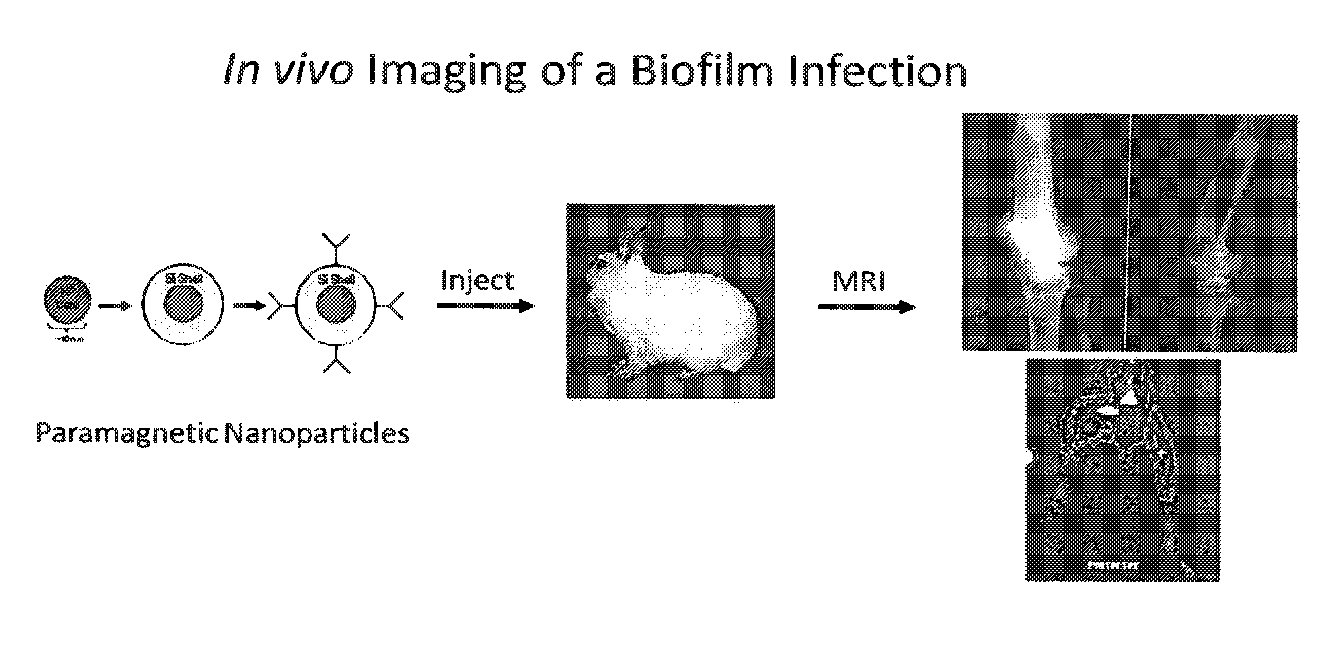
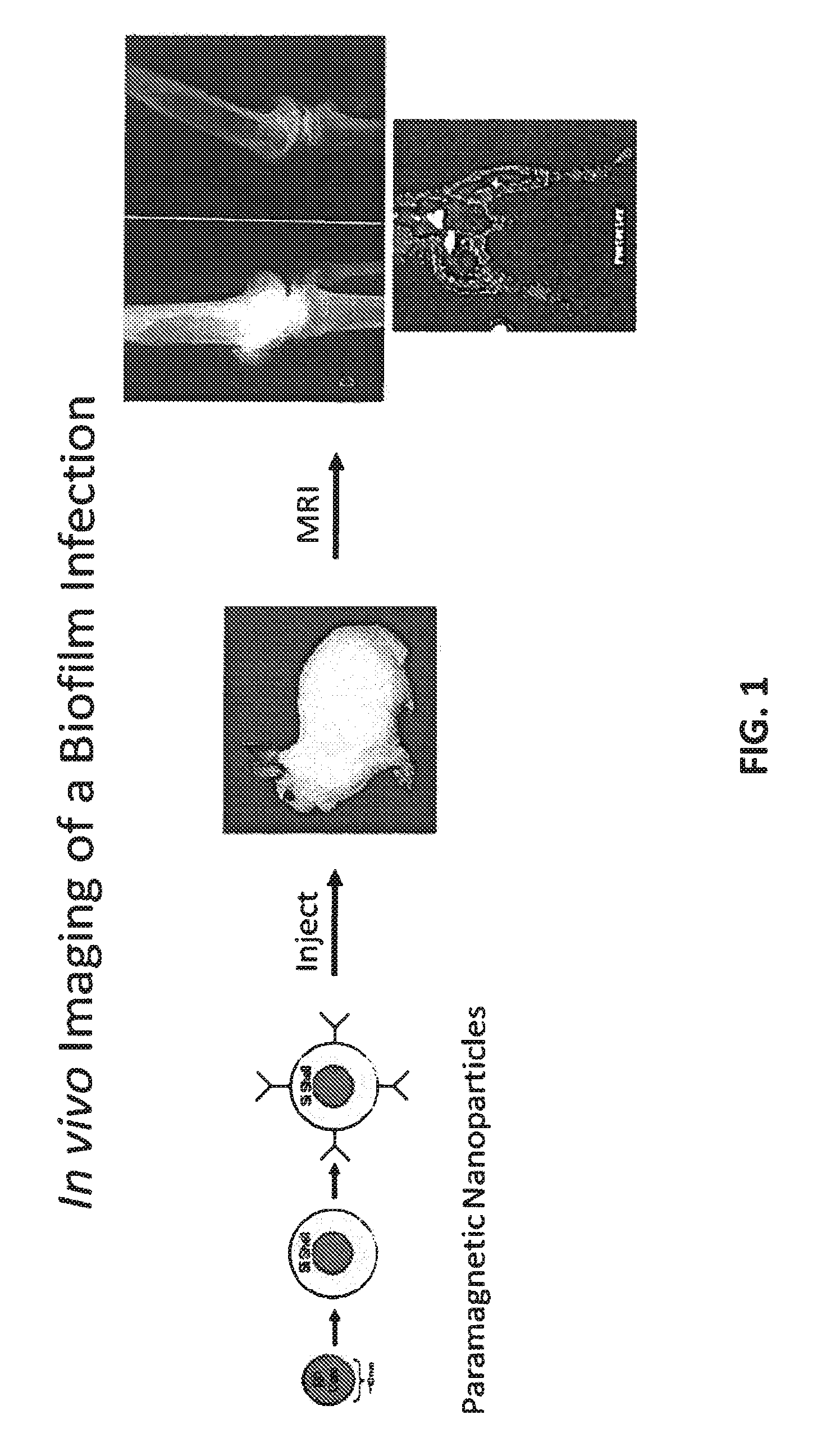
In vivo biofilm infection diagnosis and treatment
a biofilm infection and in vivo technology, applied in the field of in vivo biofilm infection diagnosis and treatment, can solve the problems of biofilm infection, high malignantity, compromised host immunity, etc., and achieve the effect of non-invasive, non-toxic, and convenient for a subj
- Summary
- Abstract
- Description
- Claims
- Application Information
AI Technical Summary
Benefits of technology
Problems solved by technology
Method used
Image

Examples
example 1
Preparation of Functionalized Paramagnetic Particles
[0109]In a variation of the Stöber process (see references below), 2-propanol, water, and washed ferrofluid are combined and sonicated in a ratio of 5:1:1.25×10⁻⁴, respectively (total volume=48.1 mL) in a round-bottom flask suspended in ultrasonic water bath (e.g., Bransonic Model B2510). After an initial dispersion period, 10 μL of tetraethyl orthosilane (TEOS) and 500 μL of concentrated NH4OH are added. The reaction proceeds with constant sonication for 90 min. before 5 μL aminopropyltriethoxysilane (APTES) in 52 μL dimethylformamide are added. Sonication is continued for an additional 60 min. or until such time as a layered coating of silica is achieved on all nanoparticles, either singly or in small clusters. The corresponding Stöber process is described in, for example, Stöber, W., et al., J. Coll. and Inter. Sci., 26: 62-69 (1968); Lu, Y., et al., Nano Letters 2: 183-186 (2002); and Niedbala R. S., et al., Anal. Biochem. 293:...
example 2
Conjugation of Avidin to Functionalized Paramagnetic Particles
[0111]The functionalized particle suspension of Example 1 (APTES-PMP) was sonicated for 10 min to ensure homogeneous dispersion of the solid phase. After sonication, the contents were transferred into a storage tube where the solid phase was gathered by magnetic aggregation and the supernatant removed. Following this, the solid was washed three times with phosphate buffer saline (PBS, pH of 7.2) solution. Then the particles were incubated with a solution containing 0.5 mg avidin / 250 μL of PBS. The avidin used was Nuetra Avidin (M.W.=1.5×10⁻⁸ mol / mg) available from Pierce. The final incubation volume was about 1 mL (250 μL avidin+450 μL PMPs+300 μL PBS). The solution was then incubated with gentle agitation (e.g., using a rocker) for about 1 hr. After incubation, the supernatant was removed using magnetic aggregation and the particles washed once in 1 ml of fresh PBS. Magnetic aggregation was again used to remove the buffe...
example 3
Conjugation of Biotinylated Antibodies to Avidinylated Paramagnetic Particles
[0112]A 10 mg portion of the avidinylated PMP from Example 2 was re-suspended in 1 mL PBS solution. Biotinylated antibody was added to the solution in a 5:1 weight ratio to PMPs. The suspension was allowed to incubate with gentle agitation at 4° C. (in a cold room) for 30 minutes. Using magnetic decantation, the excess solution was poured off and 1 mL fresh PBS added. The suspension was agitated gently by inverting the tube. The foregoing magnetic decantation, addition of 1 mL fresh PBS, and agitation was repeated three times. After final re-suspension in PBS, the particles were stored at 4° C. until use.
PUM
| Property | Measurement | Unit |
|---|---|---|
| diameter | aaaaa | aaaaa |
| diameter | aaaaa | aaaaa |
| diameter | aaaaa | aaaaa |
Abstract
Description
Claims
Application Information