Method and kit for detecting 5-hydroxymethylcytosine in nucleic acids
a technology of methylcytosine and nucleic acid, which is applied in the field of methods and kits for detecting methylcytosine in nucleic acids, can solve the problems that the position of methylcytosine in the fragment cannot be specified, and none of these methods can be applied directly to the detection of methylcytosine, so as to achieve the effect of accurately and easily detecting the presence and position of methylcytosine in the nucleic acid
- Summary
- Abstract
- Description
- Claims
- Application Information
AI Technical Summary
Benefits of technology
Problems solved by technology
Method used
Image
Examples
example 1
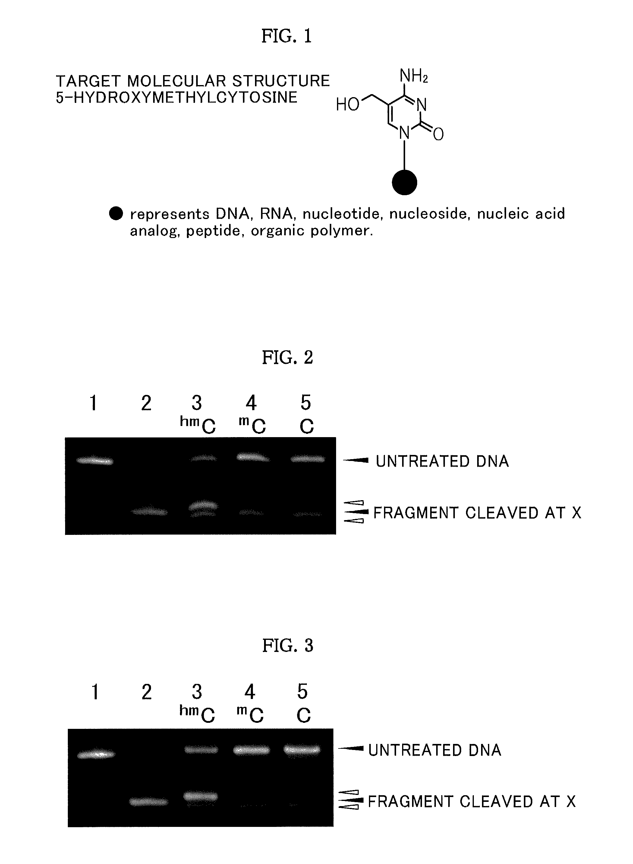
[0071]In 50 ml of water, 1.25 μg of each of fluorescently labeled DNAs having the base sequences of SEQ ID NO: 1, respectively, was dissolved, and the pH was adjusted to 7.0 with a sodium phosphate-sodium chloride buffer solution. To such a solution, 165 μg of each of the oxidizing agents shown in Table 1 was added, and a reaction was allowed to proceed at 50° C. for 120 minutes.
[0072]A desalination treatment was conducted on the reaction mixture by using Micro BioSpin Column 6 Tris (Bio-Rad Laboratories, Inc.).
[0073]Subsequently, piperidine was added to the treated material at 10% by mass, and a heat treatment was conducted at 90° C. for 120 minutes.
[0074]The treated material was subjected to a freeze dry treatment, and the residue was analyzed by polyacrylamide gel electrophoresis.
[0075]
SEQ ID NO: 1:5′-Fluorescein-AAAAAAGXGAAAAAA-3′(X = hmC, mC, C).
[0076]Hereinafter, hmC represents 5-hydroxymethylcytosine, mC represents 5-methylcytosine, and C represents cytosine.
[0077]FIG. 2 show...
example 2
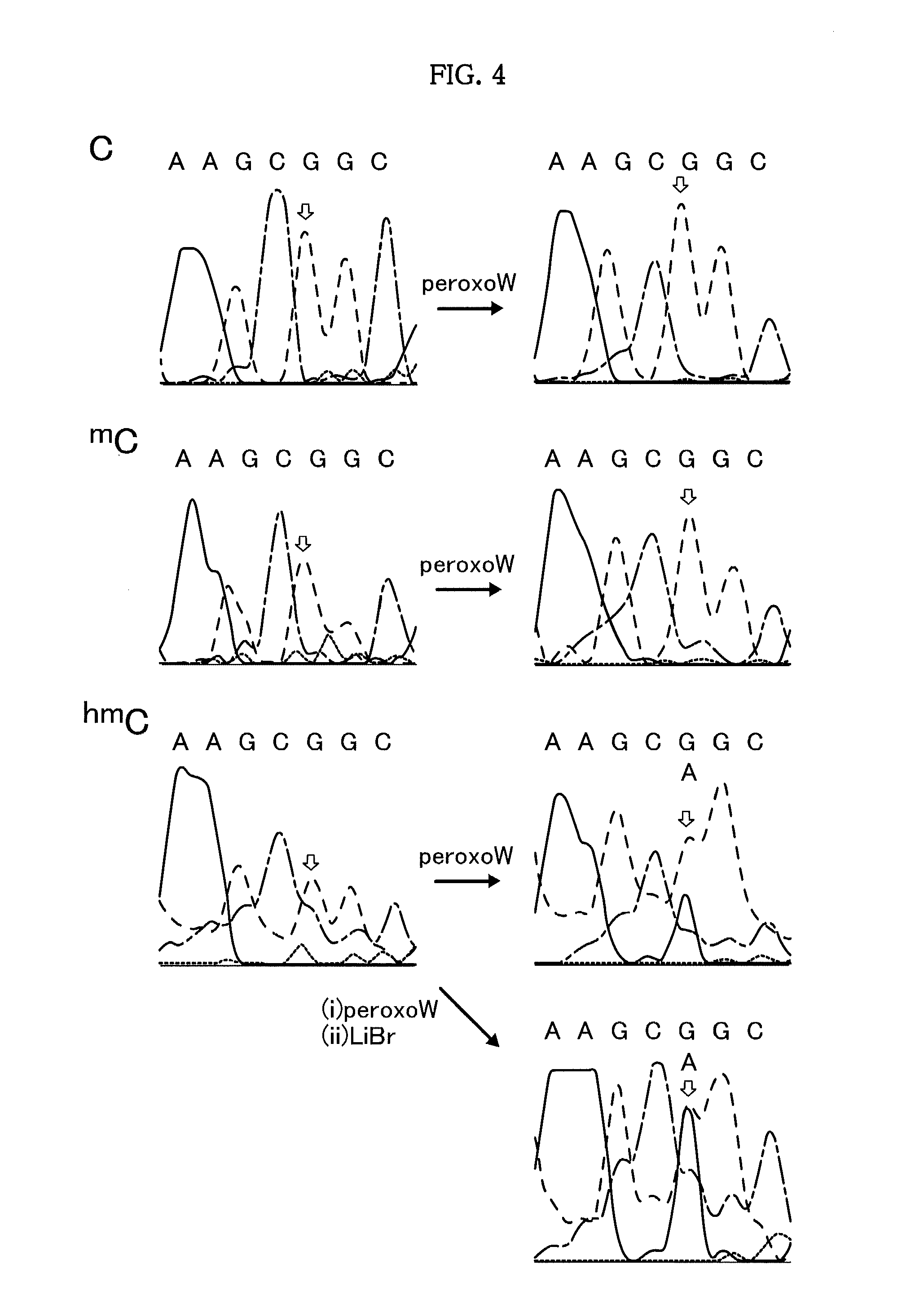
[0083]DNAs (human TNF-β putative promoter region DNA fragments containing CpG, mCpG, and hmCpG, respectively) of SEQ ID NO: 2 were synthesized by the general purpose phosphoramidite method using a NTS H-6 DNA / RNA synthesizer. In 50 μl of water, 1.25 μg of each of the DNAs having a base sequence of SEQ ID NO: 2 was dissolved, and the pH was adjusted to 7.0 with a sodium phosphate-sodium chloride buffer solution. To such a solution, 165 μg of each of the oxidizing agents shown in Table 1 was added, and a reaction was allowed to proceed at 50° C. for 120 minutes.
[0084]The reaction mixture was subjected to a desalination treatment using Micro BioSpin Column 6 Tris (Bio-Rad Laboratories, Inc.), and a treated product I was obtained.
[0085]Subsequently, the treated product I was dissolved in 50 μl of water, and 435 μg of lithium bromide was added. Then, a reaction was allowed to proceed at 50° C. for 5 hours to obtain a treated product II.
[0086]The treated product II was subjected to a desa...
example 3
[0094]Products were obtained by an oxidation treatment with K2[{W(═O)(O2)2(H2O)}2(μ-O)] and a subsequent piperidine treatment in the same manner as in Example 1, except that DNAs having the following base sequences (A) to (K) of SEQ ID NOs: 4 to 14 and being fluorescently labeled with Fluorescein were used instead of the fluorescently labeled DNAs having the base sequences of SEQ ID NO: 1 used in Example 1. The products were analyzed by polyacrylamide gel electrophoresis.
[0095]
SEQ ID NO: 4:(A) Fluo-GATACTGXGT TGCAASEQ ID NO: 5:(B) Fluo-AGTGCATXGC AAGAASEQ ID NO: 6:(C) Fluo-GACATACXGA AGTGASEQ ID NO: 7:(D) Fluo-AAGTGCAXGA TGCGASEQ ID NO: 8:(E) Fluo-CACGTTTTTT AGTGATTTXG TCATTTTCAA GTCGTCAAGTSEQ ID NO: 9:(F) Fluo-GAAAAACACA TACGTTGAAA ACXGGCATTG TAGAACAGTGSEQ ID NO: 10:(G) Fluo-GTTGTGAGGX GCTGCCCCCASEQ ID NO: 11:(H) Fluo-GCAGGGCCCA CTACXGCTTC CTCCAGATGASEQ ID NO: 12:(I) Fluo-GAGTGAGXAG TAAGASEQ ID NO: 13:(J) Fluo-AGAGCAGXTG ATGAASEQ ID NO: 14:(K) Fluo-AAGCCAGXCG TATGA
[0096]FIGS. 5 and...
PUM
| Property | Measurement | Unit |
|---|---|---|
| pH | aaaaa | aaaaa |
| pH | aaaaa | aaaaa |
| temperature | aaaaa | aaaaa |
Abstract
Description
Claims
Application Information
 - R&D
- Intellectual Property
- Life Sciences
- Materials
- Tech Scout
- Unparalleled Data Quality
- Higher Quality Content
- 60% Fewer Hallucinations
Browse by: Latest US Patents, China's latest patents, Technical Efficacy Thesaurus, Application Domain, Technology Topic, Popular Technical Reports.
© 2025 PatSnap. All rights reserved.Legal|Privacy policy|Modern Slavery Act Transparency Statement|Sitemap|About US| Contact US: help@patsnap.com


