Rapid and sensitive method for quantitative determination of the level of heparin—PF4 complex induced immunoglobulin antibodies
a sensitive method and immunoglobulin technology, applied in the field of rapid and sensitive method for quantitative determination of the level of heparin — pf4 complex induced immunoglobulin antibodies, can solve the problems of limited to simple screening applications, label may impede a radio frequency signal and the amount of impedance detected by electronic means, and is not easily quantifiable in their present form, so as to achieve cost-effective
- Summary
- Abstract
- Description
- Claims
- Application Information
AI Technical Summary
Benefits of technology
Problems solved by technology
Method used
Image

Examples
example 1
Preparation of the Complex
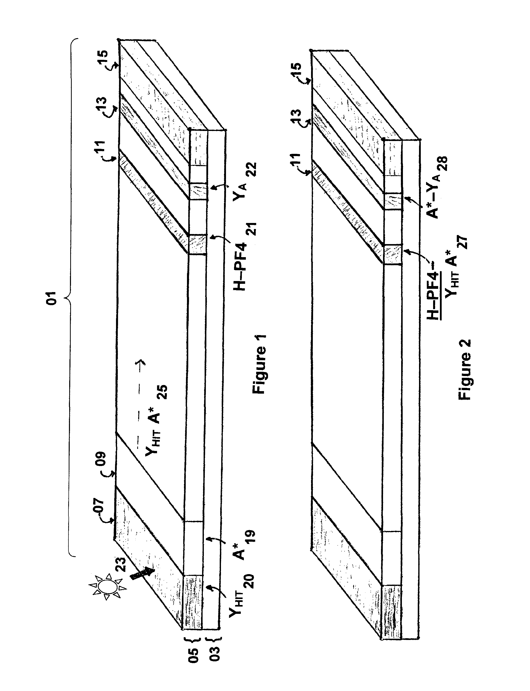
[0047]Heparin (HP), 40 ul of 1000 U / mL, in phosphate buffered saline (PBS, Fisher Scientific PN: BP665-1) is mixed with 94 ul of PF4 (1.4 mg / mL, HPF, BioMedomics) and 56 ul of water, then incubated at room temperature for 30 minutes to yield the Complex mixture. The Complex mixture is then mixed with stabilizing agents by adding 4 ul of 50% sucrose (Fisher PN: S5) with 4 ul of 25% trehalose (Sigma PN: T9449) and 2 ul of 1M TAPS buffer (Sigma PN: T5441, pH 9.0).
example 2
Preparation of the Membrane
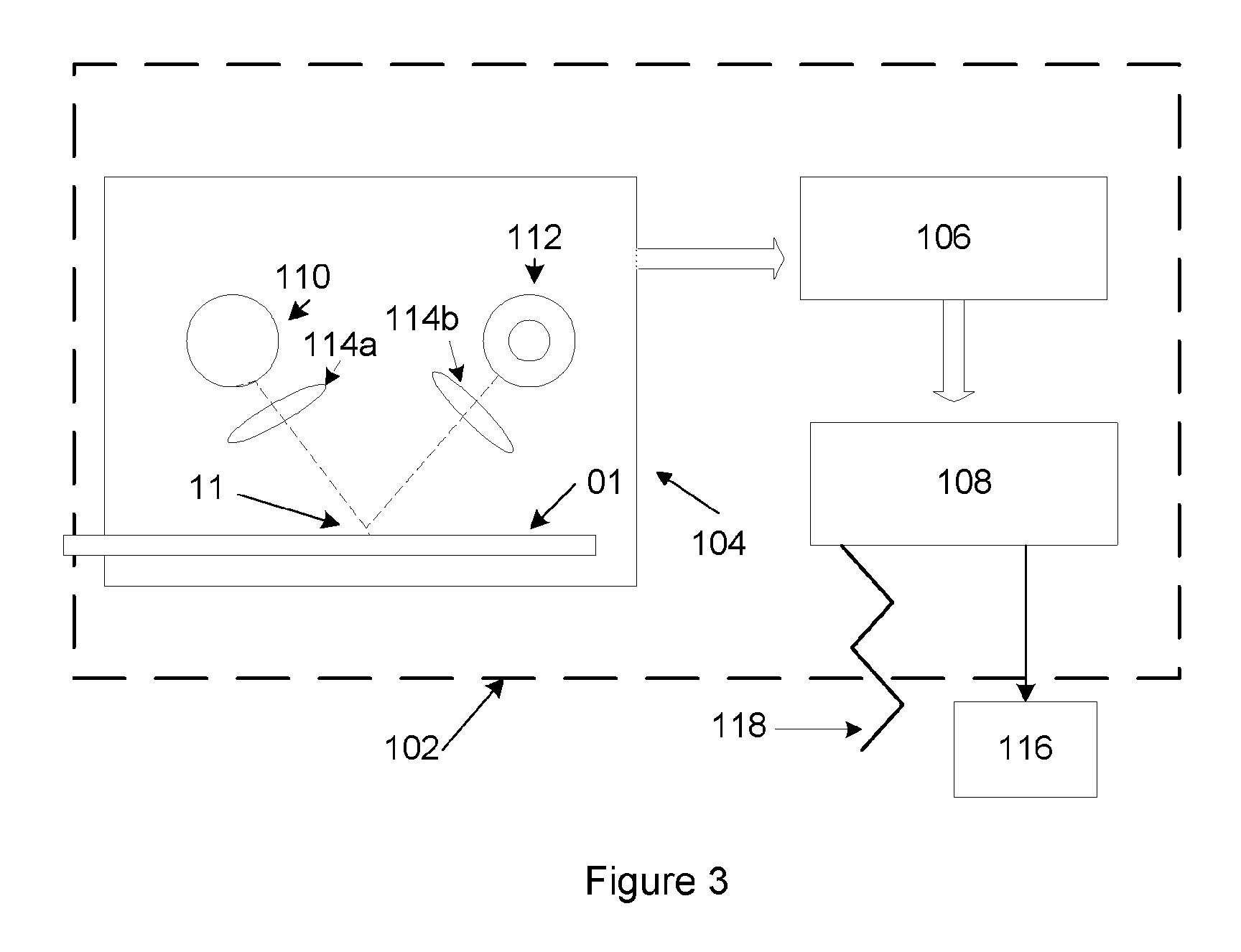
[0048]The Complex mixture described above is striped onto a Millipore Hi-Flow mylar backed nitrocellulose membrane (25 mm wide, SHF0900425) affixed to an adhesive backed card (polystyrene 59 mm by 305 mm from GML or equivalent) at 0.075 ul / mm using an Imagene Isoflow striper (or equivalent) at 45 mm / second under conditions described by the manufacturer's user manual to create a test line, The test line is proximal to the sample pad at a position to be determined by the cassette holder assembly and, if employed, corresponding position for a reader. Simultaneously, a reference reagent comprising of 0.25 mg / mL of recombinant protein A (Repligen PN: rPA-50) is mixed with 1% sucrose / 0.5% trehalose / 10 mM TAPS pH 9.0 (as was done with the Complex mixture) and striped at 0.075 ul / mm at 45 mm / second at the same time as the Complex, but at a position upstream of the test line as designated by the reader and / or cassette holder assembly to create a reference line. The...
example 3
Preparation of the Protein A Latex Particles
[0049]Dark blue carboxy-latex particles, “beads,” (Bangs Beads, DC02B, 0.33 micron or equivalent), 10% in 100 ul, are covalently coupled by washing the beads with 50 mM MES (Sigma PN: M3885) pH 4.5, adding 1 mL of a fresh preparation of 10 mg / mL EDAC (Sigma PN: E1769) in 50 mM MES, pH 4.5, incubating for 30 minutes at room temperature. The resulting material is pelleted in a centrifuge at 13.4K×g for 5 minutes, the pellets washed with 50 mM MES pH 4.5, re-suspend (with sonication in water bath using Branson desktop sonicator) with 1 ml of 0.65 mg / mL recombinant protein A in 1×PBS, vortex, sonicate, and, finally, incubated at RT overnight. Next, 100 ul of 10% BSA (Proliant Biologicals PN: 68100 or equivalent) / 100×TE (Fisher PN: BP1338), is vortex sonicated, and incubated at RT for at least 1 hour. The beads are centrifuged to form pellets, which are washed with 1 mL PBS. The wash step is repeated three times to remove unbound protein A and ...
PUM
| Property | Measurement | Unit |
|---|---|---|
| time | aaaaa | aaaaa |
| time | aaaaa | aaaaa |
| time | aaaaa | aaaaa |
Abstract
Description
Claims
Application Information
 - R&D
- Intellectual Property
- Life Sciences
- Materials
- Tech Scout
- Unparalleled Data Quality
- Higher Quality Content
- 60% Fewer Hallucinations
Browse by: Latest US Patents, China's latest patents, Technical Efficacy Thesaurus, Application Domain, Technology Topic, Popular Technical Reports.
© 2025 PatSnap. All rights reserved.Legal|Privacy policy|Modern Slavery Act Transparency Statement|Sitemap|About US| Contact US: help@patsnap.com