

Bioactive scaffolds
a bioactive and scaffold technology, applied in the field of tissue engineering, can solve the problems of poor understanding of the pathophysiology of rotator cuff injury and healing, no ideal treatment method for rotator cuff healing, and rotator cuff tears are a very common cause of pain and disability
- Summary
- Abstract
- Description
- Claims
- Application Information
AI Technical Summary
Benefits of technology
Problems solved by technology
Method used
Image

Examples
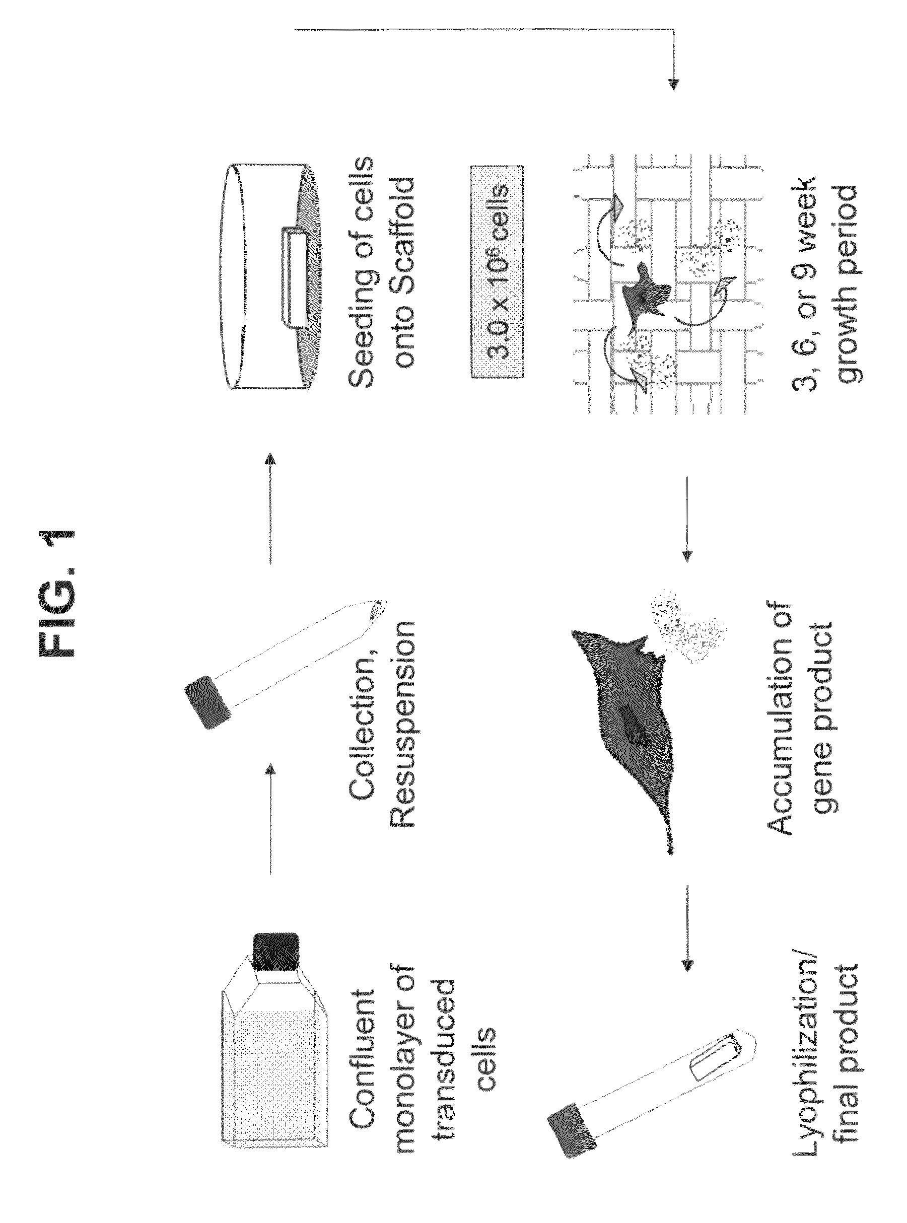
example 1
Tendon Cell-Conditioned Tissue Scaffold
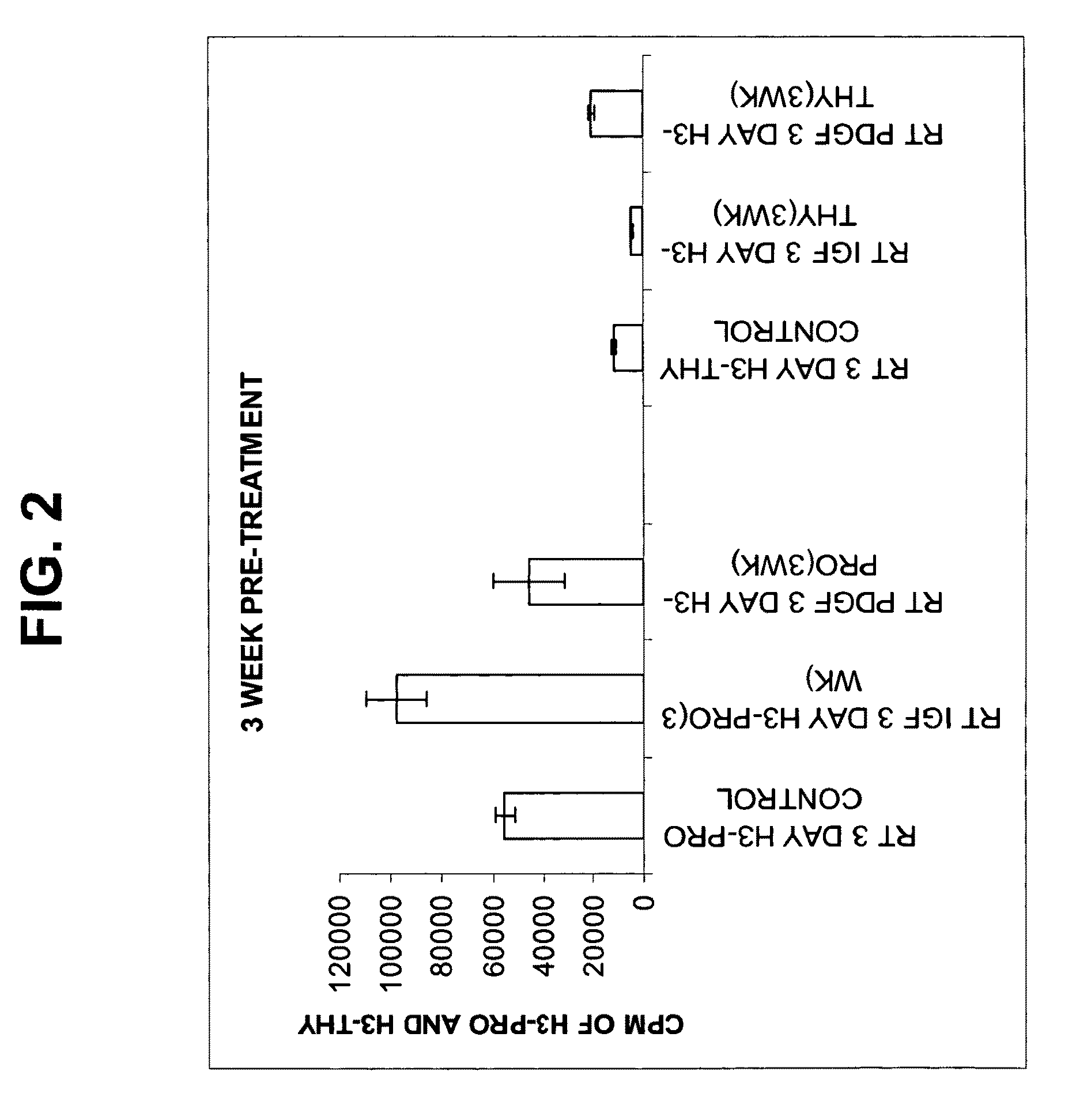
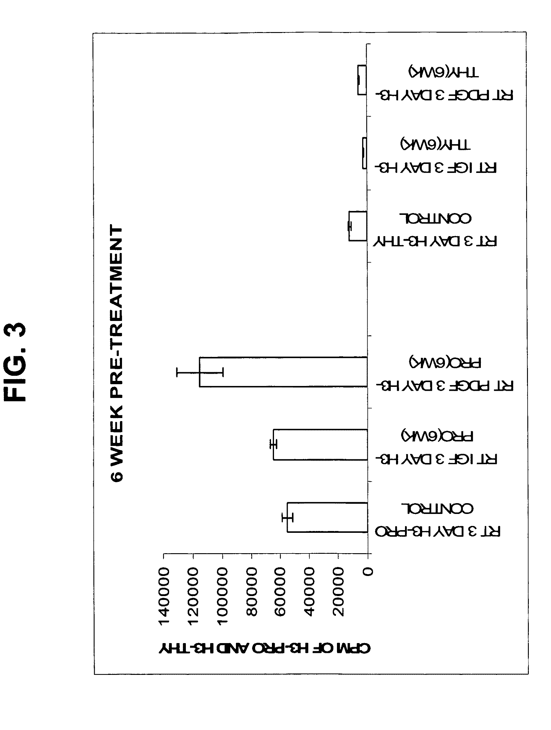
[0061]The aim of this study was to assess the metabolic response of freshly seeded tendon cells into a cell-free scaffold conditioned by IGF-1 or PDGF transduced cells.
[0062]The growth factors and their genes used in this example are extrapolated from previous in vitro studies that suggest a potential role for use in gene delivery strategies and tendon repair. PDGF-β has been shown to stimulate DNA and matrix synthesis (Kobayashi et al., 2001) especially in stretched tendon cells (Yoshikawa and Abrahamson, 2001). PDGFβ also resulted in a 2-3-fold increase in expression of cell surface integrins (Dahlgren et al., 2002). Integrins such as fibronectin are important mediators of cell attachment and angiogenesis, which would be critical to augmenting repair.
[0063]IGF-1 administered by direct injection was shown to reduce swelling and lesion size and increase cell proliferation, collagen content and stiffness in a model of collagenase-induced flexor ...
example 2
Histological Evaluation of Rat Achilles Tendon Repair Using a Bioactive Scaffold: 2 and 4 Weeks Post-op.
Introduction
[0066]Tendon injuries are encountered routinely in orthopaedic practice and cause significant disability in our society. PDGF-β and IGF-1 are potent mitogens that have been shown to augment tendon healing. This two-part study examined the effect of poly(L-lactic acid) (PLLA) scaffolds preconditioned with either wild-type rat tendon fibroblasts (RTF) or growth factor-transduced RTFs on in vitro fibroblast function and in vivo rat Achilles tendon repair.
Study Goals
[0067]The ultimate aim of this study was to determine the effect of a novel bioactive scaffold in a rat model of Achilles tendon repair. The goal was to note any increased rates of repair and also to detect any inflammatory response as a result of implantation. This was a two-part study with an in vitro component followed by an in vivo one.
Methods
[0068]Both investigations utilized preconditioned scaffolds. RTFs...
example 3
Detection of Preconditioned Scaffolds in Articular Defects Using Ultrasound Technology
Introduction
[0080]The goal of this study was to evaluate ultrasound as a safe, novel imaging modality for the non-invasive monitoring of the healing response of an osteochondral defect that was treated with implantation of a preconditioned bioactive scaffold, and to correlate the ultrasound imaging with histological observation.
Methods and Materials
[0081]Bilateral osteochondral defects were surgically created in the trochlear groove of the knee in fifteen adult male New Zealand white rabbits, under IACUC approval. Twenty-four defects were filled with PLLA scaffolds preconditioned with either IGF-1 or BMP-7 (using the transduced cell technology described above), and eight were left as unfilled controls. The knees were then harvested at 3, 6, and 12 weeks post-surgery, and evaluated with histological evaluation, as compared to ultrasound, using FlexScan image processing software.
Results
[0082]Osteocho...
PUM
| Property | Measurement | Unit |
|---|---|---|
| time | aaaaa | aaaaa |
| time | aaaaa | aaaaa |
| time | aaaaa | aaaaa |
Abstract
Description
Claims
Application Information