NMR detection of coagulation time

a technology of coagulation time and nmr, which is applied in the direction of nmr measurement, biochemistry apparatus and processes, instruments, etc., can solve the problems of uncontrollable bleeding or formation of unwanted blood clots, unsuitable or inconvenient for point of care testing or home use, and achieve high time-resolved accuracy and accuracy at the point of care setting

Active Publication Date: 2015-10-13
T2 BIOSYST
View PDF101 Cites 6 Cited by
  • Summary
  • Abstract
  • Description
  • Claims
  • Application Information

AI Technical Summary

Benefits of technology

[0012]The present invention provides non-optical methods for monitoring and measuring coagulation (e.g., blood coagulation, plasma coagulation) using nuclear magnetic resonance parameters detectable by relaxometer readings. Provided methods allow for accuracy and precision at point of care (POC) settings or at home settings, which are currently available only through central laboratory facilities. Provided methods can be used optionally without a need for additives beyond a coagulation reagent for initiating the coagulation process to be measured; can measure coagulation directly without sample interference due to non-invasive detection; allows fast determination of coagulation state changes, thereby providing real time monitoring of samples; are not limited to blood type, therapeutic window or other interfering factors; are not limited to clear samples required for optical assessment; require only small amounts of coagulating sample; and can provide highly time-resolved coagulation curves that allow for profiling of coagulation abnormalities. The present invention further provides test carriers for containing samples used in methods provided herein.

Problems solved by technology

Under normal conditions there is a delicate balance between the individual components of the hemostatic system.
Any disturbances in this balance, called the hemostatic potential, can result in either uncontrolled bleeding or formation of unwanted blood clots (thrombosis).
Most current methods have limitations which make them unsuitable or inconvenient for point of care testing or home use.
Some require special blood sample preparation and handling or sophisticated equipment, making them suitable only for central laboratory facilities having qualified staff Others, though possible for home use, are not cost effective for commercialization, or encounter implementation challenges (e.g., methods that require filtration of a sample through porous membranes pose wetting and uniform reagent impregnation difficulties).
Furthermore, besides cost and challenge of operation, a number of methods do not measure coagulation directly; and most tests do not measure coagulation without the use of an additive.
Indirect measurement has been known to pose problems of accuracy in many samples.
Other methods, while appearing to function well, can be limited to a narrow range of blood types, therapeutic windows, restricted by a long list of interfering factors or require large volumes of blood.
Thus, current blood coagulation tests are generally complex and the bulk of them are performed in a centralized clinical laboratory, at a clinic, or at a physician's office.
Required visits to a clinic or a doctor's office on a regular basis to monitor anticoagulation therapy can be both inconvenient and expensive for a patient.

Method used


Image

  • NMR detection of coagulation time
  • NMR detection of coagulation time
  • NMR detection of coagulation time

Examples


example 1

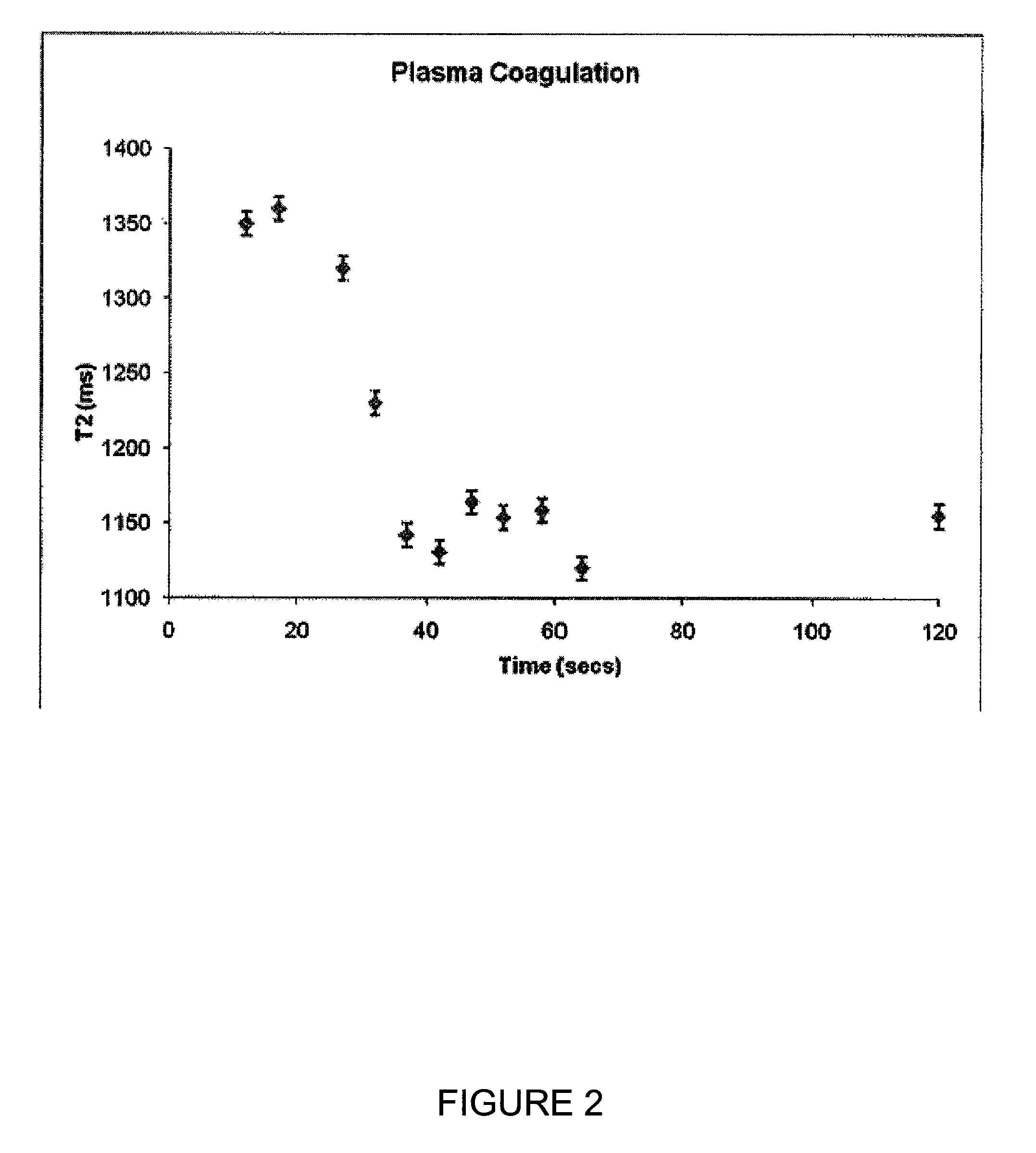
Coagulation Time Measurement in Plasma Using T2 Relaxation

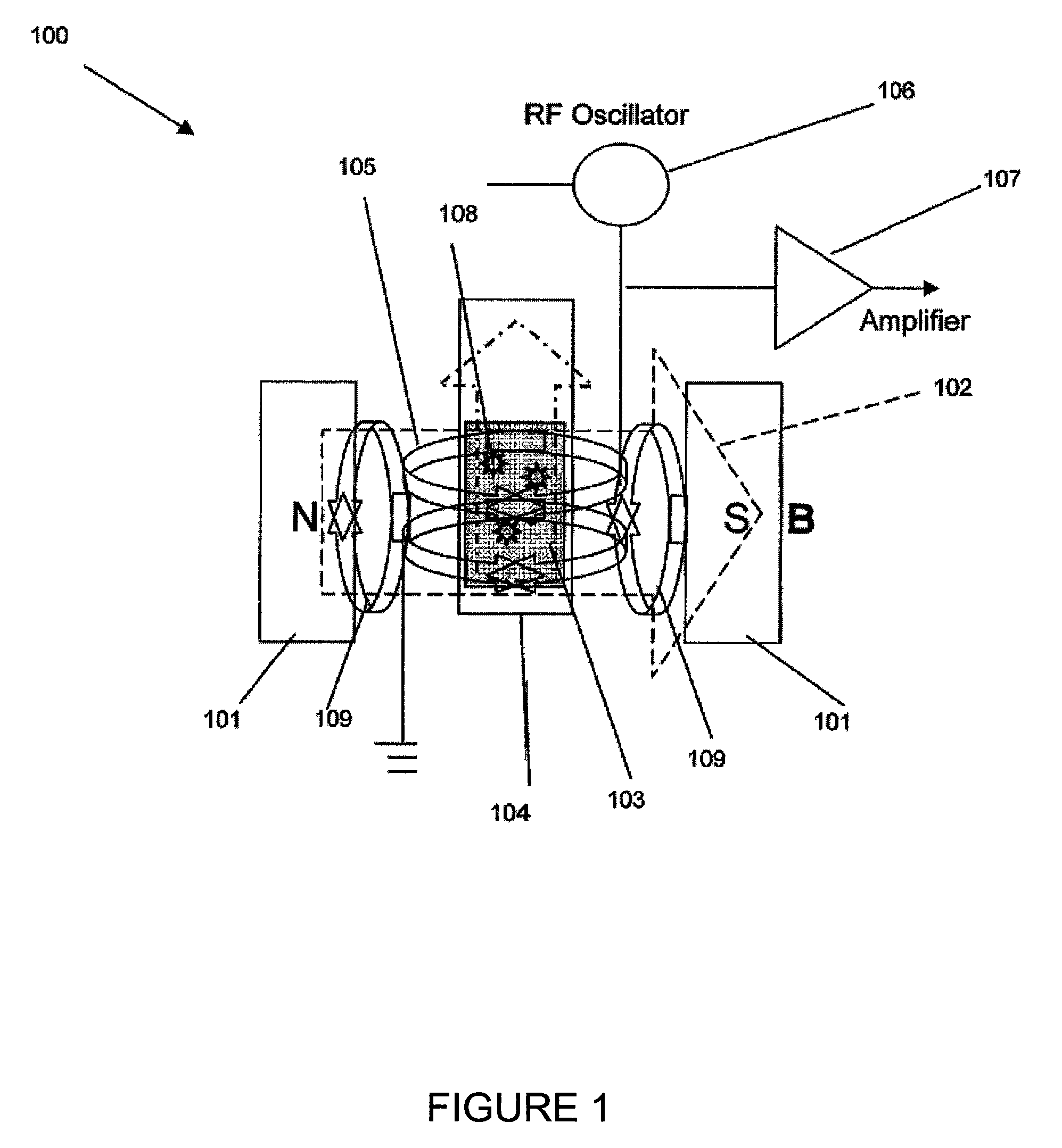
[0081]A Bruker Minispec mQseries (The Woodlands, Tex.) was adapted with pulse sequences for T2 monitoring in real time. Several effective T2 measurements made within 30 to 40 seconds and transverse relaxation times of plasma samples were measured every 5 seconds. Coagulation of a sample was induced by addition of calcium chloride to a mixture of reconstituted plasma (CITREX® I lyophilized plasma preparation, BIODATA Corporation, Horsham, PAS) and an aPTT reagent (CEPHALINEX® activated partial thromboplastin time reagent BIODATA Corporation, Horsham, Pa., USA).

[0082]T2 measurements were made kinetically on a Bruker MQ minispec using preloaded minispec software with the following CPMG settings:[0083]1. Tau=0.25[0084]2. Number of Points=1000[0085]3. Dummy Echos: 3[0086]4. Recycle Delay: 1[0087]5. 0 dB pulse at 37° C.[0088]6. Receiver gain: 75[0089]7. 1 scan

[0090]Coagulation time was estimated by curve analysis completed by midpo...

example 2

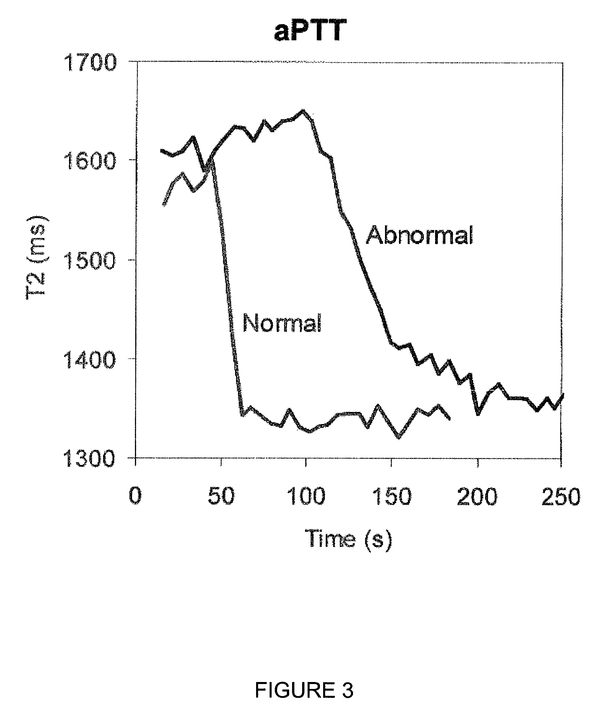
Coagulation Measurements Using Pooled Normal and Single Donor Abnormal Samples via T2 Relaxation

[0091]Real patient plasmas were purchased through George King Bio-Medical and used within 2 hours following thawing. Both normal and abnormal samples were run in duplicate to provide experimental error (averages are shown). Duplicate sampling resulted in a more precise coagulation time compared to the reference (start-4) data. (Note: standard deviation of samples controlled two factors: effective T2 values, coagulation time).

[0092]Measurement of changes in T2 relaxation time over time were taken. Measurements were made kinetically on a Bruker MQ minispec using preloaded minispec software with the following CPMG settings:[0093]1. Tau=0.25[0094]2. Number of Points=1750[0095]3. Dummy Echos: 3[0096]4. Recycle Delay: 1[0097]5. 18 dB pulse at 37° C.[0098]6. Receiver gain: 75[0099]7. 1 scan

[0100]Curve analysis was completed to generate coagulation times by midpoint determination between the T2 i...

example 3

Correlation Between Coagulation Method Results Obtained Using a Method of the Present Invention and Results Obtained with a Commercial Bench-top Coagulation Instrument

[0103]For aPTT measurements, 100 μL of patient plasma and 100 μL of PTT-A (Diagnostica Stago) were pre-warmed to 37° C. in a 5 mm NMR tube. 100 μL of calcium chloride pre-warmed to 37° C. was added to the plasma and clotting reagent activating coagulation. For PT measurements 100 μL of patient plasma and 200 μL of Neoplastine CI Plus (Stago Diagnostica, Parsippany, N.J.) pre-warmed to 37° C. were mixed activating coagulation. Measurements were made kinetically on a Bruker M Q minispec using preloaded minispec software with CPMG parameters described in Example 2. Curve analysis to generate coagulation times was completed by midpoint determination between the T2 intial and T2 final.

[0104]Prothrombin Time (PT) and Activated Partial Thromboplastin Time (aPPT) of plasma samples were measured using a Bruker Minispec mQseries...


PUM

PropertyMeasurementUnit
dwell timeaaaaaaaaaa
prothrombin timeaaaaaaaaaa

Abstract

The invention relates to detecting coagulation and coagulation-related activities including agglutination and fibrinolysis of samples. More particularly the invention relates to methods and apparatus for monitoring coagulation and / or obtaining a coagulation time of a sample using NMR-based detectors.

Description

CROSS-REFERENCE TO RELATED APPLICATIONS[0001]This application is the U.S. National Stage of International Application PCT / US2009 / 062537, filed Oct. 29, 2009, which claims benefit of U.S. Application No. 61 / 109,367, filed Oct. 29, 2008, each of which is hereby incorporated by reference.[0002]The invention relates to detecting coagulation and coagulation-related activities such as, for example, agglutination and fibrinolysis of samples (e.g., human blood samples). More particularly, the invention relates to methods and apparatus for obtaining a coagulation time of a sample (e.g., plasma, blood concentrate, citrated blood) using NMR-based detectors.BACKGROUND OF THE INVENTION[0003]Hemostasis, the physiological process of preventing excess blood loss by arresting flow via the formation of a hemostatic plug while maintaining blood in a fluid state within intact blood vessels, is maintained by tightly regulated interactions of the blood vessel wall, blood platelets, and blood plasma prote...

Claims


Application Information

Patent Timeline
Patent Type & Authority Patents(United States)
IPC IPC(8): C12Q1/56G01R33/561G01R33/50G01N33/49G01N33/86G01R33/465G01N33/48G01N33/487G01N33/483
CPCG01R33/465G01N33/4905G01N33/86G01R33/5617G01R33/50G01N33/487G01N33/48G01N33/483C12Q1/56G01N24/088
Inventor TAKTAK, SONIAMOZELESKI, BRIAN M.LOWERY, JR., THOMAS J.
Owner T2 BIOSYST
Why Eureka
Try Eureka