Biphasic osteochondral scaffold for reconstruction of articular cartilage

a cartilage layer and scaffold technology, applied in the field of synthetic osteochondral scaffolds, can solve the problems of pain in osteoarthritis and limited movement, damage to the cartilage layer, and no gold standard, and achieve the effect of reducing pain and limited movemen

Inactive Publication Date: 2015-11-10
STEVENS INSTITUTE OF TECHNOLOGY
View PDF48 Cites 564 Cited by
  • Summary
  • Abstract
  • Description
  • Claims
  • Application Information

AI Technical Summary

Problems solved by technology

Thus, the body generally cannot heal the articular cartilage on its own and the eventual degradation of the tissue leads to painful osteoarthritis and limited movement.
Unfortunately, although there are numerous treatments, none have been marked as a gold standard due to each one having its own drawbacks, especially when it comes to reproducing the exact physiological structure of articular cartilage capable of integrating with the surrounding tissue and bone.
Damage to the cartilage layer may also involve damage to the underlying subchondral bone.
However, critical defects are not able to be healed by the bone's natural regenerative processes.
But the use of autografts is plagued by supply issues and donor site morbidity issues.
Allografts, despite being osteoconductive and fairly abundant in supply, can be associated with disease transmissions and require processing, preservation, and sterilization steps that decrease the healing properties of the allograft.
When there is a partial depth defect (i.e., the defect does not penetrate through the cartilage layer), progenitor cells from the bone marrow cannot be recruited to form new cartilage, thus repair will be extremely limited without the bone marrow mesenchymal cells.
But when there is a full thickness defect (i.e., an osteochondral defect), even though the mesenchymal stem cells are released, there is no structure on which the cells can attach, proliferate, and differentiate.
In such a situation, the stem cells become fibrocartilage, which is a poor substitute for articular cartilage due to its lack of mechanical strength and lubrication properties.

Method used


Image

  • Biphasic osteochondral scaffold for reconstruction of articular cartilage
  • Biphasic osteochondral scaffold for reconstruction of articular cartilage
  • Biphasic osteochondral scaffold for reconstruction of articular cartilage

Examples


example

Fabrication and Testing of an Osteochondral Scaffold

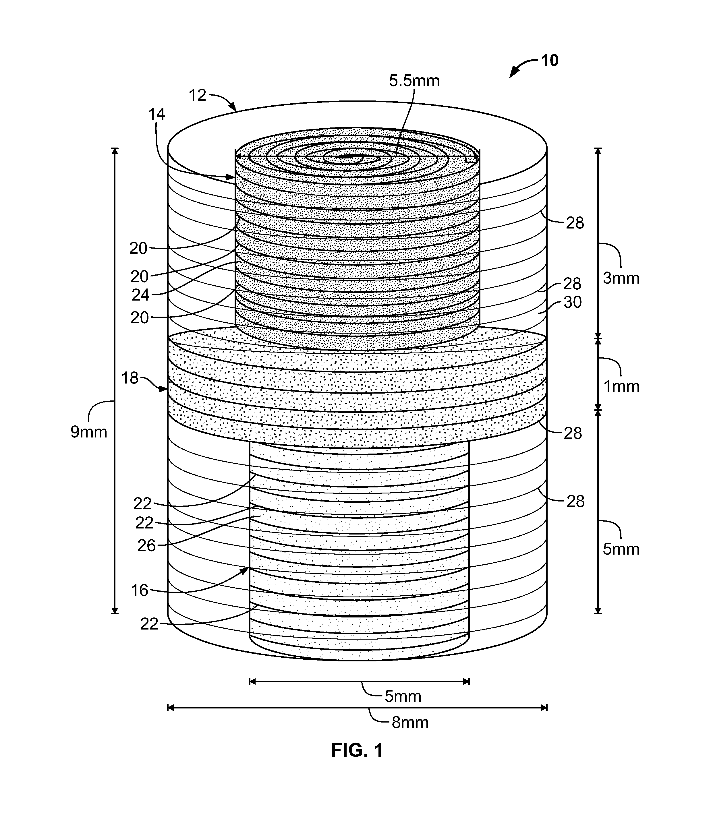
[0067]The following is a non-limiting example of the fabrication and testing of an osteochondral scaffold according to an embodiment of the present invention. This example is merely meant to show how one type of osteochondral scaffold may be made. Both the osteochondral scaffold and the method of making it are included within the scope of the present invention.

Methods

Microsphere Formation

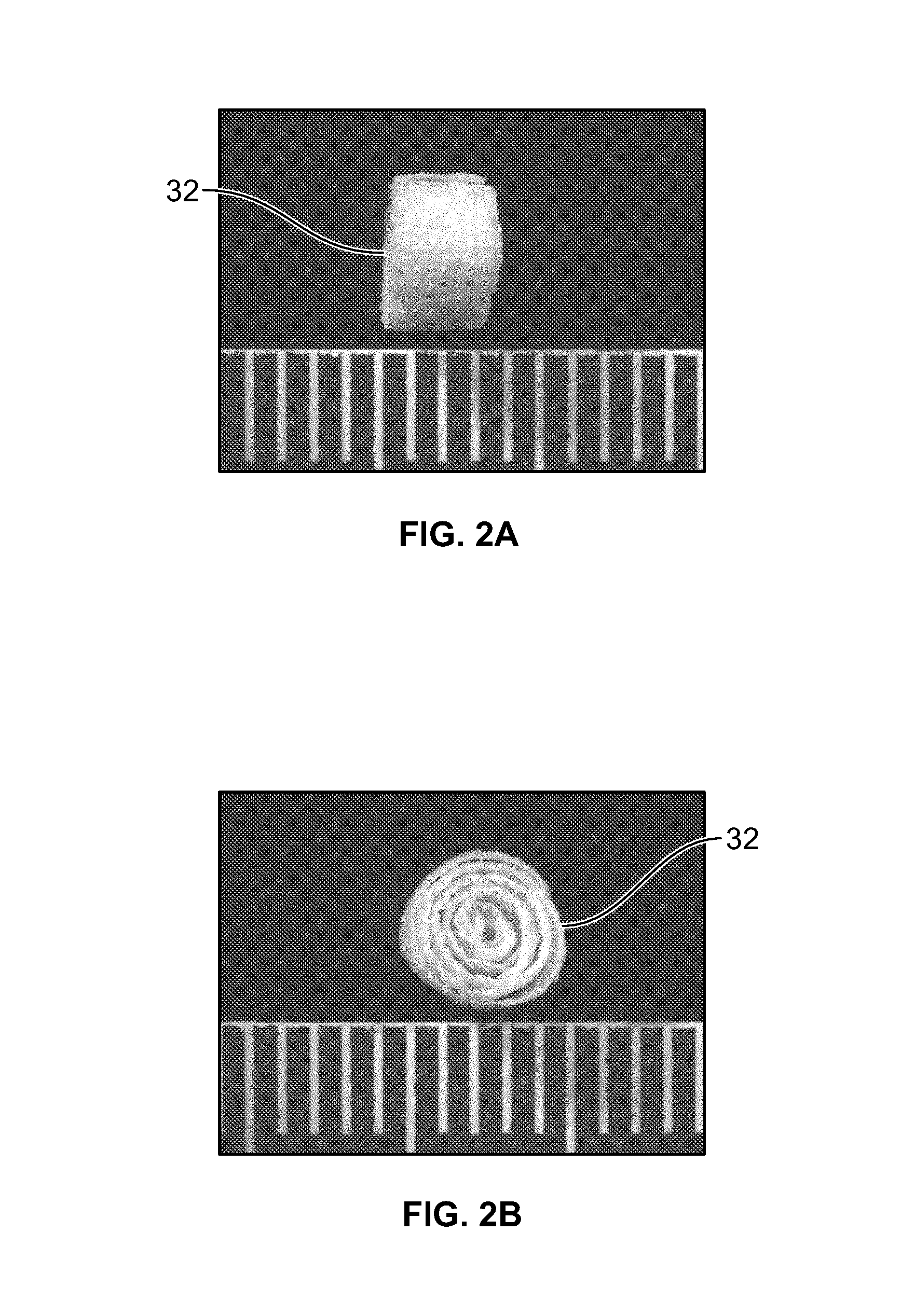
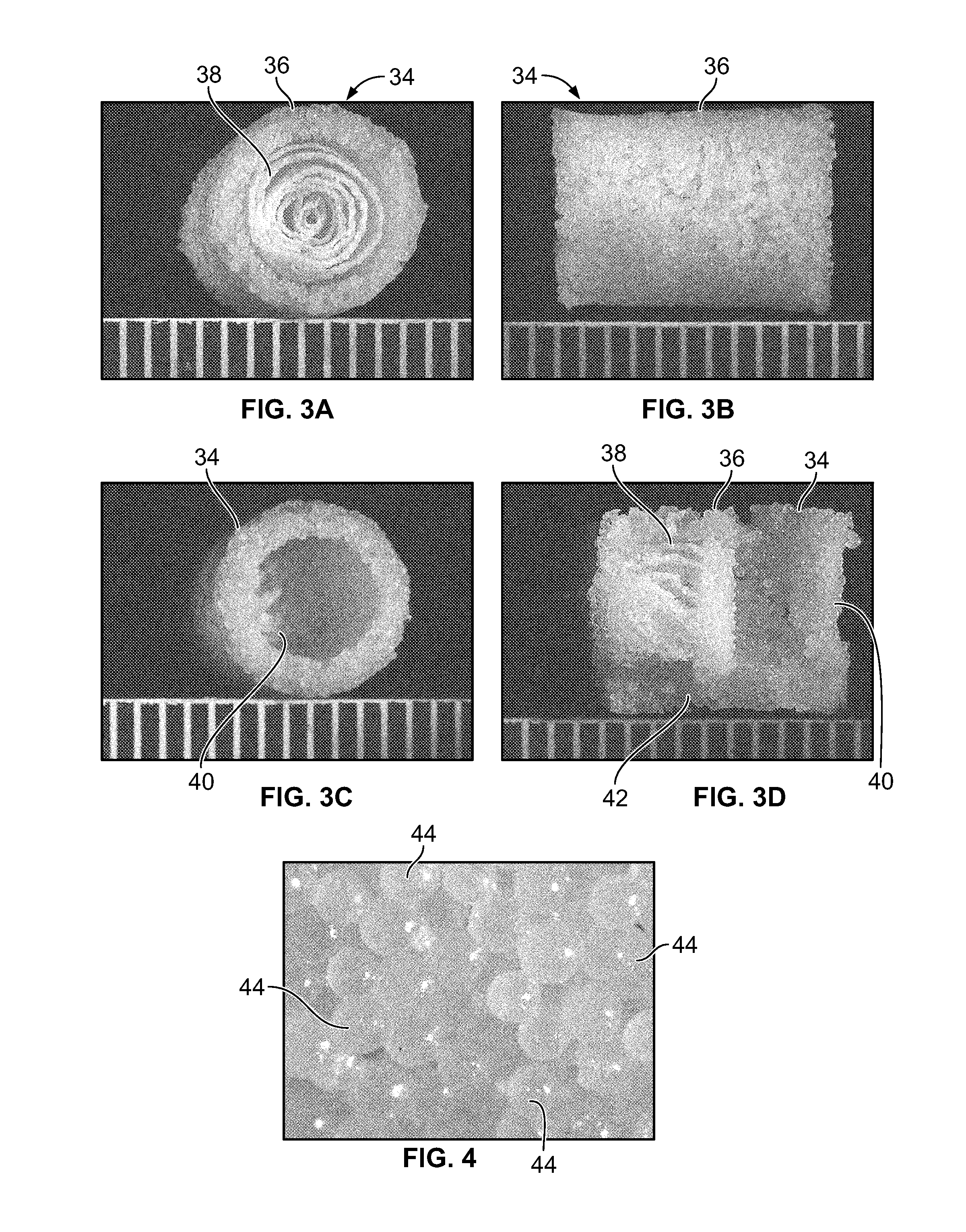
[0068]Microspheres of PLGA / PCL in proportions of 100 / 0, 75 / 25, or 50 / 50 were prepared by mixing 100% PLGA (85-15); 75% PLGA 25% PCL (MW: 80,000); or 50% PLGA 50% PCL as 10% (w / v) solutions in dichloromethylene (DCM) To create an water-in-oil emulsion effect, a 1% (w / v) poly(vinyl) alcohol (PVA) (MW: 31,000-50,000) solution was prepared and stirred at 360 RPM with an impeller. The polymer (i.e., PLGA or PLGA / PCL) solution was then loaded into a 10 ml syringe with a 16 gauge needle. The polymer solution was then forced out in a steady stream into the...


PUM

PropertyMeasurementUnit
thicknessaaaaaaaaaa
diametersaaaaaaaaaa
diametersaaaaaaaaaa

Abstract

An osteochondral scaffold has a chondrogenic spiral scaffold in one end of an outer shell made of sintered microspheres, and an osteogenic spiral scaffold in the other end of the outer shell. Each spiral scaffold has nanofibers of a composition selected to promote attachment and proliferation of the desired types of cells. The nanofibers for the chondrogenic spiral scaffold have a different composition than the nanofibers for the osteogenic spiral scaffold. The nanofibers of each spiral scaffold are aligned to orient the attached cells so as to recreate the structure of the native tissue.

Description

CROSS-REFERENCE TO RELATED APPLICATIONS[0001]The present application claims the benefit of U.S. Provisional Patent Application No. 61 / 645,319, filed on May 10, 2012, which is incorporated herein by reference.STATEMENT REGARDING FEDERALLY SPONSORED RESEARCH[0002]Not applicable.FIELD OF THE INVENTION OR TECHNICAL FIELD[0003]The present invention is directed to a synthetic osteochondral scaffold for promoting articular cartilage regeneration.BACKGROUND OF THE INVENTION[0004]Articular cartilage covers the end of all diarthroidal joints, allowing the bones to slide against each other without actually coming into contact with each other. Due to the lack of vascularity above the subchondral region, healing of damaged cartilage is very rare. Thus, the body generally cannot heal the articular cartilage on its own and the eventual degradation of the tissue leads to painful osteoarthritis and limited movement.[0005]Current treatments for osteoarthritis include joint replacement, microfracturin...

Claims


Application Information

Patent Timeline
Patent Type & Authority Patents(United States)
IPC IPC(8): A61L27/26A61F2/28A61L27/38B29C53/56A61F2/30C08L5/08A61L27/36A61L27/54
CPCA61L27/26A61F2/28A61F2/30756A61L27/365A61L27/3852A61L27/54B29C53/562C08L5/08A61F2002/30016A61F2002/3084A61F2002/3092A61F2002/30224A61F2002/30293A61L2430/02A61L2430/06
Inventor YU, XIAOJUNLEE, PAUL
Owner STEVENS INSTITUTE OF TECHNOLOGY
Why Eureka
Try Eureka