A biomimetic cell-containing bulk osteochondral bioscaffold and preparation method thereof

A bio-scaffold and osteochondral technology, applied in the field of biomedical tissue engineering, can solve the problems of surrounding tissue fit, singleness, support, vascular network channels and cell-induced differentiation, so as to speed up repair and promote integrated repair and reconstruction. Effect

Active Publication Date: 2022-08-09
SHANGHAI UNIV
View PDF5 Cites 0 Cited by
  • Summary
  • Abstract
  • Description
  • Claims
  • Application Information

AI Technical Summary

Problems solved by technology

However, the current osteochondral scaffold structure is relatively simple and single, which cannot meet the cartilage surface structure requirements of natural osteochondral and the functional requirements of large osteochondral tissue, and cannot fit with the surrounding tissue after implantation, and there is support for large osteochondral tissue. , vascular network channels and the interaction of cell-induced differentiation

Method used


Image

  • A biomimetic cell-containing bulk osteochondral bioscaffold and preparation method thereof
  • A biomimetic cell-containing bulk osteochondral bioscaffold and preparation method thereof

Examples


preparation example Construction

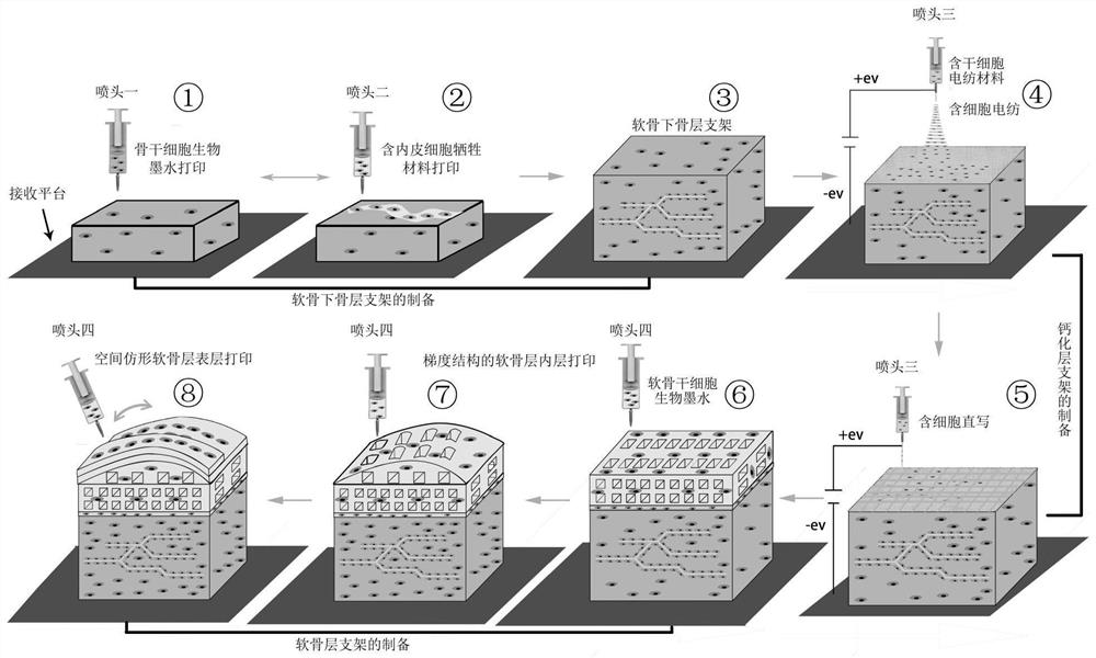
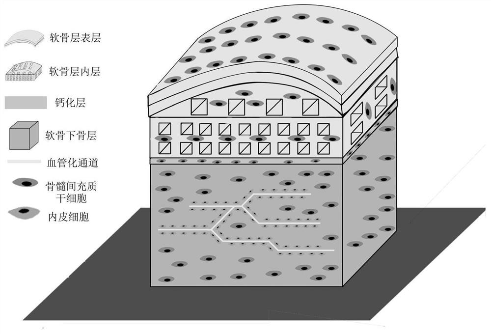
[0031] The invention provides a method for preparing a biomimetic cell-containing bulk osteochondral biological scaffold, comprising the following steps:

[0032] The subchondral bone material containing bone marrow mesenchymal stem cells is subjected to the first extrusion 3D printing, and the sacrificial material containing human umbilical vein endothelial cells is used for the second extrusion 3D printing in the formed subchondral bone scaffold structure, alternately in turn. performing the first extrusion 3D printing and the second extrusion 3D printing to obtain a subchondral bone layer containing a prefabricated vascularized network structure;

[0033] Using electrospinning material containing bone marrow mesenchymal stem cells, electrospinning is performed on the subchondral bone layer, and a cell-containing nanofiber isolation membrane is formed on the upper surface of the subchondral bone layer;

[0034] Continue to use the electrospinning material containing the bone...

Embodiment 1

[0077] according to figure 1 The flow shown carries out the scheme of embodiment 1:

[0078] a. Preparation of bone stem cell bioink: using PBS buffer as solvent, gelatin (10%), sodium alginate (5%), bone marrow mesenchymal stem cells (5×10 6 cells / g) were mixed and prepared, and BMP-2 protein 5% (w / w, relative to the total mass of the bone stem cell bioink) was added; 10 mL of the prepared bone stem cell bioink was added to a medical syringe, and the syringe Installed on the first print nozzle;

[0079] Preparation of cell sacrificial material: PBS buffer was used as a solvent, and collagen (5%, w / v), PVA (10%, w / v) and human umbilical vein endothelial cells (5×10 6 cells / g) mixed and prepared; then add 10 mL of the prepared cell-containing sacrificial material into a medical syringe, and install it on the second printing nozzle;

[0080] b. Preparation of electrospinning materials containing stem cells: using PBS buffer as solvent, PEO (15%), sodium alginate (5%) and huma...

Embodiment 2

[0088] The difference between this embodiment and Embodiment 1 is only:

[0089] Bone stem cell bioink, the solvent is HBSS solution (Hank's balanced salt solution), using methacrylated gelatin (12% w / v), hydroxyapatite (5% w / w) and bone marrow mesenchymal stem cells (5% w / w) ×10 6 cells / g) were prepared by mixing and adding LAP photoinitiator 0.2% (w / w, relative to the total mass of the bone stem cell bioink);

[0090] The cartilage stem cell bioink solvent is HBSS solution (Hank's balanced salt solution), using methacrylated gelatin (10% w / v), hyaluronic acid (10% w / v) and bone marrow mesenchymal stem cells (5 × 10 6 cells / g) were prepared by mixing and adding LAP photoinitiator 0.2% (w / w, relative to the total mass of the cartilage stem cell bioink);

[0091] The prepared bulk osteochondral scaffold was placed under blue light with a wavelength of 405 nm for cross-linking;

[0092] Other steps are the same as in Example 1.


PUM

PropertyMeasurementUnit
diameteraaaaaaaaaa
diameteraaaaaaaaaa
porosityaaaaaaaaaa

Abstract

The invention provides a biomimetic cell-containing bulk osteochondral biological scaffold and a preparation method thereof, belonging to the technical field of biomedical tissue engineering. The invention adopts the composite process of cell-containing extrusion 3D printing technology, cell-containing electrospinning technology, cell-containing electrohydrodynamic direct writing technology and sacrificial material method, which can not only realize the integrated osteochondral structure with bionic human osteochondral tissue structure The scaffold is formed, and the preparation of the osteochondral scaffold containing cells can be realized, which provides a solution for the construction of a large osteochondral bioscaffold, and promotes the integrated repair and reconstruction of the osteochondral tissue.

Description

technical field [0001] The invention relates to the technical field of biomedical tissue engineering, in particular to a biomimetic cell-containing bulk osteochondral biological scaffold and a preparation method thereof. Background technique [0002] Due to trauma, accident, obesity, strain, tumor, joint congenital anomaly and joint deformity, and senile degenerative osteoarthropathy, the number of patients with articular osteochondral defect is increasing, and it has become one of the main causes of limb disability. Among them, articular cartilage damage can easily lead to joint lesions of subchondral bone, which makes self-repair of damaged articular cartilage tissue very difficult. At present, autologous or allogeneic tissue, artificial scaffolds and other implants are mainly used for clinical repair. Among them, the source of autologous osteochondral tissue is limited, it is difficult to repair large tissue defects, and it is easy to cause secondary damage. Allogeneic os...

Claims


Application Information

Patent Timeline
Patent Type & Authority Patents(China)
IPC IPC(8): A61L27/38A61L27/12A61L27/16A61L27/18A61L27/20A61L27/22A61L27/24A61L27/56B33Y70/10
CPCA61L27/3834A61L27/3852A61L27/24A61L27/222A61L27/20A61L27/12A61L27/16A61L27/18A61L27/56B33Y70/10A61L2430/06A61L2400/12
Inventor 胡庆夕刘随红张海光
Owner SHANGHAI UNIV
Why Eureka
Try Eureka