
Osteochondral scaffold

a scaffold and osteochondral technology, applied in the field of osteochondral scaffolds, can solve the problems of reduced joint function, limited availability, and individual pain of oa, and achieve the effects of easy delivery to the osteochondral defect, stable physical environment, and healthy growth of the overlying cartilag

Active Publication Date: 2022-08-16
UCL BUSINESS PLC
View PDF13 Cites 0 Cited by
  • Summary
  • Abstract
  • Description
  • Claims
  • Application Information

AI Technical Summary

Benefits of technology

This patent is about a new type of scaffold that can help repair damaged cartilage. It can mimic the natural environment in which cartilage heals, providing support and reducing pain. This scaffold can be used to treat small cartilage defects before resorting to joint replacement. It can improve quality of life by keeping people active.

Problems solved by technology

Individuals with OA often experience joint pain and reduced joint function unless appropriate treatment is adopted.
However, donor site morbidity, limited availability of matching donor tissues, a high complication rate as well as early degenerative changes of the transplanted cartilage are well-known disadvantages.
The excessive bone formation caused by the healing leads to subchondral stiffening which leads to the degeneration of the overlying cartilage.
This will happen when the loss of quality of cartilage and bone at the joint interface has significantly reduced the quality of life of the patient, and non-surgical treatments are no longer effective.
These are major surgical procedures, which are only undertaken when the disease has progressed to the stage where there is no alternative but a severely limited lifestyle.
This can mean that patients will have suffered long periods of pain, discomfort and reduced mobility before an operation is performed.
However, implants do not come without risk or complications.
A growing number of implant recipients have experienced implant failure and other severe complications associated with their joint replacements.
The treatment of cartilage and osteochondral defects remains a challenge because treatments to date have failed to achieve a complete restoration of the joint cartilage surface and its properties.
However, none of these products promotes satisfactory durable regeneration of large cartilage and osteochondral defects.
The main issues for current commercially available osteochondral defect treatments are poor cartilage fill and associated fibrocartilaginous repair rather than hyaline cartilage, as well as poor osteochondral repair (Christensen, B. B., et al.
This results in an inappropriate physical environment for cartilage healing.

Method used


Image

  • Osteochondral scaffold
  • Osteochondral scaffold
  • Osteochondral scaffold

Examples


example 1

oxyapatite Coating Improves Cell Growth on Titanium Matrix

[0110]Titanium scaffold is used for the bone section of the osteochondral scaffold. Sheep bone marrow derived stem cells were used for evaluation of the cellular performance. Examination revealed that the cells attached to the titanium surface and can proliferate in the titanium scaffold over the 28 days culture period, as demonstrated by Alamar blue analysis shown in FIG. 6.

[0111]To improve the cells growth and bone formation within the titanium matrix, the titanium scaffold could be coated with a thin layer (1-10 micro meter) of nano-hydroxyapatite by electro-chemical deposition technique, as shown in FIG. 7a. The in vivo evaluation using sheep bone marrow derived stem cells indicated an increase in number of cells seeded on the scaffolds within 21 days, and presence of metabolically active and proliferating cells on both HA coated and non-coated samples, as shown in FIG. 7b Alamar blue analysis indicated that the cells on ...

example 2

Evaluation of the Osteochondral Scaffold

[0112]Sheep bone marrow mesenchymal stem cells were used to evaluate the in vitro performance of the tri-layered osteochondral scaffold based on the titanium / PLGA / collagen system. Tri-layered collagen-hydroxyapatite osteochondral scaffold was used as control. The tissue culture was performed in static culture in normal culture medium. Alamar blue analysis indicated an increase in number of cells seeded on the scaffolds within 21 days (FIG. 8). The presence of metabolically active and proliferating cells on both the invented osteochondral scaffold and on the control scaffold were shown within 21 days culture. The titanium / PLGA / collagen osteochondral scaffold exhibited persistent higher Alamar blue activity than the control scaffold over 21 days' culture.

example 3

chondral Scaffold Achieves Stable Mechanical Fixation

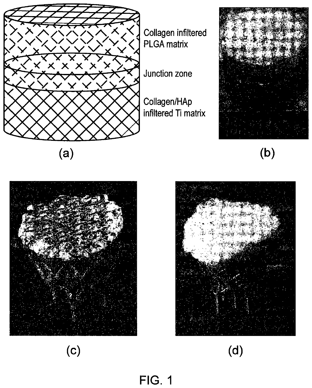
[0113]Sheep condyle and Sawbone were used to test the mechanical fixation of the scaffold. For this test, cylindrical osteochondral scaffold comprised of a lower titanium section and an upper PLA section. Multi-layered collagen-hydroxyapatite composite scaffolds were used as control. The mechanical fixation was determined in terms of interface shear strength and push-in and push-out strength. It was observed that both PLA and titanium-PLA scaffold have higher interface shear strength than the control scaffold in both sheep condyle and Sawbone model tests. In both cases, titanium-PLA double layers composite scaffold exhibited the higher interface shear strength, as shown in FIG. 9a.

[0114]The titanium scaffold was further evaluated in sheep chodyle model by push-in and push-out tests. It revealed that the push-in strength for a 10 mm diameter 10 mm thick cylindrical scaffold reached 2.43 MPa for titanium scaffold and 2.53 MPa for t...


PUM

PropertyMeasurementUnit
unit cell sizeaaaaaaaaaa
porosityaaaaaaaaaa

Abstract

There is described a multiphasic osteochondral scaffold for osteochondral defect repair, the scaffold comprising a bone phase and a cartilage phase, wherein the bone phase comprises a support matrix and the cartilage phase comprises a polymeric matrix, and the scaffold comprises a non-porous layer between the bone phase and the cartilage phase. Also described is a multiphasic osteochondral scaffold for osteochondral defect repair, the scaffold comprising a bone phase and a cartilage phase, wherein the bone phase comprises a support matrix and the cartilage phase comprises a polymeric matrix, and wherein the support matrix is tapered so that the dimensions of the support matrix are less at the lower end of the support matrix than at the upper end of the support matrix.

Description

FIELD OF THE INVENTION[0001]The invention relates to an osteochondral scaffold, a method of producing an osteochondral scaffold and its use in orthopaedic therapy. The scaffold is a functionally biomimetic osteochondral scaffold which can be used for cartilage and osteochondral defect (OCD) repair.BACKGROUND TO THE INVENTION[0002]Osteoarthritis (OA) is a degenerative joint disease (Litwic, A., et al. (2013). British Medical Bulletin 105: 185-199). Individuals with OA often experience joint pain and reduced joint function unless appropriate treatment is adopted. It affects over 18% of the population when they reach their mid-50s. This figure rises to over 52% by the time they reach their mid-70s. More than 650,000 people in the UK have painful OA in one or both hips, and more than 6 million people have OA in one or both knees. It may start slowly with mild to moderate symptoms; pain typically may occur on motion or after inactivity.[0003]Cartilage transplantation has been explored as...

Claims


Application Information

Patent Timeline
Patent Type & Authority Patents(United States)
IPC IPC(8): A61L27/36A61L27/06A61L27/24A61L27/12A61L27/56A61F2/30A61L27/58A61L27/18A61L27/22
CPCA61L27/365A61F2/30A61L27/06A61L27/12A61L27/18A61L27/24A61L27/3654A61L27/56A61L27/58A61F2002/30214A61L27/222A61L27/225A61L27/227A61L2430/02A61L2430/06A61L2430/24C08L67/04
Inventor LIU, CHAOZONGBLUNN, GORDON
Owner UCL BUSINESS PLC
Learn More