Isolation, expansion and use of autologous pluripotent stem cells

a technology autologous expansion, applied in the field of autologous pluripotent stem cells, can solve the problems of inability to faithfully recapture disease processes, methods using expensive technologies, and relatively expensive and time-consuming generation of ips cells, and achieves low risk of cancer, high nuclear to cytoplasm ratio, and small size

Active Publication Date: 2016-08-23
THE UNIV OF NORTH CAROLINA AT CHAPEL HILL
View PDF20 Cites 0 Cited by
  • Summary
  • Abstract
  • Description
  • Claims
  • Application Information

AI Technical Summary

Benefits of technology

[0007]Autologous pluripotent stem (aPS) cells are proposed to be primitive stem cells in the postnatal animal and to play a role in tissue regeneration. aPS cells have been isolated from human peripheral blood and cerebrospinal fluid, and are proposed to exist in other and in animal tissues. These cells exhibit phenotypic similarity to embryonic stein cells and induced pluripotent stem (iPS) cells, including their protein and RNA expression (e.g., OCT4 (POU5F1), NANOG, SSEA4, TRA-1-60, TRA-1-80), and their high nuclear to cytoplasm ratio. However, as compared with ESC and iPS cells, the aPS cell size is smaller, generally from about one to about three microns, and by virtue of the small size, the nucleus contains densely packed heterochromatin. The nucleus of aPS cells has been observed to stain with DNA specific (e.g. DRAQ5™) and non-specific (e.g. 4′,6-diamidino-2-phenylindole [DAPI]) nucleic acid stains. In general, aPS cells have a density that is typically greater than 1.1 g / mL, a density greater than that of most other cells, including red blood cells. APS cells also differ from iPS cells in that do they generally do not form teratomas, and thus may have a low risk of cancer with regenerative medication applications.
[0008]The present invention is based, in part, on the discovery that aPS cells possess a high specific gravity relative to other cells, which can be exploited to isolate aPS cells. APS cells are smaller and denser than previously identified stem cell populations and can be isolated and distinguished from previously identified stem cell populations using these characteristics. The isolation methods of the invention improve on conventional methods of stein cell isolation by: (1) optimizing the number of aPS stem cells recovered per mL of whole blood, (2) using relatively simple technologies, making the methods less costly to implement and potentially usable in settings where FACS sorting of cells is not currently available; and / or (3) simplifying procedures to a protocol that in some embodiments can take as little as 2 to 6 hours and that is easily scalable so that aPS cells can be isolated from relatively large amounts of blood (e.g., a pint of blood or more), other animal body fluids or tissues, or other cell mixtures.
[0010]As one aspect, the present invention provides methods for isolating a very small (e.g., 1 to 5 micron diameter), primitive stem cell expressing OCT4 in the nucleus from a biological sample such as an animal body fluid (including blood, plasma, serum, bone marrow, lymph, tears, urine, breast milk, seminal fluid, saliva, amniotic fluid, bronchial lavage, cerebrospinal fluid, pleural fluid, peritoneal fluid and / or colostrum) and / or animal tissue (including, but not limited to brain and / or olfactory epithelium). In representative embodiments, the cell separation method is based on cell density. In embodiments, the method includes use of Stokes' Equation to determine optimal centrifugation parameters, which may minimize cell loss, avoid insufficient centrifugation time that could result in contamination of the final aPS cell product with other cell types, and avoid excessive centrifugation speeds and time that may damage cells and / or result in low yields of aPS cells. In representative embodiments, the method includes using a density barrier, continuous flow cell separation, and / or other methods that separate cells based on specific gravity.

Problems solved by technology

However, obstacles to clinical use of iPS cells include that iPS cells may be prone to cancer or other pathologies, that the iPS cells, as “artificial”, may not faithfully recapitulate disease processes (e.g., due to epigenetic factors), and that generation of iPS cells is relatively expensive and time consuming.
Disadvantages of these methods include that the methods use expensive technologies and may take an unrealistic amount of time (for example days) to isolate sufficient cells for clinical use3.

Method used


Image

  • Isolation, expansion and use of autologous pluripotent stem cells
  • Isolation, expansion and use of autologous pluripotent stem cells
  • Isolation, expansion and use of autologous pluripotent stem cells

Examples


example 1

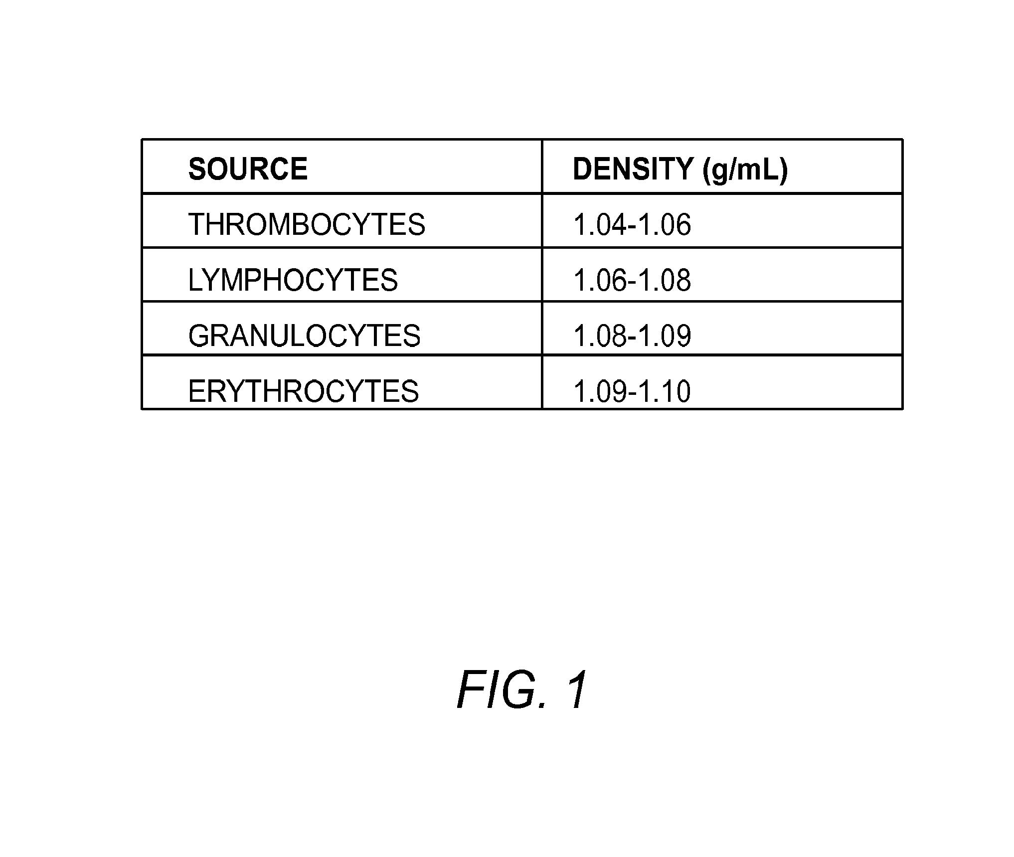
Isolation of Stem Cells

[0187]In the example describe herein, 10 mL of peripheral blood was collected by routine venipuncture into sodium citrate (green top) tubes. If more blood is collected the processing steps are scaled up accordingly. The blood was kept at 37° C. in a water bath for less than 2 hours.

[0188]Contaminating red blood cells were removed by lysis by mixing 2.5 mL of whole blood with 47.5 mL of 37° C. hypotonic lysis buffer (BD Pharm, containing ammonium chloride, potassium carbonate, and EDTA) in a 50 mL centrifuge tube. Any other red blood cell lysis buffer may also be used. The tubes were gently vortexed for about 15 seconds, and then incubated in a 37° C. water bath protected from light for 15 minutes.

[0189]Alternatively, other body fluids and tissues can be obtained and processed as appropriate to that tissue using methods known to others familiar with the art.

[0190]The tubes were centrifuged at 1000 g for 30 minutes. The supernatant was decanted, and the pellet r...

example 2

Alternate Protocol for Isolating aPS Cells

[0203]As an alternative embodiment to isolate aPS cells from animal body fluids or tissues, a high throughput immunomagnetie separation method such as the Quadrasep QMC™ quadrupole magnetic sorter (Ikotech) may be used directly on whole blood, after lysis of red blood cells, or after density barrier separation.

[0204]In this embodiment, cells are labeled with appropriate antibodies against contaminating cell types, such as white blood cells, platelets, and red blood cells, or cells specific to the tissue used, but that will not substantially interact (or will not interact at all) with aPS cells. In embodiments, the antibodies also have small magnetic particles attached. The cell suspension is subject to a high throughput immunomagnetic separation method such as Quadrasep QMC™ quadrupole magnetic sorter (Ikotech) using methods known to those skilled in the art, and the appropriate fraction containing aPS cells is collected.

[0205]An alternative...

example 3

Alternative Embodiment for Isolation of aPS Cells

[0207]In the example describe herein, 1 mL of peripheral blood was collected by routine venipuncture into sodium citrate (green top) tubes. If more blood is collected the processing steps can be scaled up accordingly. The blood was kept at 37° C. in a water bath for less than 2 hours.

[0208]In this example a sterile Ultrafree CL centrifuge filter tube fitted with a 5 micron filter is filled with 5.5 mL of PERCOLL™ with a specific gravity of 1.12 g / mL and briefly centrifuged so that the PERCOLL™ fills the outer tube and inner tube to the same level, and comes up to slightly above the filter of the inner tube. The 5 micron filter is large enough to allow passage of the aPS cells, and minimizes any mixing of the cell suspension with the density barrier material. One mL of whole blood is diluted with an equal volume of PBS, and the cell suspension was layered over the PERCOLL™ in the inner centrifuge tube, and the inner centrifuge tube was...


PUM

PropertyMeasurementUnit
specific gravityaaaaaaaaaa
mean diameteraaaaaaaaaa
sizeaaaaaaaaaa

Abstract

The present invention relates to methods of isolating and culturing autologous pluripotent stem (aPS) cells. The present invention also provides isolated aPS cells, populations of aPS cells and cultures of aPS cells. Further provided are culture media for expanding aPS cells and methods of culturing aPS cells. The invention also provides for the use of aPS cells, e.g., for diagnostics, drug evaluation and screening, and regenerative medicine.

Description

RELATED APPLICATION INFORMATION[0001]This application claims the benefit of U.S. Provisional Application No. 61 / 496,892, filed Jun. 14, 2011, the disclosure of which is incorporated by reference herein in its entirety.FIELD OF THE INVENTION[0002]The present invention relates to methods of isolating and culturing autologous pluripotent stem (aPS) cells as well as isolated aPS cells and populations of aPS cells and cultures of aPS cells.BACKGROUND OF THE INVENTION[0003]Stem cells derived from an animal are potentially useful for a variety purposes, including regeneration of damaged tissues, reproduction, and as cellular models that could inform personal medicine, including diagnoses, treatments to alleviate a condition of disease or disorder, or warnings of adverse reaction to a potential treatment. Currently, induced pluripotent stem cells (iPS cells) are the dominant model system. iPS cells are derived from dividing multipotent or committed cells (such as fibroblasts, fat stem cells...

Claims


Application Information

Patent Timeline
Patent Type & Authority Patents(United States)
IPC IPC(8): C12N5/074C12N5/00C12N5/0789C12N5/0775
CPCC12N5/0087C12N5/0647C12N5/0696C12N5/0665C12N2509/00
Inventor PERKINS, DIANAJEFFRIES, CLARKTURAGA, VANDANA
Owner THE UNIV OF NORTH CAROLINA AT CHAPEL HILL
Why Eureka
Try Eureka