Diagnostic devices, methods and systems for detecting platelet factor 4 (PF4)/heparin antibodies

a technology of platelet factor 4 and detection method, applied in the field of immunodeficiency diagnosis, can solve the problem of providing a very limited dynamic range, and achieve the effect of facilitating the visibility of antibodies and facilitating the visibility of antibodies

Active Publication Date: 2017-03-21
IMMUCOR GTI DIAGNOSTICS
View PDF7 Cites 0 Cited by
  • Summary
  • Abstract
  • Description
  • Claims
  • Application Information

AI Technical Summary

Benefits of technology

[0014]The numerous technical advantages of using an immobilized PF4 / polyanion complex instead of an immobilized PF4 / heparin complex for PF4 / heparin antibody capture are discussed in detail in U.S. Pat. No. 5,972,718, which discussion is incorporated herein by reference. Further, the device and method described herein provide superior assay performance by virtue of employing non-particulate fluorescent labels, which is contrary to the popular practice of using particulate labels for lateral flow detection. This design feature addresses the strongly negative charge profile of the immobilized PF4 / polyanion complex. In the course of development, the inventors found that positively charged latex particles tend to aggregate and bind non-specifically to the negatively charged polyanion. Although neutrally charged gold particles do not have the specificity problem, they provide a very limited dynamic range for the purposes of detecting PF4 / heparin antibodies. The present inventors subsequently discovered that non-particulate fluorescent labels, particularly those without a significant positive charge, provide an excellent dynamic range while demonstrating minimal non-specific binding to the PF4 / polyanion complex.
[0016]In a further aspect, the invention provides a lateral flow assay device for detecting the presence of human platelet factor 4 (PF4) / heparin antibodies in a fluid sample, such as a blood sample. The device comprises a test strip which includes a conjugate pad and a porous membrane positioned downstream of, and in fluid communication with, the conjugate pad. The conjugate pad includes a conjugate zone, a sample zone, and a sample buffer zone. The conjugate zone contains anti-human antibodies, e.g., goat anti-human immunoglobulins, releasably attached thereto and labeled with a non-particulate fluorescent dye, e.g., an amine-reactive dye. The sample zone is positioned downstream of the conjugate zone and configured to receive a fluid sample, such as a whole blood, serum or plasma sample. The sample buffer zone is positioned intermediate to the conjugate zone and the sample zone and configured to receive a sample buffer that facilitates lateral transport of the fluid sample. The porous membrane is positioned downstream of, and in fluid communication with, the conjugate pad and includes a detection zone and a control zone. The detection zone contains a PF4 / polyanion complex immobilized thereon. The control zone is positioned downstream of the detection zone and contains a capture reagent, e.g., mouse anti-goat immunoglobulins, immobilized thereon and configured to bind with the labeled anti-human antibodies.
[0017]In this aspect of the invention, the conjugate pad may further include a conjugate buffer zone positioned upstream of the conjugate zone and configured to receive a conjugate buffer that solubilizes the labeled anti-human antibodies and facilitates their lateral transport. In other embodiments, the conjugate buffer zone may reside on a separate pad, herein referred to as a “wet pad,” which is positioned upstream of, and in fluid communication with, the conjugate pad. In preferred embodiments, the test strip may also include a wicking pad in fluid communication with the porous membrane, such that the membrane is positioned intermediate to the conjugate pad and the wicking pad. The purpose of the wicking pad is to enhance lateral transport by withdrawing fluid from the porous membrane. Although the conjugate pad is preferably embodied in a single integral pad, it is further contemplated that the conjugate pad may include a discrete conjugate portion, containing the conjugate zone, and a discrete sample portion, containing the sample zone and the sample buffer zone, such that the conjugate and sample portions are in fluid communication with each other.
[0018]In addition to its use of a non-particulate fluorescent label in combination with an immobilized PF4 / polyanion complex, the lateral flow assay configuration described herein has a further advantageous design feature that provides superior assay performance. Namely, the conjugate zone containing the labeled anti-human antibody is located upstream of the sample zone and sample buffer zone. This is contrary to the conventional lateral flow assay arrangement, wherein the conjugate zone is positioned downstream of the sample zone and / or sample buffer zone in order to solubilize the detection label and bind it to an analyte of interest as the analyte flows through the conjugate zone.
[0028]In yet another aspect, the invention provides a point-of-care lateral flow immunoassay system for determining the presence and / or quantity of PF4 / heparin complex induced immunoglobulin antibodies in a body fluid of a patient. The system includes a linear membrane of an inert, fibrous material capable of supporting a movement of liquids by capillary action and which in turn, is supported by a backing; a means for binding a light absorbing label to PF4 / heparin complex induced immunoglobulin antibodies present in a sample of the body fluid passing through the conjugate / sample pad; and a means for immobilizing on the membrane in the detection zone, a PF4 / polyanion complex. The linear membrane is zoned, from proximal end to distal end, into a conjugate zone, a sample pad, a detection zone, a control zone, and a wicking pad, each optionally separated from its adjacent zone by a flow zone. The PF4 / polyanion complex is capable of conjugatively binding to PF4 / heparin complex induced immunoglobulin antibodies bound to the light absorbing label passing into the detection zone, wherein the light absorbing label facilitates the visibility of antibodies bound to the PF4 / polyanion complex immobilized in the detection zone.
[0029]In this aspect of the invention, the body fluid may be blood or an immunoglobulin-containing component thereof, such as plasma or serum. The polyanion is preferably selected from the group consisting of polyvinyl sulfonate, polystyrene sulfonate, polyanetholesulfonate, polyvinyl phosphate, polyvinyl phosphonate, polyvinyl sulfate, and a combination thereof. In particularly preferred embodiments, the polyanion is polyvinyl sulfonate (PVS). In some embodiments, the PF4 / heparin complex induced immunoglobulin antibodies are selected from the group consisting of IgG, IgA, IgE, IgM, and a combination thereof. In certain preferred embodiments, the light absorbing label is a fluorescent label, and the visibility of the antibodies is facilitated by an electro-optical reading device, such as a fluorometer.

Problems solved by technology

Although neutrally charged gold particles do not have the specificity problem, they provide a very limited dynamic range for the purposes of detecting PF4 / heparin antibodies.

Method used


Image

  • Diagnostic devices, methods and systems for detecting platelet factor 4 (PF4)/heparin antibodies
  • Diagnostic devices, methods and systems for detecting platelet factor 4 (PF4)/heparin antibodies
  • Diagnostic devices, methods and systems for detecting platelet factor 4 (PF4)/heparin antibodies

Examples


example 1

Preparation of PF4 / Heparin Lateral Flow Assay Device

[0084]A polyester conjugate pad was blocked with a blocking buffer containing 10 mM borate, 3% bovine serum albumin (BSA), 1% polyvinylpyrrolidone (MW 40,000, PVP-40), 0.25% Triton X-100 (octylphenol ethylene oxide), and 0.05% 2-methyl-4-isothiazolin-3-one and 5-chloro-2-methyl-4-isothiazolin-3-one (ProClin®), pH 8.0. An amine-reactive fluorescent dye label (DyLight™ N-hydroxysuccinimide (NHS) ester, Thermo Fisher Scientific Inc., Rockford, Ill.), was directly conjugated to a mixture of goat anti-human IgA, IgG and IgM antibodies according to the manufacturer's instructions (Instructions for DyLight™ Microscale Antibody Labeling Kits, Thermo Fisher Scientific Inc., Rockford, Ill.). The amine-reactive fluorescent dye labeled goat anti-human IgG antibody conjugate was dissolved in a buffer containing phosphate buffered saline (PBS), 0.05% ProClin®, 7% sucrose, and 3% trehalose, applied and dried on the conjugate pad.

[0085]A nitrocell...

example 2

PF4 / Heparin Lateral Flow Assay Protocol

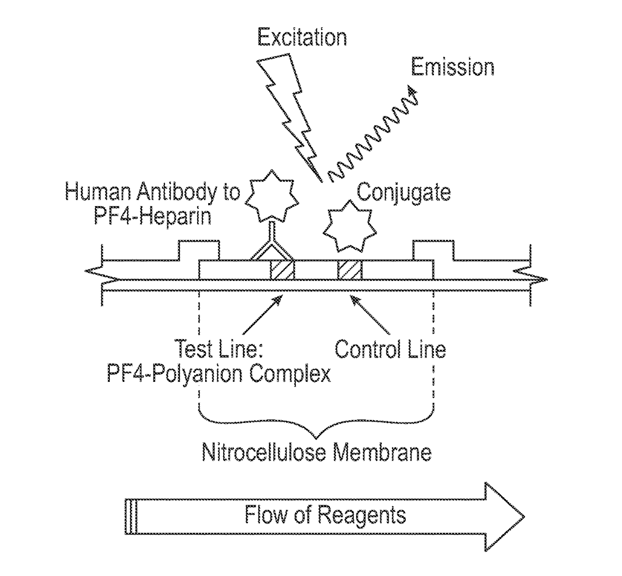
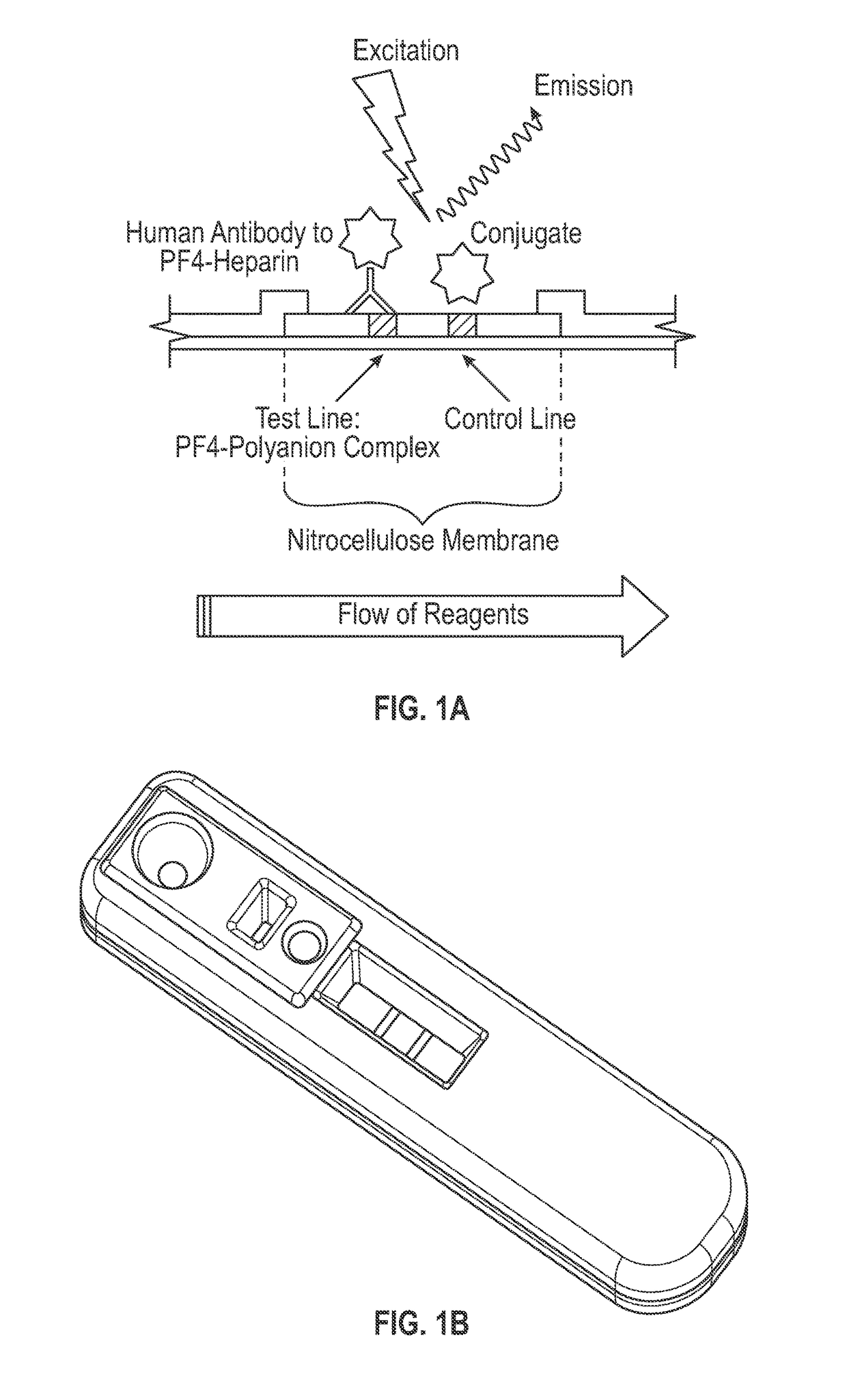
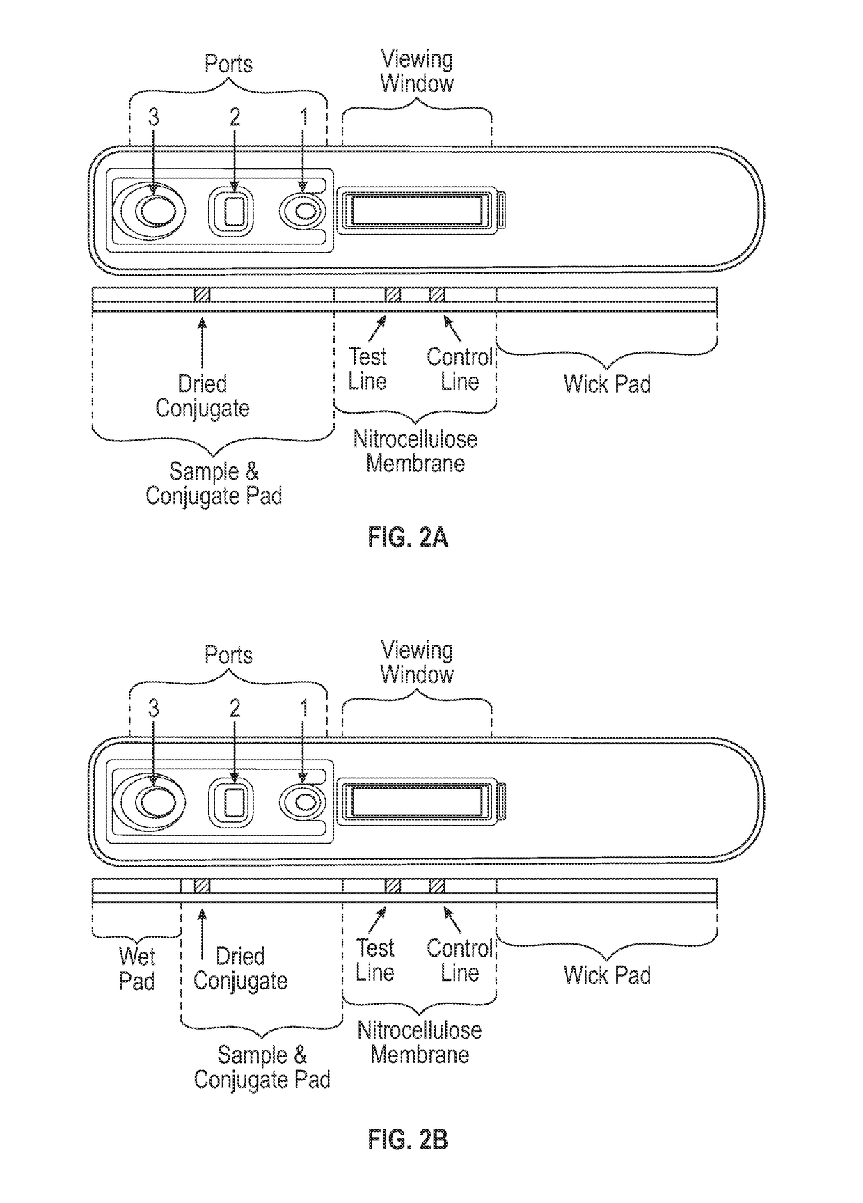
[0087]The lateral flow assay protocol was as follows. A 5 μl serum sample was applied to the sample port, shown as port 1 in FIGS. 1A-1C, and 15 μl of sample buffer (1×PBS, 1% BSA, and 0.1% polysorbate 20 (Tween 20™), pH 7.4) was immediately applied to the sample buffer zone, shown as port 2 in FIGS. 2A-2C. After approximately 3 minutes, 90 μl of conjugate buffer (1×PBS, 1% BSA, and 0.1% polysorbate 20 (Tween 20™), pH 7.4) was applied to the conjugate buffer zone, shown as port 3 in FIGS. 2A-2C. After approximately 12 minutes, the assay cassette was placed into a fluorescence reader, and fluorescent signals from the amine-reactive fluorescent label were measured at the detection and control zones. Each sample was tested in the PF4 / heparin lateral flow assay using replicates of 4.

example 3

Comparative Performance of PF4 / Heparin Lateral Flow Assay

[0088]To compare the relative performance of PF4 / heparin lateral flow assay with that of a reference PF4 / heparin enzyme immunoassay, a sample set of 83 sera from patients on heparin therapy was tested in a PF4 / heparin lateral flow assay according to the present invention and a commonly used ELISA test, which is described in detail in U.S. Pat. No. 5,972,718. In the ELISA, a PF4 / PVS complex immobilized in microwells reacts with PF4 / heparin antibodies from a patient sample with subsequent detection via an alkaline phosphatase conjugate (PF4 Enhanced®, Gen-Probe GTI Diagnostics, Inc., Waukesha, Wis.). Both assays detect IgG, IgA, and IgM antibodies. The PF4 / heparin lateral flow assay was carried out as described in Examples 1 and 2 above, with the exception that the labeled anti-human antibody conjugate was applied to the conjugate pad in liquid form shortly after the sample buffer was applied to the sample buffer zone. Because t...


PUM

PropertyMeasurementUnit
molecular weightaaaaaaaaaa

Abstract

The present invention provides a novel assay for detecting human antibodies specific for a platelet factor 4 (PF4) / heparin complex in a fluid sample. The assay utilizes an immobilized PF4 / polyanion complex and an anti-human antibody conjugated to a non-particulate fluorescent dye to capture and detect human PF4 / heparin antibodies. Various devices, methods and systems based on the disclosed PF4 / heparin assay are also provided.

Description

CROSS-REFERENCE TO RELATED APPLICATIONS[0001]This application is a national phase filing under 35 U.S.C. §371 of International Application No. PCT / US2012 / 040398 filed Jun. 5, 2012, which claims priority under 35 U.S.C. §119(e) to provisional patent application Ser. No. 61 / 495,171 filed Jun. 9, 2011, the disclosures of which are hereby incorporated by reference in their entirety.FIELD OF THE INVENTION[0002]This invention generally relates to the field of immunodiagnostics. More specifically, the invention relates to the detection of antibodies against the platelet factor 4 (PF4, also known as C—X—C motif chemokine ligand 4 or CXCL4) and heparin complex, which are associated with heparin-induced thrombocytopenia (HIT). In particular, the invention provides novel lateral flow assay devices, methods and systems for detecting antibodies specific for PF4 / heparin or PF4 / polyanion complexes in fluid samples.BACKGROUND OF THE INVENTION[0003]Heparin-induced thrombocytopenia (HIT), the most fr...

Claims


Application Information

Patent Timeline
Patent Type & Authority Patents(United States)
IPC IPC(8): G01N33/558G01N33/68G01N33/86
CPCG01N33/6854G01N33/558G01N33/6863G01N33/86G01N2800/32G01N33/54388
Inventor CHANCE, SUZETTEVISENTIN, GIAN PAOLO
Owner IMMUCOR GTI DIAGNOSTICS
Why Eureka
Try Eureka