Method for antigen delivery

a bioactive molecule and antigen technology, applied in the field of new antigen and/or bioactive molecule delivery methods, can solve the problems of short shelf life of both liposomes and hydrogels, inability to fusion hapten and klh coding sequences by molecular cloning methods, and inability to express klh protein in bacteria or hapten and klh coding sequences, etc., to prolong the time period of antigens

Active Publication Date: 2017-08-08
BOGAZICI UNIVERSITY
View PDF6 Cites 0 Cited by
  • Summary
  • Abstract
  • Description
  • Claims
  • Application Information

AI Technical Summary

Benefits of technology

[0017]A further objective of the present invention is to slow down the degradation of antigens endocytosed or phagocytosed together with the ASC speck carrier by enzymes inside cells and to lengthen the time period antigens stay in the environment and thereby enhance the antigen presentation capacity of APCs to T cells.
[0019]Yet another objective of the present invention is to increase the shelf-life of antigens and / or bioactive molecules carried by the ASC speck carrier.

Problems solved by technology

Due to its big size, expression of KLH protein in bacteria or fusion of hapten and KLH coding sequences by molecular cloning methods is not practical.
Besides, liposomes also tend to fuse with each other.
Thus, both liposomes and hydrogels have short shelf-lives.
Methods that are used in the synthesis of polymer based microparticles (organic solvents, high temperature and freeze-thaw cycles) may damage the structure and activity of the antigen or bioactive molecule payload.
However, no suggestion was made about achieving antigen delivery by binding antigen peptides to ASC protein or ASC specks (Ellebedy et al., 2011).

Method used


Image

  • Method for antigen delivery
  • Method for antigen delivery
  • Method for antigen delivery

Examples


specific embodiments

Example 1

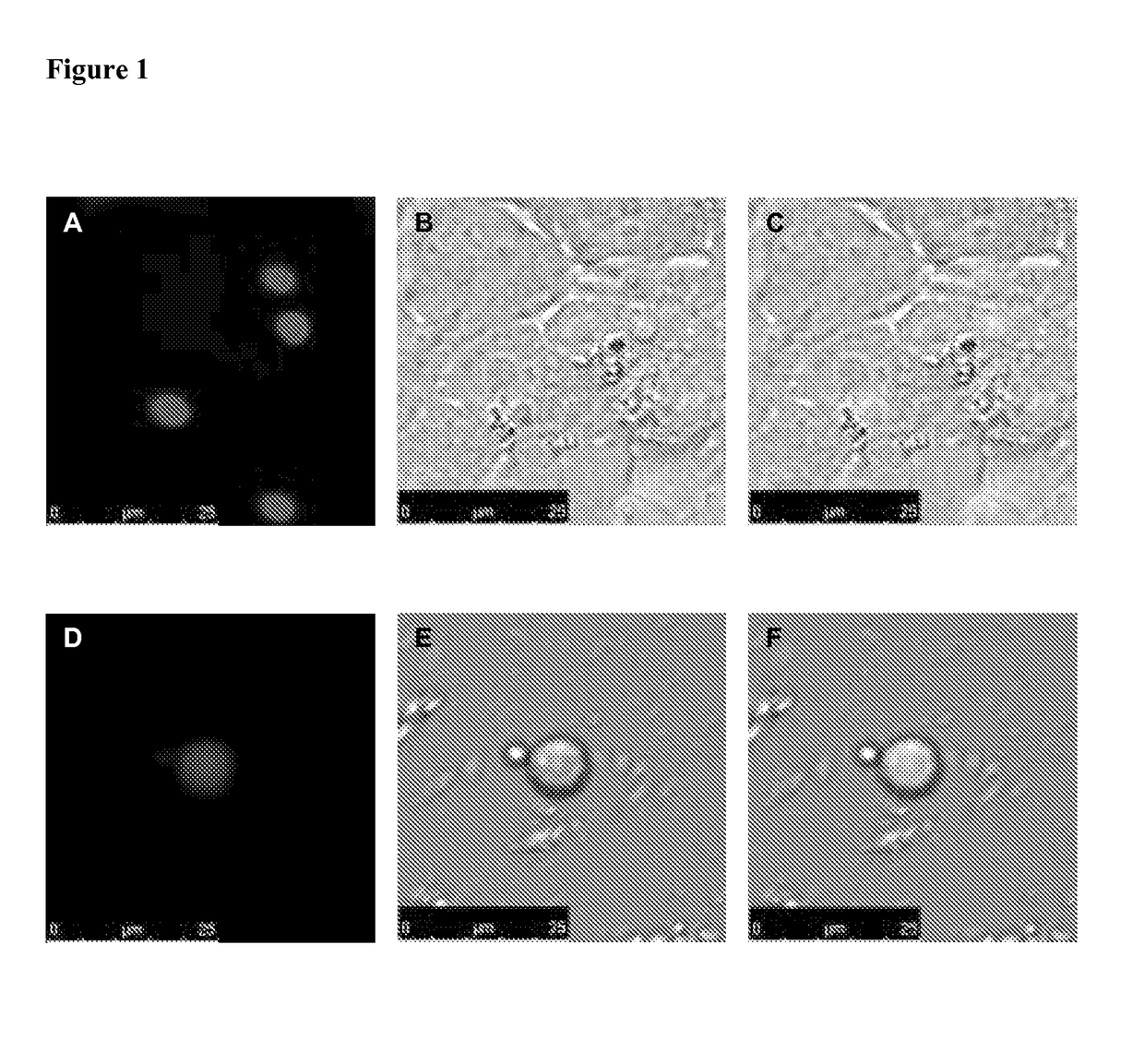
[0071]Synthesis of the composition containing the ASC speck carrier and mCherry proteins fused to ASC proteins forming the ASC speck carrier in HEK 293 FT cells and its purification from HEK 293 FT cell culture.

[0072]The DNA sequence encoding an antigen or a bioactive molecule in peptide or protein nature, is cloned into a plasmid containing ASC protein (SEQ ID NO: 1) coding sequence in a way that they will form a fusion protein. The plasmid should preferably contain an SV40 origin of replication (e.g. pcDNA3 or pEGFP-C3 vector backbone). The plasmid created accordingly is transfected into a selected cell line in order to overexpress the ASC-antigen (or bioactive molecule) fusion protein. HEK 293 FT cell line was used in this invention because it multiplies SV40 origin of replication containing plasmids in high copy numbers inside the cells.

[0073]pmCherry-C3.1-ASC plasmid encoding mCherry-ASC fusion protein was transfected into HEK 293 FT cells by calcium-phosphate method. ...

example 2

[0079]Synthesis of a fused DNA sequence encoding the mCherry-ASC fusion protein.

[0080]In order to clone pmCherry-C3.1-ASC plasmid that encodes for mCherry-ASC fusion protein, the DNA sequence encoding the human ASC protein was obtained from pcDNA3-ASC plasmid by digesting with HindIII-EcoRI enzymes and ligated to the multiple cloning site of pmCherry-C3.1 vector digested with same enzymes to create an mCherry-ASC fusion protein coding sequence. The pcDNA3-ASC plasmid was a gift from Gabriel Nunez (University of Michigan, Ann Arbor, USA).

[0081]In order to clone pmCherry-C3.1 plasmid, the mCherry protein coding sequence was PCR amplified from pcDNA3 IFP 1.4 plasmid using primers NheI_mCherry_F (gctagcaccatggtgtctaagggcgaaga) and XhoI_mCherry_R (ctcgagtttcttgtacagctcgtccat). The EGFP protein coding sequence was digested and removed from the pEGFP-C3 plasmid by NheI-XhoI enzymes and PCR product digested with same enzymes was ligated to its place. pcDNA3 IFP 1.4 was a gift from Tsien Lab...

example 3

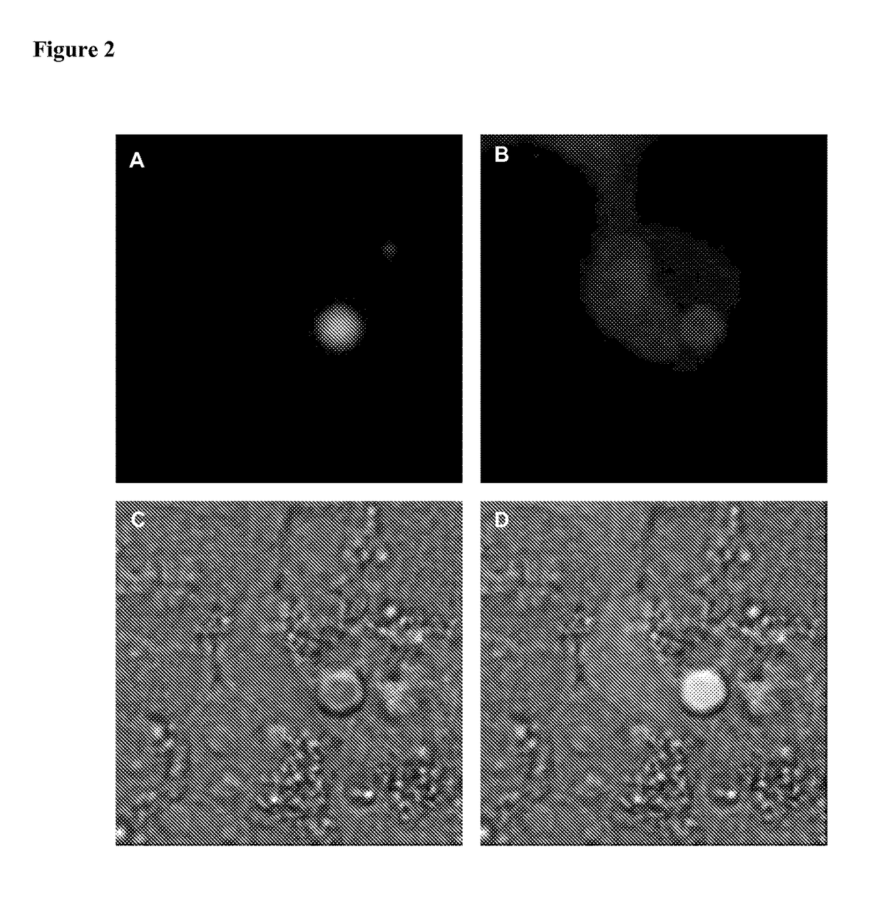
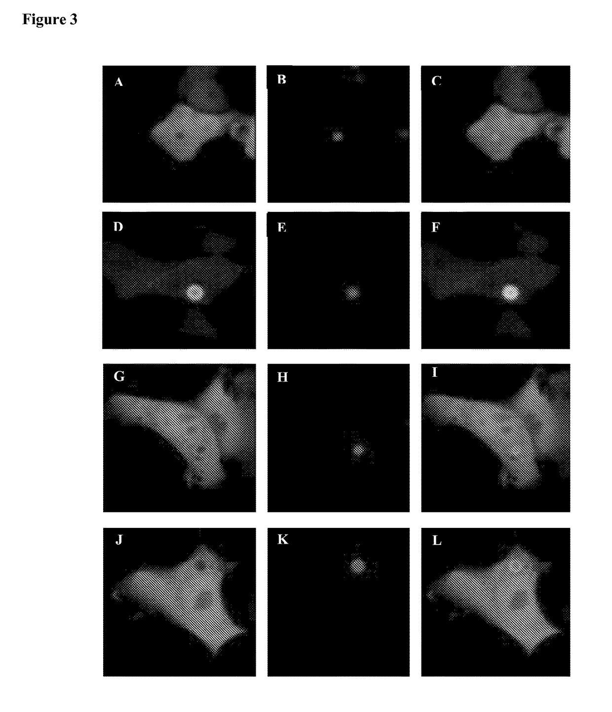
[0083]Alternative methods of loading antigens and / or bioactive molecules to the ASC speck carrier.

[0084]FGF2 (fibroblast growth factor 2, SEQ ID NO: 10) loaded to the ASC speck carrier was given as an example of bioactive molecules which can be loaded to the ASC speck carrier. The FGF2 protein is a growth factor known to have role in angiogenesis, embryonic development and wound healing.

[0085]When it is desirable to load an antigen or a bioactive molecule to ASC specks without creating a fusion protein with the ASC protein (SEQ ID NO: 1), the plasmid containing the DNA sequence encoding the antigen and / or the bioactive molecule and the plasmid containing the DNA sequence encoding the ASC protein are co-transfected into the preferred cell line.

[0086]As an example, the plasmid encoding mCherry-FGF2 fusion protein was co-transfected with the plasmid encoding EGFP-ASC fusion protein. A mixture of mCherry-FGF2 and EGFP-ASC encoding plasmids, 1 μg each, were mixed in a total 219 μl volume...


PUM

PropertyMeasurementUnit
molecular weightaaaaaaaaaa
temperatureaaaaaaaaaa
volumeaaaaaaaaaa

Abstract

The present invention provides methods for antigen and / or bioactive molecule delivery to antigen presenting cells utilizing ASC specks as vehicles; 1) the ASC speck composition is made of ASC protein(s) and the antigen and / or bioactive molecule(s) purified from cells as a compact micro-spherical structure; 2) purified micro-spherical ASC speck delivery vehicles are stable at 37° C. for more than 30 days; and 3) antigen and / or bioactive molecules carried by ASC specks can be efficiently phagocytosed by antigen presenting cells and their degradation in the lysosome allows antigen processing and presentation.

Description

FIELD OF THE INVENTION[0001]The present invention relates to a novel method for antigen and / or bioactive molecule delivery.BACKGROUND[0002]Traditionally, vaccines are based on the principle of body's antibody production against killed or live-attenuated pathogens. Recently, there are studies about developing subunit vaccines based on injecting particular components of a pathogen rather than injecting the whole pathogen itself, in order to improve biosafety. Subunit vaccines contain antigens in peptide or protein nature encoded by pathogens. In order to facilitate engulfment of these peptides and proteins by antigen presenting cells (APCs), antigens are loaded to nanometer or micrometer-sized particles. These types of vaccines are called particulate vaccines (De Temmerman et al., 2011).[0003]There are ongoing studies about numerous antigen delivery methods to be used as particulate vaccines. Emulsions, liposomes, immunostimulant complexes, virus-like particles, gold, silica particles...

Claims


Application Information

Patent Timeline
Patent Type & Authority Patents(United States)
IPC IPC(8): C07K14/00C07K14/50C07K14/47
CPCC07K14/4747A61K47/646C07K14/503C07K2319/50C07K2319/70A61K47/64
Inventor SAHILLIOGLU, ALI CANOZOREN, NESRIN
Owner BOGAZICI UNIVERSITY
Why Eureka
Try Eureka