Exosomes and micro-ribonucleic acids for tissue regeneration

a technology of exosomes and microribonucleic acids, applied in the field of repair or regeneration, can solve the problems of reducing the quality of life, reducing work efficiency, and reducing the productivity of workers, and achieves the effects of improving tissue function, reducing arrhythmias, and contractility

Active Publication Date: 2017-11-28
CEDARS SINAI MEDICAL CENT
View PDF368 Cites 18 Cited by
  • Summary
  • Abstract
  • Description
  • Claims
  • Application Information

AI Technical Summary

Benefits of technology

[0008]As such, there are provided herein methods for regenerating tissue in an individual having damaged tissue, comprising, identifying an individual having damaged tissue and administering a plurality of exosomes to the individual, wherein the exosomes are secreted from regenerative cells, wherein the exosomes comprise one or more microRNA fragments, and wherein after administration of the plurality of exosomes, the one or more microRNA fragments alter gene expression in the damaged tissue, improve the viability of the damaged tissue, and / or facilitate the formation of new tissue in the individual. In several embodiments, administration of the exosomes results in functional improvement in the tissue, in combination with one or more of the above-mentioned positive results. In several embodiments, the exosomes are synthetic in origin. In some such embodiments, the synthetic exosomes are generated in order to replicate, substantially, or closely mimic exosomes that are secreted from regenerative cells.
[0020]Administration can be via a variety of routes, depending on the embodiment. For example, in some embodiments, delivery is locally to the tissue. In some embodiments, delivery is systemically. In one embodiment, delivery is via an intramyocardial route, while in other embodiments, delivery is via an intracoronary route. Combinations of delivery routes are used, in certain embodiments, in order to improve the speed with which positive effects are realized and or improve the duration of treatment. For example, in some embodiments, miRNAs are delivered directly to a target tissue and exosomes are delivered via a systemic route.

Problems solved by technology

Just these non-limiting examples are the source of substantial medical costs, reduced quality of life, loss of productivity in workplaces, workers compensation costs, and of course, loss of life.
Even among those who survive the MI, many will still die within one year, often due to reduced cardiac function, associated side effects, or progressive cardiac disease.
Regardless of the etiology, most of those afflicted with coronary heart disease or heart failure have suffered permanent heart tissue damage, which often leads to a reduced quality of life.
In some embodiments, administration of the exosomes results in an increase in cardiac wall thickness in the area subjected to the infarction.
In additional embodiments, the tissue is damaged due to chronic disease or ongoing injury.
For example, progressive degenerative diseases can lead to tissue damage that propagates over time (at times, even in view of attempted therapy).
However, in several embodiments, ultracentrifugation is not used.
The proteins or molecules themselves need not be desirable per se (e.g., the protein or molecule may have an overall beneficial effect in the context of the damage to the tissue, but in other contexts would not yield beneficial effects).
However, in several embodiments, the cells of the damaged tissue that are influenced by the disclosed methods are damaged cells.

Method used


Image

  • Exosomes and micro-ribonucleic acids for tissue regeneration
  • Exosomes and micro-ribonucleic acids for tissue regeneration
  • Exosomes and micro-ribonucleic acids for tissue regeneration

Examples


example 1

and Characterization of Exosomes

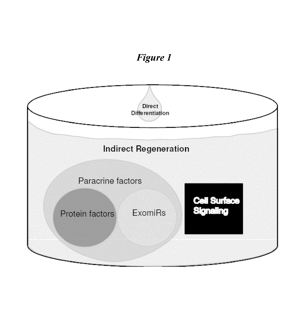
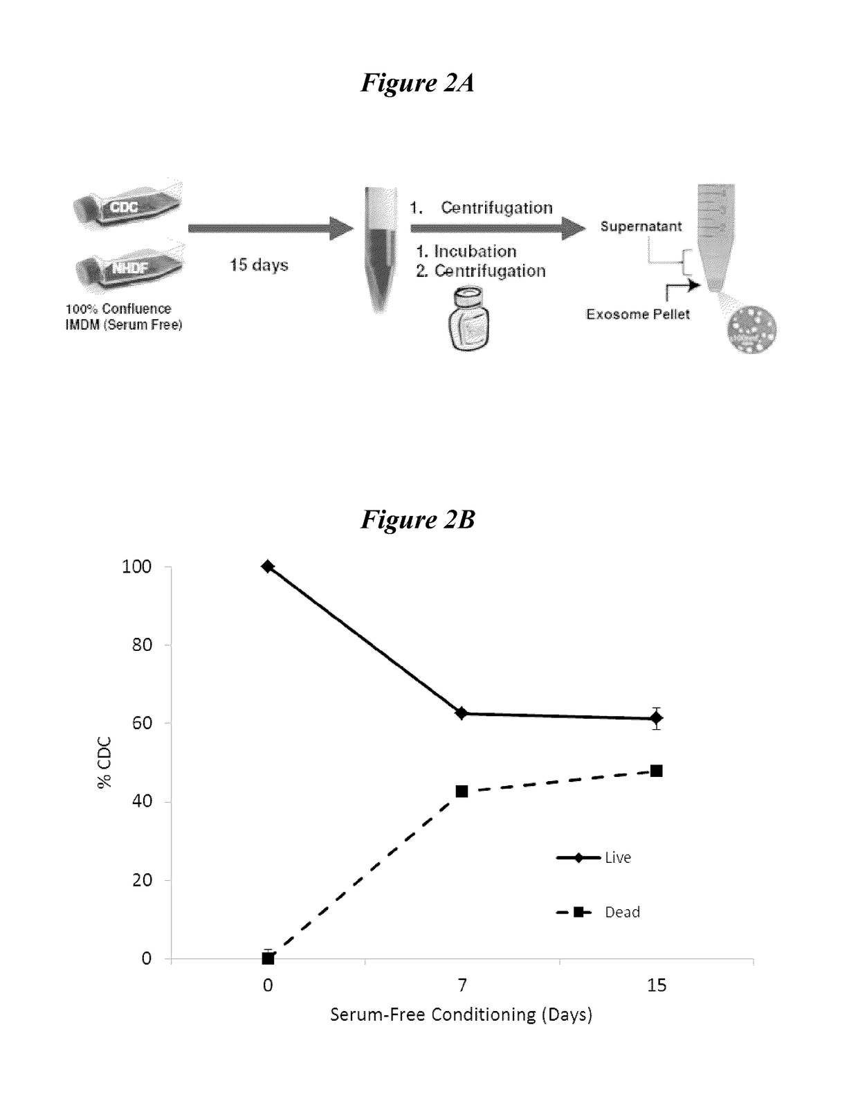
[0083]Prior studies in the area of cardiac tissue repair and regeneration have demonstrated that the repair and / or regeneration of cardiac tissue is a result of both direct and indirect factors. For example, it has been shown that CDCs account for approximately 10% of regenerated cardiac tissue. Such studies suggest that alternative mechanisms, such as indirect effects, are at play. As discussed above, exosomes and their nucleic acid content may be involved, at least in part, in providing cellular or tissue repair and / or regeneration via indirect mechanisms. The present example was designed to characterize exosomes and their nucleic acid content.

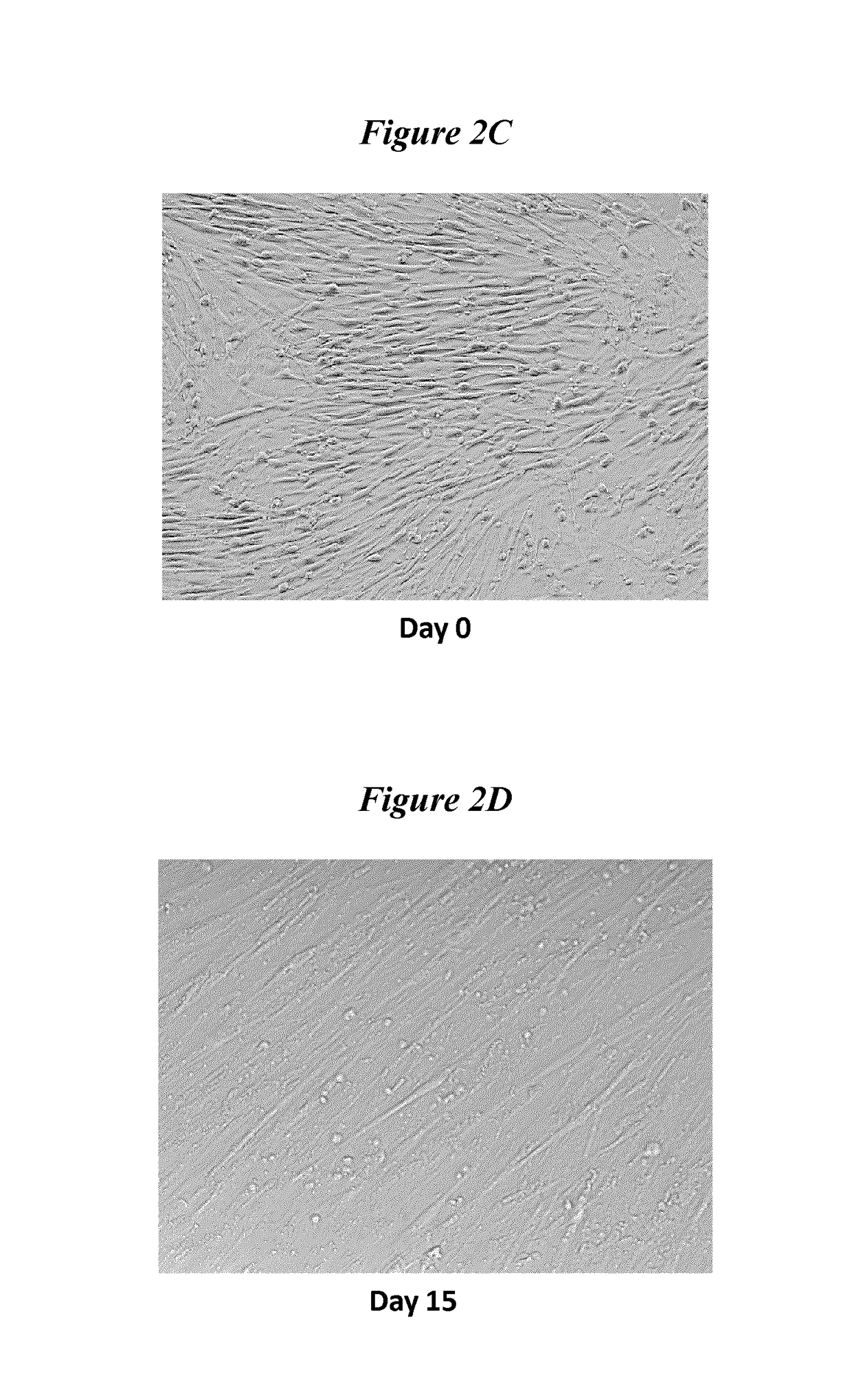
[0084]In order to isolate exosomes, cultured cells were grown to 100% confluence in serum free media. For this experiment, exosome yield and RNA content was compared between cultured CDCs and normal human dermal fibroblast (NHDF) cells. It shall be appreciated that, in several embodiments, exosomes may be isol...

example 2

Promote Survival and Proliferation of Other Cells

[0086]In vitro experiments were undertaken to evaluate the pro-regenerative and anti-apoptotic effects of exosomes on other cell types. Exosomes were isolated from CDCs or NHDF cells as discussed above. A portion of the exosome pellet fraction was then co-incubated with cultured neonatal rat ventricular myocytes (NRVM) in chamber slides for approximately 7 days. At the end of seven days, the co-cultures were evaluated by immunohistochemistry for changes in indices of proliferation or cell death (as measured by markers of apoptosis). A schematic for this protocol is shown in FIG. 4. FIG. 5A shows data related to death of the NRVM cells, as measured by TUNEL staining. Incubation of NRVM cells with exosomes isolated from CDCs resulted in a significantly lower degree of apoptosis, as compared to both control cells and cells incubated with exosomes from NHDF cells (CDC: 25.2±0.04%; NHDF: 45.1±0.05%, p<0.01); Control: 41.4±0.05%, n=4, p<0.0...

example 3

Promote Angiogenesis

[0087]In addition to increased proliferation and / or reduced death of cells or tissue in a region of damage or disease, reestablishment or maintenance of blood flow may play a pivotal role in the repair or regeneration of cells or tissue. As such, the ability of exosomes to promote angiogenesis was evaluated. Human umbilical vein endothelial cells (HUVEC) were subjected to various co-incubation conditions. These conditions are depicted in FIG. 6. Briefly, HUVEC cells were grown in culture dishes on growth factor reduced MATRIGEL. The cells were grown in either neonatal rat cardiomyocyte media (NRCM), MRCM supplemented with CDC-derived exosomes, MRCM supplemented with NHDF-derived exosomes, vascular cell basal media (VCBM), or vascular cell growth media (VCGM). As shown in FIG. 7, VCGM induced robust tube formation as compared to VCBM (CDC: 9393±689; NHDF: 2813±494.5, control, 1097±116.1, n=3, p<0.05). Media from NRCM resulted in tube formation similar to VCBM (dat...


PUM

PropertyMeasurementUnit
diameteraaaaaaaaaa
diameteraaaaaaaaaa
diameteraaaaaaaaaa

Abstract

Several embodiments relate to methods of repairing and / or regenerating damaged or diseased tissue comprising administering to the damaged or diseased tissues compositions comprising exosomes. In several embodiments, the exosomes comprise one or more microRNA that result in alterations in gene or protein expression, which in turn result in improved cell or tissue viability and / or function.

Description

RELATED CASES[0001]This application is the U.S. National Phase under 35 U.SC. 371 of International Application No. PCT / US2013 / 054732, filed Aug. 13, 2013, which claims the benefit of U.S. Provisional Application No. 61 / 682,666, filed Aug. 13, 2012 the entire disclosure of each of which is incorporated by reference herein.STATEMENT REGARDING GOVERNMENT SPONSORED GRANT[0002]The inventions disclosed herein were made with Government support under the Research Project Grant (R01 HL083109) by the National Institutes of Health. The United States Government has certain rights in this invention.BACKGROUND[0003]Field[0004]The present application relates generally to methods and compositions for the repair or regeneration of damaged or diseased cells or tissue. Several embodiments relate to administration of exosomes (or protein and / or nucleic acids from the exosomes) isolated from cells or synthetic surrogates in order to repair and / or regenerate damage or diseased tissues. In particular, sev...

Claims


Application Information

Patent Timeline
Patent Type & Authority Patents(United States)
IPC IPC(8): C12N15/113A61K31/7105C12N15/11A61K47/46A61K35/34C12N5/077A61K47/42A61K9/00
CPCC12N15/113A61K31/7105A61K35/34A61K47/46C12N5/0657C12N15/111A61K9/0019C12N2502/28A61K47/42C12N2310/141C12N2320/30C12N2502/1329A61K2300/00A61P29/00A61P35/00A61P43/00A61P9/00A61P9/10A61P9/14Y02A50/30
Inventor MARBAN, EDUARDOCHENG, KEIBRAHIM, AHMED
Owner CEDARS SINAI MEDICAL CENT
Why Eureka
Try Eureka