Positron emitting radionuclide labeled peptides for human uPAR PET imaging

a radionuclide and peptide technology, applied in the direction of peptide/protein ingredients, radiation therapy, therapy, etc., can solve the problems of local invasion or metastasis, and achieve the effect of superior in vivo characteristics of upar pet ligands, high tumor-to-background ratio, and high tumor uptak

Active Publication Date: 2018-02-06
CURASIGHT APS
View PDF10 Cites 10 Cited by
  • Summary
  • Abstract
  • Description
  • Claims
  • Application Information

AI Technical Summary

Benefits of technology

[0015]The present inventors have surprisingly found that [68Ga]-, [64Cu]- and [Al18F]-NOTA-AE105 have superior in vivo characteristics as a uPAR PET ligand, with high and specific tumor uptake, thus resulting in a high tumor-to-background ratio and thereby superior contrast as a PET ligand for uPAR expression tumors. The inventors have found that both [68Ga]- and [64Cu]-NOTA-AE105 was able to specifically detect uPAR expressing human-derived brain tumor lesions in a orthotropic human cancer mouse model. Moreover, [Al 18F]-NOTA-AE105 was useful to detect uPAR positive human prostate cancer lesions after subcutaneously inoculation in mice. Overall, the radiolabeling of NOTA-AE105 with 18F, 68Ga and 64Cu, thus enable the visualization and quantification of uPAR using PET Imaging. This is a major improvement in PET Imaging.
[0046]The present conjugates for use in accordance with the present invention can discriminate between uPAR expression levels in the primary tumor and metastases. Also, the use of quantification e.g. SUVmean and especially SUVmax can predict prognosis, progression and recurrence. The positron-emitting radionuclide labelled peptides of the present invention specifically target uPAR-positive cancer cells and / or uPAR positive stroma cells surrounding the cancer such as neutrophils and macrophages, and in particular the most aggressive (metastatic) cells. Moreover, the peptides of the present invention can be used for non-invasive detection and quantification of the expression level of uPAR using PET imaging. No current methods measuring uPAR is capable of this non-invasively in humans.

Problems solved by technology

Malignant tumors are capable of degrading the surrounding extracellular matrix, resulting in local invasion or metastasis.

Method used


Image

  • Positron emitting radionuclide labeled peptides for human uPAR PET imaging
  • Positron emitting radionuclide labeled peptides for human uPAR PET imaging
  • Positron emitting radionuclide labeled peptides for human uPAR PET imaging

Examples


example 1

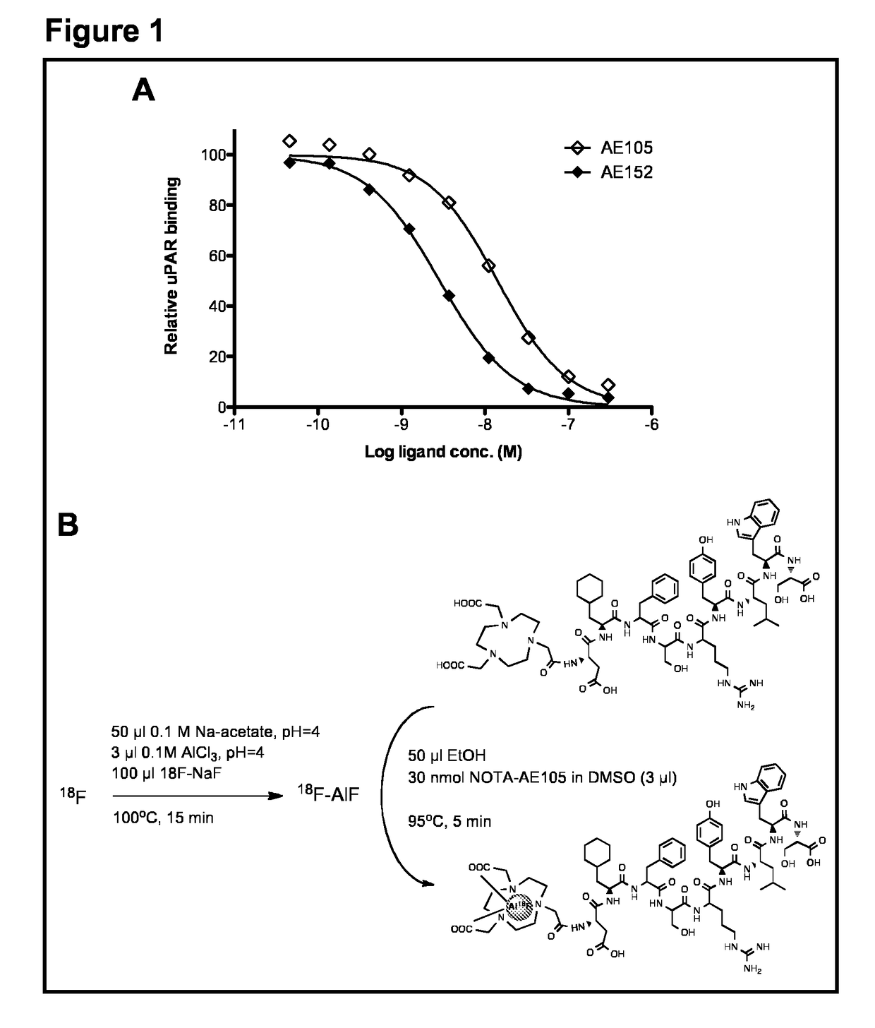
[0064]The aim of the present study was to synthesize a NOTA-conjugated peptide and use the Al18F method for development for the first 18F-labeled PET ligand for uPAR PET imaging and to perform a biological evaluation in human prostate cancer xenograft tumors. To achieve this, the present inventors synthesized high-affinity uPAR binding peptide denoted AE105 and conjugated NOTA in the N-terminal. 18F-labeling was done according to a recently optimized protocol26. The final product (18F—AlF-NOTA-AE105) was finally evaluated in vivo using both microPET imaging in human prostate tumor bearing animals and after collection of organs for biodistribution study.

Chemical Reagents

[0065]All chemicals obtained commercially were of analytical grade and used without further purification. No-carrier-added 18F-fluoride was obtained from an in-house PETtrace cyclotron (GE Healthcare). Reverse-phase extraction C18 Sep-Pak cartridges were obtained from Waters (Milford, Mass., USA) and were pretreated w...

example 2

[64Cu]NOTA-AE105 (NOTA-Asp-Cha-Phe-Ser-Arg-Tyr-Leu-Trp-Ser-OH)

[0087]64CuCl2 dissolved in 50 ul metal-free water was added to a solution containing 10 nmol NOTA-AE105 and 2.5 mg gentisic acid dissolved in 500 ul 0.1 M NH4OAc buffer (pH 5.5) and left at room temperature for 10 minutes resulting in 375 MBq [64Cu]NOTA-AE105 20 with a radiochemical purity above 99%. The radiochemical purity decreased to 94% after 48 hours storage.

example 3

In Vivo uPAR PET Imaging with [64Cu]NOTA-AE105 in a Orthotropic Human Glioblastoma Mouse Model

[0088]A mouse was inoculated with human derived glioblastoma cells in the brain. 3 weeks later a small tumor was visible using microCT scan A microPET images was recorded 1 hr post i.v. injection of approximately 5 MBq [64Cu]NOTA-AE105. Uptake in the tumor and background brain tissue was quantified. Moreover, was a control mouse (with no tumor inoculated) also PET scanned using the same procedure, to investigate the uptake in normal brain tissue with intact blood brain barrier. See FIG. 6.


PUM

PropertyMeasurementUnit
pore sizeaaaaaaaaaa
v/vaaaaaaaaaa
tissue densityaaaaaaaaaa

Abstract

There is provided a positron-emitting radionuclide labelled peptide for non-invasive PET imaging of the Urokinase-type Plasminogen Activator Receptor (uPAR) in humans. More specifically the invention relates to human uPAR PET imaging of any solid cancer disease for diagnosis, staging, treatment monitoring and especially as an imaging biomarker for predicting prognosis, progression and recurrence.

Description

[0001]This application is a National Stage Application of PCT / DK2013 / 050402, filed 29 Nov. 2013, which claims benefit of Serial No. 2012 70751, filed 3 Dec. 2012 in Denmark, and which claims benefit of Ser. No. 61 / 732,443, filed 3 Dec. 2012 in the United States, and which application(s) are incorporated herein by reference. To the extent appropriate, a claim of priority is made to each of the above disclosed applications.FIELD OF THE INVENTION[0002]The present invention relates to a positron-emitting radionuclide labelled peptide for noninvasive PET imaging of the Urokinase-type Plasminogen Activator Receptor (uPAR) in humans. More specifically the invention relates to human uPAR PET imaging of any solid cancer disease for diagnosis, staging, treatment monitoring and especially as an imaging biomarker for predicting prognosis, progression and recurrence.BACKGROUND OF THE INVENTION[0003]Urokinase-type plasminogen activator receptor (uPAR) is over-expressed in a variety of human cance...

Claims


Application Information

Patent Timeline
Patent Type & Authority Patents(United States)
IPC IPC(8): A61K51/00A61K38/08A61K51/08A61M36/14
CPCA61K51/088A61K51/083A61K51/08A61K38/08
Inventor KJAER, ANDREASPERSSON, MORTENMADSEN, JACOB
Owner CURASIGHT APS
Why Eureka
Try Eureka