PSMA binding ligand-linker conjugates and methods for using

a technology of psma and conjugates, which is applied in the field of compounded and methodological treatments of prostate cancer, can solve the problems of affecting the quality of life of patients, osteoporosis and liver damage, and the use of hormonal drugs is often accompanied by side effects

Active Publication Date: 2018-04-24
PURDUE RES FOUND INC
View PDF312 Cites 28 Cited by
  • Summary
  • Abstract
  • Description
  • Claims
  • Application Information

AI Technical Summary

Benefits of technology

[0025]In another embodiment, linkers are described having at least one hydrophobic side chain group, such as an alkyl, cycloalkyl, aryl, arylalkyl, or like group, each of which is optionally substituted. In one aspect, the hydrophobic group is included in the linker by incorporating one or more Phe or Tyr groups, including substituted variants thereof, and analogs and derivatives thereof, in the linker chain. It is appreciated that such Phe and / or Tyr side chain groups may form positive pi-pi (π-π) interactions with Tyr and Phe residues found in the funnel of PSMA. In addition, it is appreciated that the presence of large side chain branches, such as the arylalkyl groups found on Phe and Tyr may provide a level of conformational rigidity to the linker, thus limiting the degrees of freedom, and reducing coiling and promoting extended conformations of the linker. Without being bound by theory, it is appreciated that such entropy restrictions may increase the overall binding energy of the bound conjugates described herein. In addition, it is appreciated that the rigidity increases that may be provided by sterically hindered side chains, such as Phe and Tyr described herein, may reduce or prevent coiling and interactions between the ligand and the imaging agent.
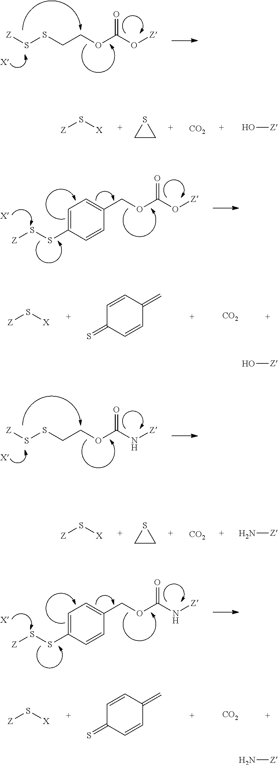
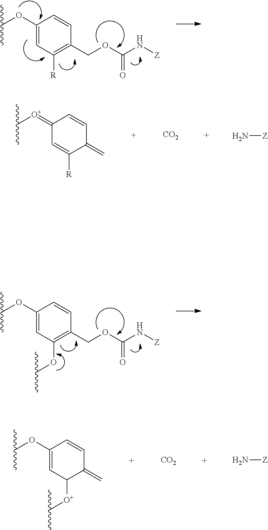
[0033]The term “releasable linker” as used herein refers to a linker that includes at least one bond that can be broken under physiological conditions (e.g., a pH-labile, acid-labile, oxidatively-labile, or enzyme-labile bond). The cleavable bond or bonds may be present in the interior of a cleavable linker and / or at one or both ends of a cleavable linker. It should be appreciated that such physiological conditions resulting in bond breaking include standard chemical hydrolysis reactions that occur, for example, at physiological pH, or as a result of compartmentalization into a cellular organelle such as an endosome having a lower pH than cytosolic pH. Illustratively, the bivalent linkers described herein may undergo cleavage under other physiological or metabolic conditions, such as by the action of a glutathione mediated mechanism. It is appreciated that the lability of the cleavable bond may be adjusted by including functional groups or fragments within the bivalent linker L that are able to assist or facilitate such bond breakage, also termed anchimeric assistance. The lability of the cleavable bond can also be adjusted by, for example, substitutional changes at or near the cleavable bond, such as including alpha branching adjacent to a cleavable disulfide bond, increasing the hydrophobicity of substituents on silicon in a moiety having a silicon-oxygen bond that may be hydrolyzed, homologating alkoxy groups that form part of a ketal or acetal that may be hydrolyzed, and the like. In addition, it is appreciated that additional functional groups or fragments may be included within the bivalent linker L that are able to assist or facilitate additional fragmentation of the PSMA binding drug linker conjugates after bond breaking of the releasable linker.
[0039]In this embodiment, the hydroxyalkanoic acid, which may cyclize, facilitates cleavage of the methylene bridge, by for example an oxonium ion, and facilitates bond cleavage or subsequent fragmentation after bond cleavage of the releasable linker. Alternatively, acid catalyzed oxonium ion-assisted cleavage of the methylene bridge may begin a cascade of fragmentation of this illustrative bivalent linker, or fragment thereof. Alternatively, acid-catalyzed hydrolysis of the carbamate may facilitate the beta elimination of the hydroxyalkanoic acid, which may cyclize, and facilitate cleavage of methylene bridge, by for example an oxonium ion. It is appreciated that other chemical mechanisms of bond breakage or cleavage under the metabolic, physiological, or cellular conditions described herein may initiate such a cascade of fragmentation. It is appreciated that other chemical mechanisms of bond breakage or cleavage under the metabolic, physiological, or cellular conditions described herein may initiate such a cascade of fragmentation.
[0139]It is appreciated that the urea and thiourea compounds described herein may also be advantageous in the preparation of the ligands also described herein due to the sub-nanomolar potency, water solubility, and / or long term stability of these compounds. The thiourea compounds described herein may generally be prepared from commercially available starting materials as described herein.
[0171]In another illustrative aspect, any effective regimen for administering the binding ligand drug delivery conjugates can be used. For example, the binding ligand drug delivery conjugates can be administered as single doses, or can be divided and administered as a multiple-dose daily regimen. In other embodiments, a staggered regimen, for example, one to three days per week can be used as an alternative to daily treatment, and for the purpose of defining this invention such intermittent or staggered daily regimen is considered to be equivalent to every day treatment and within the scope of this invention. In one embodiment, the host is treated with multiple injections of the binding ligand drug delivery conjugate to eliminate the population of pathogenic cells. In another embodiment, the host is injected multiple times (preferably about 2 up to about 50 times) with the binding ligand drug delivery conjugate, for example, at 12-72 hour intervals or at 48-72 hour intervals. In other embodiments, additional injections of the binding ligand drug delivery conjugate can be administered to the patient at an interval of days or months after the initial injections(s) and the additional injections prevent recurrence of the disease state caused by the pathogenic cells.
[0270]General Method for Metastatic Prostate Cancer Imaging. Male athymic nu / nu mice (4-5 weeks of age) are anesthetized using 2-3% isfluorane in oxygen prior to injection of 1×10⁶ 22 RV1 cells suspended in 100 μL cell culture medium into the left ventricle of the heart. Once the mouse is fully anethetized, the animal is placed on its back securing both arms down with tape. The chest is then wiped with 70% ethanol, allowing for the external visualization of the external anatomy of the chest. The injection is made at the second intercostal rib space just along side of the sternum using a 25 gauge needle. The needle is inserted slowly into the heart (6 mm) and cell suspension is injected into the left ventricle of the heart slowly over a period of 30 seconds to 1-minute. After the completion of injection, the needle is quickly withdrawn to prevent cells leaking from the heart.

Problems solved by technology

However, many of these treatments affect the quality of life of the patient, especially those men who are diagnosed with prostrate cancer over age 50.
For example, the use of hormonal drugs is often accompanied by side effects such as osteoporosis and liver damage.
Though PSMA is expressed in brain, that expression is minimal, and most ligands of PSMA are polar and are not capable of penetrating the blood brain barrier.

Method used


Image

  • PSMA binding ligand-linker conjugates and methods for using
  • PSMA binding ligand-linker conjugates and methods for using
  • PSMA binding ligand-linker conjugates and methods for using

Examples


examples

[0197]The compounds described herein may be prepared by conventional organic synthetic methods. In addition, the compounds described herein may be prepared as indicated below. Unless otherwise indicated, all starting materials and reagents are available from commercial supplies. All amino acid starting materials were purchased from Chem-Impex Int (Chicago, Ill.). 1H NMR spectra were obtained using a Bruker 500 MHz cryoprobe, unless otherwise indicated.

Example

[0198]General synthesis of PSMA imaging agent conjugates. Illustrated by synthesis of a 14-atom linker compound, where B is a PSMA binding ligand as described herein.

[0199]

[0200]Compounds are synthesized using standard fluorenylmethyloxycarbonyl (Fmoc) solid phase peptide synthesis (SPPS) starting from Fmoc-Cys(Trt)-Wang resin (Novabiochem; Catalog #04-12-2050). Compounds are purified using reverse phase preparative HPLC (Waters, xTerra C18 10 μm; 19×250 mm) A=0.1 TFA, B=Acetonitrile (ACN); λ=257 nm; Solvent gradient: 5% B to 80...

example

[0270]General Method for Metastatic Prostate Cancer Imaging. Male athymic nu / nu mice (4-5 weeks of age) are anesthetized using 2-3% isfluorane in oxygen prior to injection of 1×10⁶ 22 RV1 cells suspended in 100 μL cell culture medium into the left ventricle of the heart. Once the mouse is fully anethetized, the animal is placed on its back securing both arms down with tape. The chest is then wiped with 70% ethanol, allowing for the external visualization of the external anatomy of the chest. The injection is made at the second intercostal rib space just along side of the sternum using a 25 gauge needle. The needle is inserted slowly into the heart (6 mm) and cell suspension is injected into the left ventricle of the heart slowly over a period of 30 seconds to 1-minute. After the completion of injection, the needle is quickly withdrawn to prevent cells leaking from the heart.

[0271]The development of metastasis is imaged and diagnosed from fourth week onward in vivo by tail vein injec...


PUM

PropertyMeasurementUnit
lengthaaaaaaaaaa
total volumeaaaaaaaaaa
temperatureaaaaaaaaaa

Abstract

Described herein are prostate specific membrane antigen (PSMA) binding conjugates that are useful for delivering therapeutic, diagnostic and imaging agents. Also described herein are pharmaceutical compositions containing them and methods of using the conjugates and compositions. Also described are processes for manufacture of the conjugates and the compositions containing them.

Description

CROSS-REFERENCE TO RELATED APPLICATIONS[0001]This application is a continuation-in part of U.S. application Ser. No. 13 / 580,436, filed Aug. 22, 2012, which is a U.S. national application under 35 U.S.C. § 371(b) of International Application Serial No. PCT / US2011 / 026238 filed Feb. 25, 2011, and claims priority under 35 USC § 119(e) to U.S. Provisional Application Ser. No. 61 / 308,190, filed on Feb. 25, 2010, the entire disclosures of each of which are incorporated herein by reference.STATEMENT REGARDING PRIOR DISCLOSURES BY THE INVENTOR OR A JOINT INVENTOR[0002]International Publication Number WO2009 / 026177, published on Feb. 26, 2009, was made by the inventor or joint inventor.TECHNICAL FIELD[0003]The invention described herein pertains to compounds and methods for treating diseases of the prostate, such as prostate cancer and related diseases. More specifically, embodiments of the invention described herein pertain to conjugates of biologically active agents conjugated to PSMA bindi...

Claims


Application Information

Patent Timeline
Patent Type & Authority Patents(United States)
IPC IPC(8): A61K38/00C07K5/00C07K7/00C07K14/00C07K16/00C07K17/00C12N9/48A61K31/454A61K31/4745C07K4/00C07K2/00A61K47/54A61K51/04A61K49/00C12N9/96A61K31/475
CPCC12N9/485A61K31/454A61K31/475A61K31/4745A61K47/542A61K49/0052A61K51/0402C12N9/96A61K49/0032C12Y304/17021A61K38/00A61K51/0497
Inventor LOW, PHILIP STEWARTCHELVAM, VENKATESHKIM, YOUNGSOONKULARATNE, SUMITH A.
Owner PURDUE RES FOUND INC
Why Eureka
Try Eureka