Methods and composition related to in vivo imaging of gene expression
A technology of receptors and tissues, applied in preparations for in vivo experiments, chemical instruments and methods, biochemical equipment and methods, etc., can solve problems such as restrictions, non-targeting target tissues, radiopharmaceuticals that are not known to be safe, etc.
- Summary
- Abstract
- Description
- Claims
- Application Information
AI Technical Summary
Problems solved by technology
Method used
Image
Examples
Embodiment 1
[0257] Through functional and anatomical imaging to
[0258] Non-invasively quantify exogenous gene expression
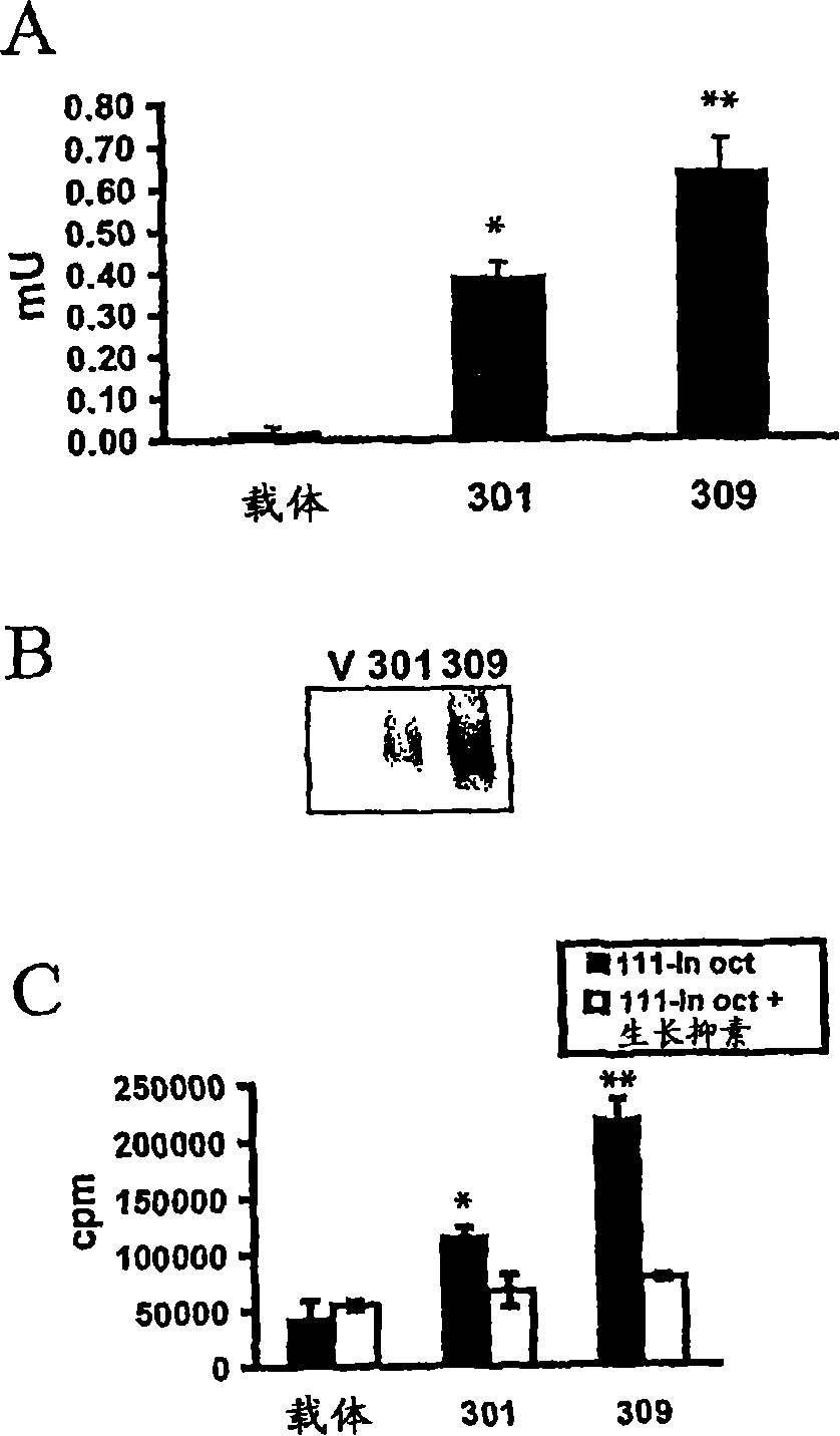
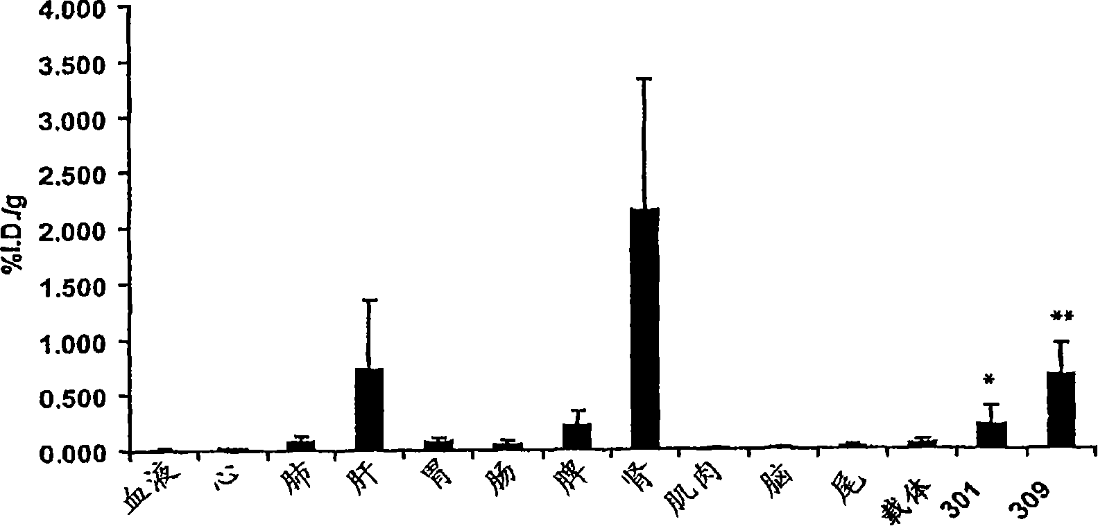
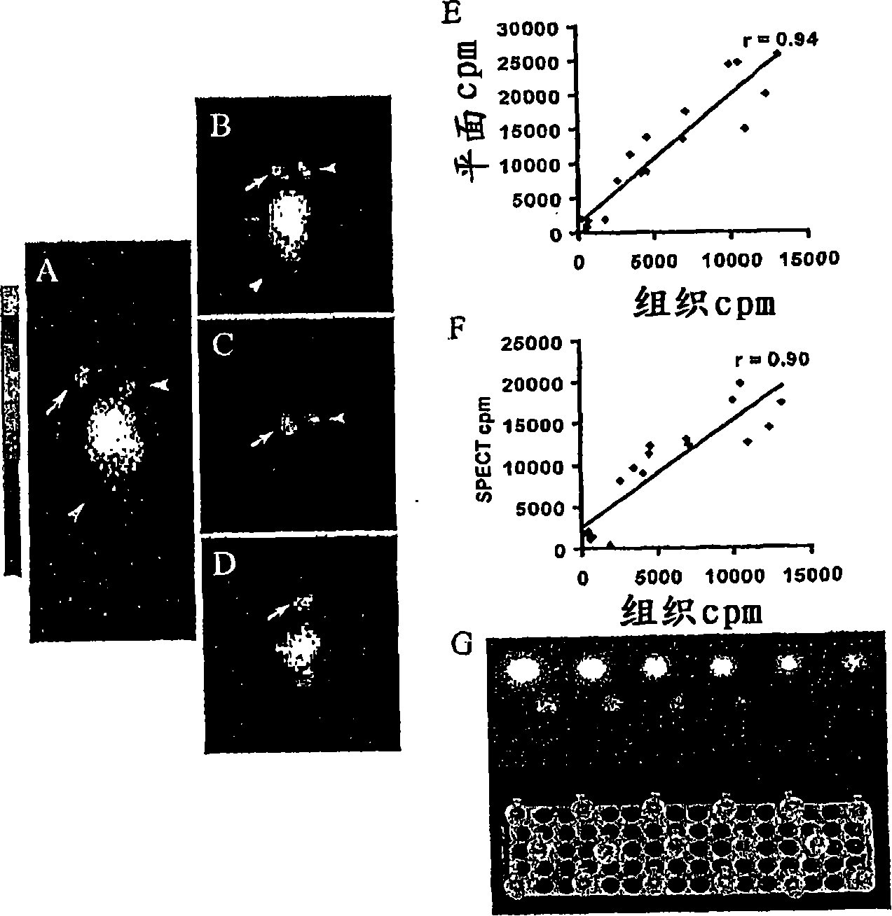
[0259]The aim of this study was to evaluate whether a combination of functional (planar and SPECT imaging) and anatomical (MR) techniques could be used to non-invasively quantify tumor expression of somatostatin receptor type 2 (SSTR2) gene chimeras in vivo .
[0260] Materials and methods
[0261] plasmid
[0262] Full-length human SSTR2A was inserted into the pDisplay vector (Invitrogen, Carlsbad, CA) as described in Kundra et al., 2002 (the entire contents of which are expressly incorporated herein by reference for this section as well as the entire patent application), at the membrane localization sequence and Downstream of the hemagglutinin A (HA) epitope tag sequence.
[0263] Cell line generation and characterization
[0264] HT1080 cells (human fibrosarcoma, ATCC, Rockville, MD) were grown in Dulbecco's modified Eagle's medium (DMEM) containing 1x glu...
Embodiment 2
[0297] for therapeutic gene expression and molecular imaging
[0298] Development of Tumor Selective hTMC Plasmid Vector
[0299] A major obstacle to successful cancer gene therapy is the lack of effective delivery systems that can specifically target tumors. One approach for targeting transgene expression in tumors is to control gene expression via tumor-specific promoters. Human telomerase reverse transcriptase (hTERT), the catalytic subunit of telomerase, is highly active in immortalized cells and in more than 85% of human cancers, but is quiescent in most normal somatic cells (Shay and Bacchetti, 1997; Shay et al., 2001). The hTERT promoter has been cloned and shown to selectively promote transgene expression in tumors but not in normal cells (Nakayama et al., 1998). However, like most other intrinsic mammalian promoters, the transcriptional promotion strength of the hTERT promoter is much weaker than commonly used viral promoters such as CMV and SV40 early promoters. ...
Embodiment 3
[0302] γ-camera imaging of gene transfer in vivo
[0303] Many molecular imaging methods for monitoring gene transfer in vivo rely on the sensitivity of gamma-camera, SPECT, or PET imaging to detect intravenously injected, radiolabeled compounds localized to the products of transferred genes, but most are both The gene of interest is not detected directly nor is FDA-approved radiopharmaceuticals for systemic use (Yamada et al., 1992; Kundra et al., 2002). radiopharmaceutical 111 In-octreotide has successfully detected cell surface membrane localization of SSTR2A product in HT1080 cells. The cell membrane localization of the product in HT1080 cells transfected with a plasmid expressing HA-SSTR2A (pHA-SSTA2A) was confirmed by immunofluorescence analysis using an anti-HA antibody (Fig. 9A). Injection into mice bearing HT1080 tumors generated by pHA-SSTR2A-transfectants 111 In-octreotide was used for biodistribution and imaging studies by gamma-camera (Fig. 9B). SSTR2A express...
PUM
 Abstract
Description
Claims
Application Information
 - R&D
- Intellectual Property
- Life Sciences
- Materials
- Tech Scout
- Unparalleled Data Quality
- Higher Quality Content
- 60% Fewer Hallucinations
Browse by: Latest US Patents, China's latest patents, Technical Efficacy Thesaurus, Application Domain, Technology Topic, Popular Technical Reports.
© 2025 PatSnap. All rights reserved.Legal|Privacy policy|Modern Slavery Act Transparency Statement|Sitemap|About US| Contact US: help@patsnap.com


