
Ultra-high pressure preparation method of pig valved blood vessel acellular bracket

An ultra-high pressure, valved technology, applied in the field of medical materials, can solve the problems of inability to remove cell components, change biomechanical properties, residues, etc., to achieve the elimination of zoonotic diseases, good biocompatibility, and conducive to colonization and growth Effect

Inactive Publication Date: 2008-05-28
NANJING CHILDRENS HOSPITAL AFFILIATED TO NANJING MEDICAL UNIV
View PDF0 Cites 13 Cited by
  • Summary
  • Abstract
  • Description
  • Claims
  • Application Information

AI Technical Summary

Problems solved by technology

The German Haverich research group and the British Ingham research group used various surfactants and proteolytic enzymes to remove the donor cells on the scaffold, and found that there were many problems: (I) low concentration of trypsin could not remove the deep cell components of the arterial wall , a high concentration of trypsin can cause the destruction of the scaffold structure
For a tissue of a certain thickness, it is difficult to remove the cells inside; (2) these surfactants are chemical substances themselves, which are also toxic to the human body, and there will still be a certain concentration of residue after washing; (3) due to the need for washing Over a period of several weeks, may alter biomechanical properties and may increase the chance of infection, among other defects

Method used


Image

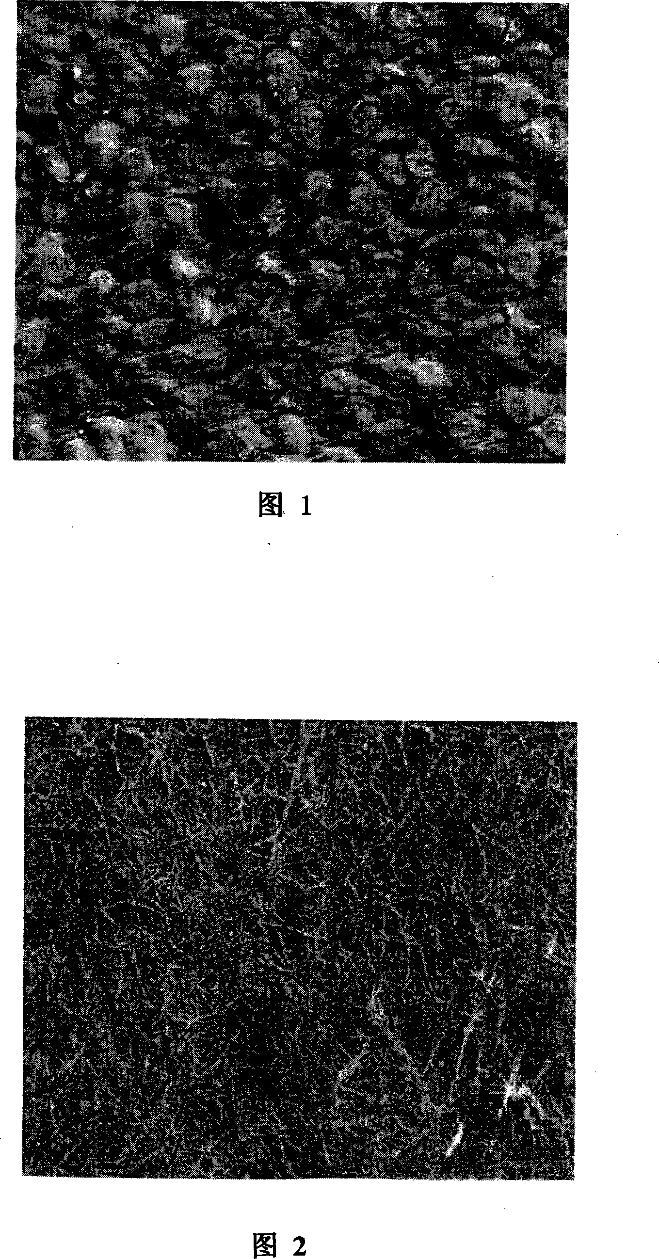
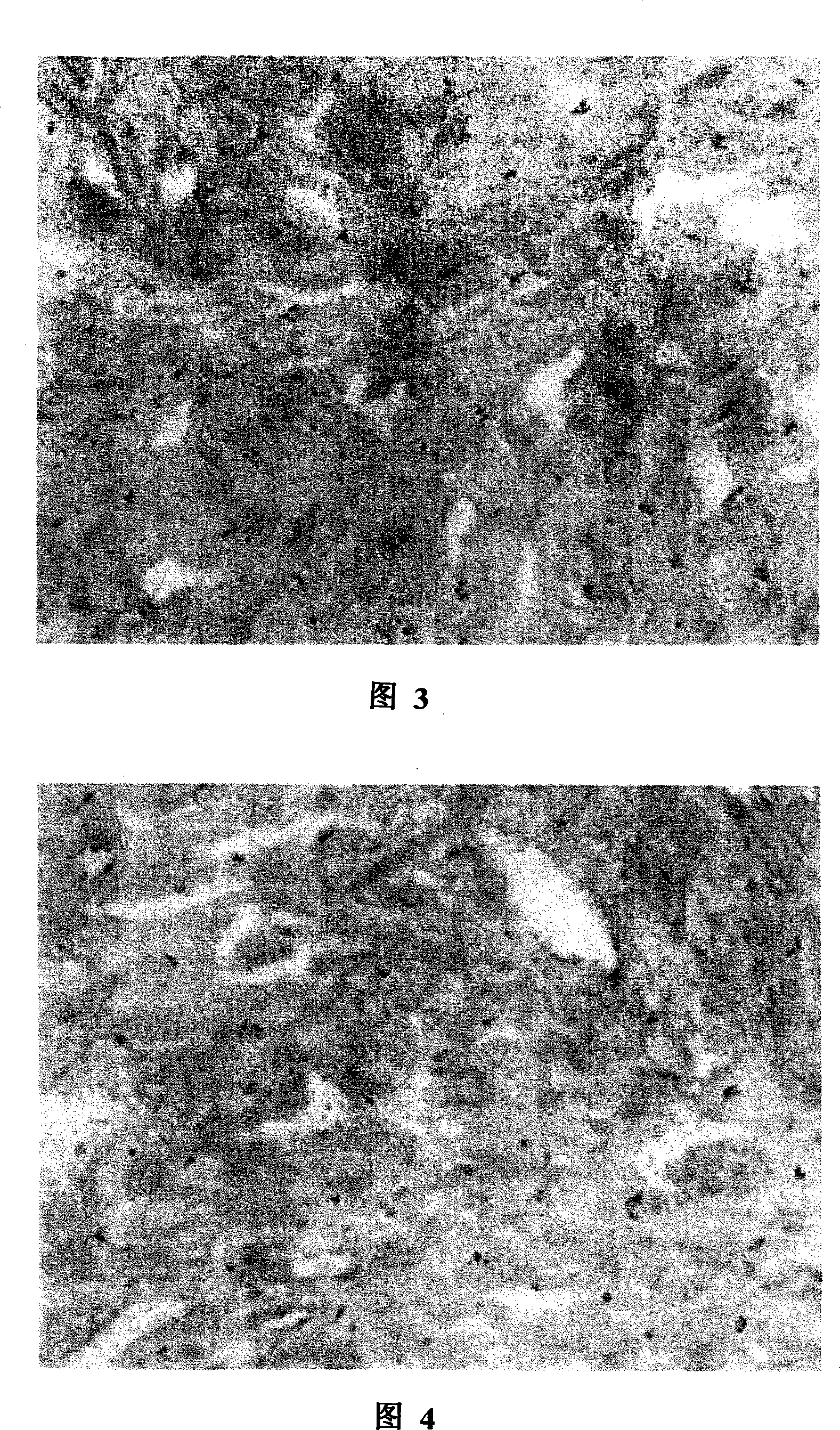
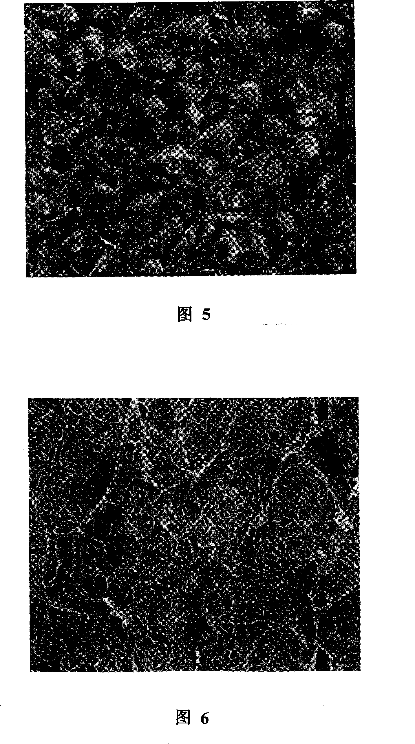
  • Ultra-high pressure preparation method of pig valved blood vessel acellular bracket
  • Ultra-high pressure preparation method of pig valved blood vessel acellular bracket
  • Ultra-high pressure preparation method of pig valved blood vessel acellular bracket

Examples


Embodiment Construction

[0019] The ultra-high pressure preparation method of the porcine valve-vessel decellularized scaffold of the present invention, through long-term experiments such as using different pressures and pressurization times, found that when the pressure of one pressurization reaches 1000Mpa and the pressurization time is longer, although the cells can also be Completely removed, but at the same time destroys laminin (Ln) and fibronectin (Fn). When the pressure is lower than 1000MPa, the cells cannot be completely removed even though the pressure is longer, but the laminin (Ln) and fibronectin (Fn) will not be damaged. In order to obtain both cells can be completely removed, and laminin (Ln) and fibronectin (Fn) are not damaged, making it more suitable for transplantation needs, the best preparation method of the decellularized scaffold of the present invention is as follows step:

[0020] A) Preparation: After successful anesthesia of healthy pigs, take out the ascending aorta, aort...


PUM


Abstract

The invention relates to an ultra high pressure preparation method of a cell enucleating stent of porcine blood vessel valve, which mainly includes the two steps of material preparation and ultra high pressure cell enucleating process; by adopting a plurality of times of gradual pressure increase, the invention can not only remove cells completely in relatively short time by ultra high pressure, but can also protect laminin protein (Ln) and fibronectin (Fn) from being damaged, so as to meet the requirements of transplantation. The invention can inactivate all kinds of bacteria, viruses, spores and other microbes simultaneously, so as to eliminate the possibility of zoonotic disease. At the same time, as the ultra high pressure method is a physical method, the invention can completely avoid a plurality of adverse consequences caused by the residues of chemicals caused by chemical methods. The cell enucleating stent of porcine blood vessel with valve prepared by the method of the invention has the advantages of good biocompatibility, appropriate degree of biodegradation rate, high mechanical strength, easy suture and fixation, etc., so the invention is conductive to the colonization and growth of the human cells and can be gradually degraded and absorbed along with the regeneration of real valve or blood vessel.

Description

technical field [0001] The invention relates to a medical material, in particular to prepare pig blood vessels with valves as substitute materials for human valves and blood vessels. The invention belongs to the field of medical prosthesis materials. Background technique [0002] Complex heart and great vessel malformations often require the use of biological blood vessels or valved conduits. At present, valved vessels are more commonly used in clinical practice, which are allogeneic or heterogeneous. The latter mainly comes from purebred pigs. Biological valve vessels have a natural stent structure, optimal hemodynamics, and do not require anticoagulation. Their preservation technology and clinical application have become a hot spot in complex cardiac surgery. Among them, the allogeneic preservation and application technology is relatively mature, but there is no regulation of brain death law in my country, coupled with the traditional concept of the Orientals, it is almos...

Claims


Application Information

Patent Timeline
Patent Type & Authority Applications(China)
IPC IPC(8): A61L27/00A61L27/50A61F2/82A61F2/06A61F2/24
Inventor 莫绪明张永生朱建宝顾海涛束亚琴
Owner NANJING CHILDRENS HOSPITAL AFFILIATED TO NANJING MEDICAL UNIV
Learn More