Mass spectrometry reagent kit and method for rapid tuberculosis diagnosis

A kit and mass spectrometry technology, applied in the field of protein detection, can solve the problems of high cost, low positive rate and long time-consuming of tuberculosis polymerase chain reaction

Inactive Publication Date: 2008-06-11
许洋
View PDF0 Cites 7 Cited by
  • Summary
  • Abstract
  • Description
  • Claims
  • Application Information

AI Technical Summary

Problems solved by technology

However, when we performed protein mass spectrometry analysis, we found that there was no standardized kit for clinical tuberculosis mass spectrometry diagnosis
[0005] Tuberculosis is a major respiratory infectious disease caused by Mycobacterium tuberculosis. The etiological diagnosis mainly relies on sputum smear and bacterial culture, but the positive rate of the former is low, and the latter takes 4 to 8 weeks, which takes a long time.
Gene rapid diagnosis that has appeared in recent years: tuberculosis polymerase chain reaction (PCR), but the specificity is poor
In addition, due to the high cost of tuberculosis polymerase chain reaction and the high requirements of laboratory conditions, it is not easy to carry out widely

Method used


Image

  • Mass spectrometry reagent kit and method for rapid tuberculosis diagnosis
  • Mass spectrometry reagent kit and method for rapid tuberculosis diagnosis
  • Mass spectrometry reagent kit and method for rapid tuberculosis diagnosis

Examples


Embodiment 1

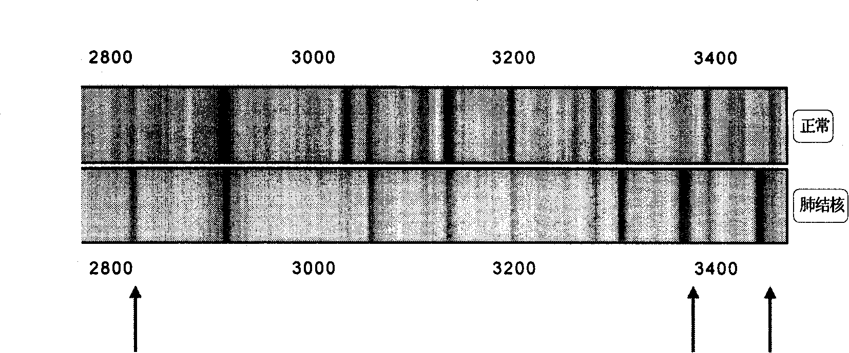
[0086] Example 1 The distinction between normal and tuberculosis and the preparation of a mass spectrometry kit

[0087] (1) Experimental method

[0088] 1. Materials

[0089] Specimen source: Standardized quality control serum (plasma) prepared by mass spectrometry. The definition meets the following criteria. The blood donors are half male and half male, and the blood type is type 0; the age is 18-30 years old; ethnicity, Han. The biochemical indicators were normal, including: total cholesterol, triglycerides, fasting blood sugar, hepatitis B surface antigen, liver function test, kidney function test; no family history of genetic diseases; no history of major infectious diseases. Women without pregnancy, men with no history of smoking. Fresh blood was drawn from 10 healthy subjects with type 0 blood (half male and half female), and centrifuged at 10,000 rpm for 5 minutes at 4°C immediately after the blood coagulated; the samples were stored at -80°C.

[0090] Take 10 μl o...

Embodiment 2

[0105] The double-blind test of embodiment 2 kit

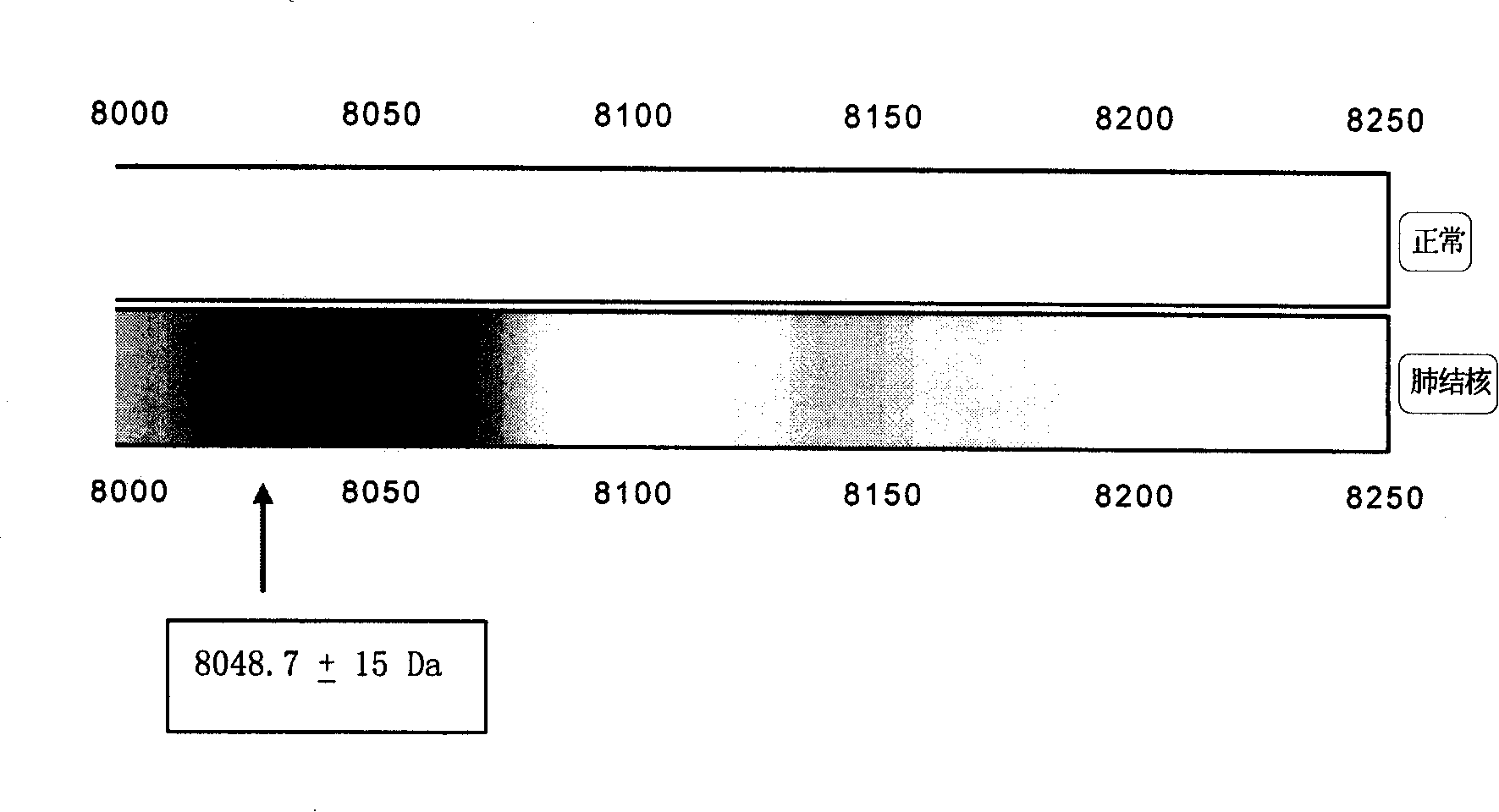
[0106] Several characteristic protein peaks 2873.3±15Da, 3379.7±15Da, 3449.3±15Da, 5808.4±15Da, 7568.9±15Da, 8048.7±15Da, 11439.2±15Da, 11526.7±15Da, 11683.0±15Da, 15114.9 were screened out from Example 1 ±15Da, 15312.8±15Da, double-blind test of 100 cases of normal and 100 cases of tuberculosis:

[0107] double blind test

Tuberculosis (100)

normal(100)

tuberculosis

100

0

normal

0

100

[0108] Sensitivity 100% Specificity 100%

[0109] The experimental results using C8 and C18 hydrophobic matrix beads are consistent with the experimental results of the above-mentioned WCX anion matrix beads.

[0110] Experimental results

[0111] The kit of the method can distinguish between normal people and tuberculosis, with 100% sensitivity and 100% specificity.

Embodiment 3

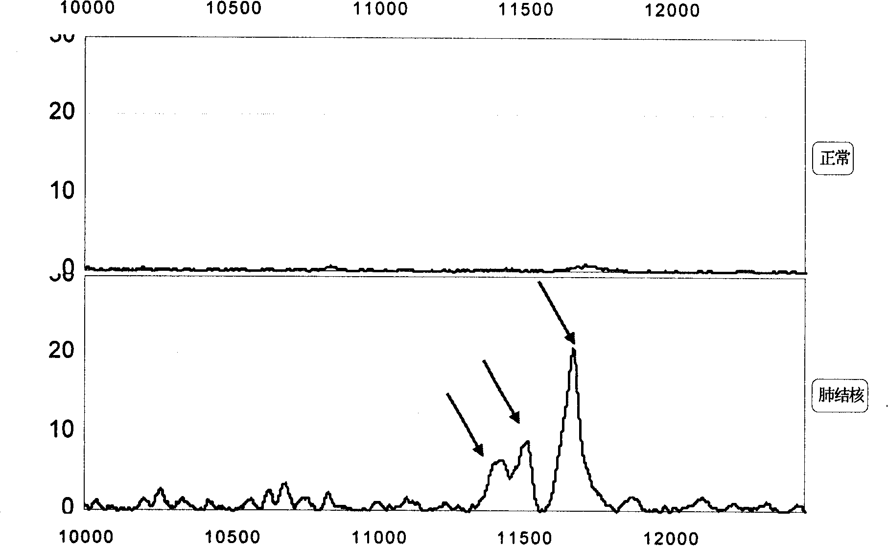
[0112] Example 3 Sorting identification of a group of characteristic peaks of tuberculosis protein fingerprint

[0113] A group of 11439.2±15Da, 11526.7±15Da, 11683.0±15Da biomarkers ( image 3 ) by multiple-stage mass spectrometry (MS / MS), post-source fragmentation (PSD) and protein ladder sequencing (protein ladder sequencing) for sequencing and other methods. By breaking molecules into pieces, protein ladders can be generated. This gradient is then analyzed by mass spectrometry. The 11439.2±15Da, 11526.7±15Da, and 11683.0±15Da biomarkers were identified as variant serum amyloid A. Its chemical structure is (104 amino acids arranged from N-terminal to C-terminal):

[0114] Chemical structures of 11683 ± 15 Da biomarkers:

[0115] N-terminal

[0116] RSFFSFLGEAFDGARDMWRAYSDMREANYIGSDKYFHARGNYDAAKRGPG

[0117] GVWAAEAISDARENIQRFFGHGAEDSLADQAANEWGRSGKDPNHFRPAGL

[0118] PEKY

[0119] C-terminal.

[0120] Chemical structure of the 11527±15 Da biomarker:

[0121] N-termi...


PUM

PropertyMeasurementUnit
molecular weightaaaaaaaaaa

Abstract

The invention relates to a method for capturing the phthisic Proteome in biological samples by supporting the stroma with the bead and separating the bead from the samples by the magnetic separator without the centrifuged samples. Then conduct the protein fingerprinting by the analysis of mass spectrography. The reagent kit tests the sensibility and the specificity of the normal person with the tuberculosis by Double Blind Test, which are all 100percent, among which the deviant SAA A can be used as blood serum in vitro for testing the normal person, minor tuberculosis patient, recrudescent tuberculosis patient, severe tuberculosis patient and tuberculosis patient after effective treatment. The elevated deviant SAA A shows poor treatment effectiveness, severe tuberculosis and the recrudescent tuberculosis, thus being a protein fingerprinting group for early warning of the early detection on tuberculosis worsening and prediction on tuberculosis recrudescence. The method provided in the invention can be used as the reagent kit to distinguish the protein fingerprinting or mass spectrum mapping between the normal person and the tuberculosis patient. The method is correct, convenient and fast.

Description

technical field [0001] The invention relates to a new kit for the protein analysis method in tuberculosis biological samples. One captures biomarkers via a protein-binding matrix and detects tuberculosis biomarkers using quantitatively controlled mass spectrometry. The invention mentioned here relates to the field of protein detection and is a new non-invasive in vitro mass spectrometry detection method. The present invention can be applied to the detection method or kit of the tuberculosis biomarker combination in the body fluid that has been separated from the human body. Background technique [0002] With the implementation and completion of the Human Genome Project, scientists proposed the concept of the post-genome (post-genome) project, and the focus of research shifted to functional genomics, and the main substance of biological function is protein. In 1994, Wilkins and Williams of Macquarie University in Australia first proposed the concept of proteome, which refer...

Claims


Application Information

Patent Timeline
Patent Type & Authority Applications(China)
IPC IPC(8): G01N33/68G01N30/88G01N33/577
Inventor 许洋
Owner 许洋
Why Eureka
Try Eureka