
Molecular spectrum imager

A technology of molecular spectroscopy and imager, applied in the direction of color/spectral characteristic measurement, etc., can solve the problems of unfavorable use of general equipment, low reliability, cumbersome operation, etc., to achieve detection and analysis, wide imaging spectral range, imaging fast effect

Active Publication Date: 2008-10-15
江苏华创高新医疗科技有限公司
View PDF1 Cites 12 Cited by
  • Summary
  • Abstract
  • Description
  • Claims
  • Application Information

AI Technical Summary

Problems solved by technology

In the actual use process, this push-broom method brings two problems: one is that push-broom is a kind of mechanical movement, and the speed is relatively slow, which makes the image acquisition time longer; the other is that push-broom movement makes the operation cumbersome and reliable. Low performance, not conducive to use as general equipment

Method used


Image

  • Molecular spectrum imager
  • Molecular spectrum imager

Examples


Embodiment

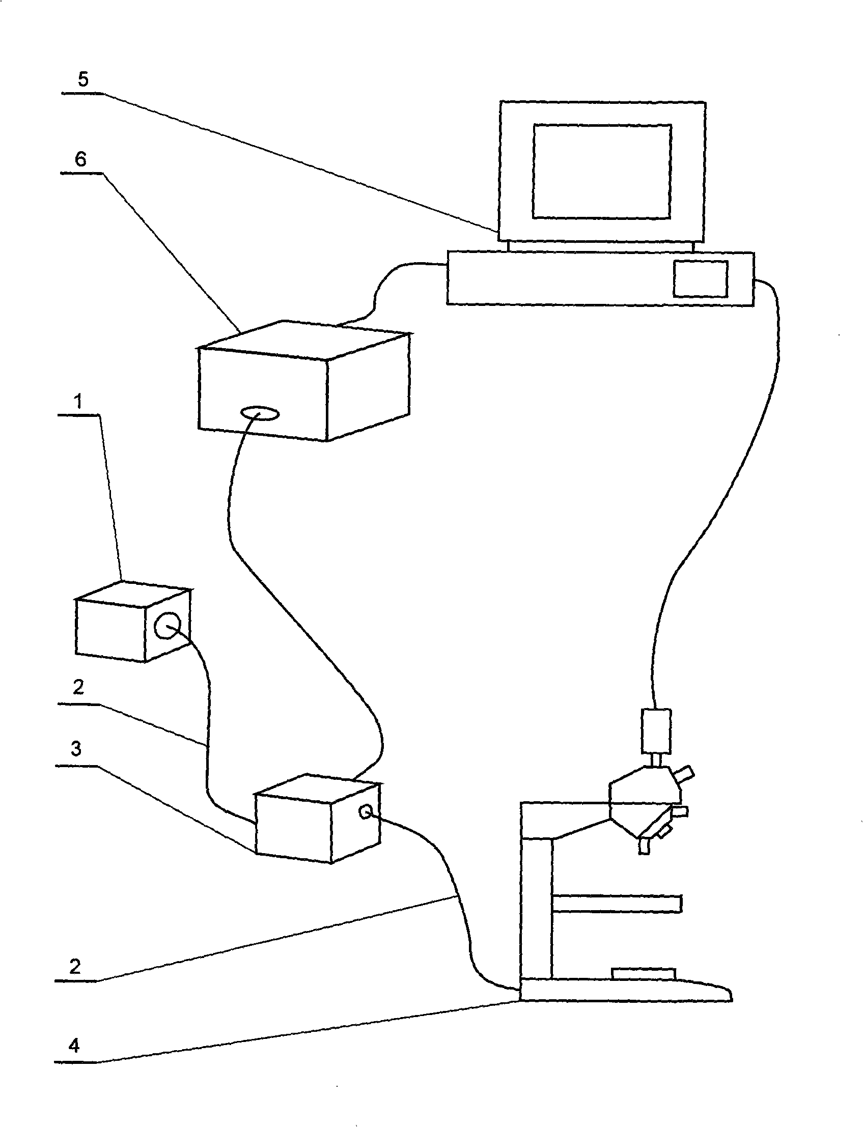
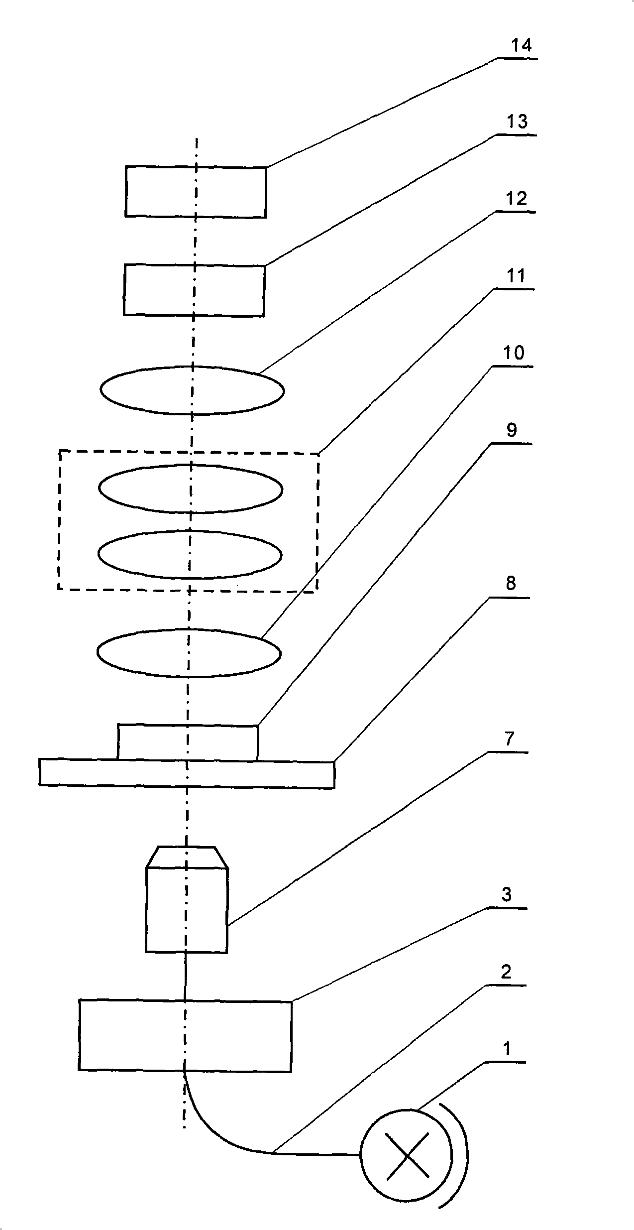
[0025] Below to figure 1 , figure 2 It is an example to illustrate the structural features, technical performance and effects of the present invention, but not to limit the scope of the present invention.

[0026] In this embodiment, the microscope 4 includes a dark field condenser 7, an object stage 8, an objective lens 10, a zoom lens 11 and an imaging lens 12, which are sequentially connected in an optical path, and the used microscope 4 is an ordinary optical microscope, or a fluorescence microscope Or an inverted microscope. The light source 1 is first connected to the dispersion unit 3 through the optical fiber 2, and then passes through the dark field condenser 7 as the illumination of the sample 9, and the illumination component can be a transmissive structure or a reflective structure. The dispersion unit controller 6 is connected to the data acquisition and control card of the computer 5, and controls the dispersion unit 3 according to the control signal of the co...


PUM


Abstract

The invention discloses a molecular spectrum imaging device applied in clinical medicine, biology, hylology, microelectronics and other subject fields. The invention is characterized in that: light sources are connected to dispersion units through optical fibers; the dispersion units are connected to a dark field optical collector through optical fibers and used for illumination of a microscope; a signal of an image sensor CCD is connected to a computer through a cable; a control wire of the computer is connected to a dispersion unit controller which is connected to the dispersion units; the computer changes the wavelength of the light sources through control of the dispersion units, acquires image data through the image sensor CCD and finally obtains molecular spectrum data of a sample. The molecular spectrum imaging device has high technical index and quick imaging speed, does not need a push-broom component, is easy to be combined with the general microscope for use and has wide imaging spectrum range covering visible light and near infrared light, thereby being favorable for detection and analysis of biological tissues.

Description

technical field [0001] The invention relates to a sample analysis device, in particular to a molecular spectrum imager. Used in medicine, materials and other fields. Background technique [0002] In the field of medical diagnosis, traditional medical imaging diagnosis shows the anatomical changes of biological tissue lesions. This imaging method can no longer meet the new requirements of the development of life sciences and pathology. How to study the mechanism of disease occurrence and development at the cellular level or even the molecular level and explore effective methods for diagnosis and treatment of diseases has become a hot spot in imaging, biology and clinical medicine. [0003] As a part of molecular imaging, molecular spectral imaging technology combines two traditional optical diagnostic techniques of spectral analysis and optical imaging. Spectral analysis can usually obtain the complete spectral data of a certain point on the sample in a certain wavelength r...

Claims


Application Information

Patent Timeline
IPC IPC(8): G01N21/25
Inventor 李庆利薛永祺
Owner 江苏华创高新医疗科技有限公司
Learn More