

Method for separating and identifying disseminated hepatoma cells
A separation method and technology of liver cancer cells, applied in the direction of tumor/cancer cells, biochemical equipment and methods, animal cells, etc., can solve the problem of lack of monoclonal antibodies on the surface of liver cancer cells, etc., and achieve simple operation, high sensitivity and specificity strong effect
- Summary
- Abstract
- Description
- Claims
- Application Information
AI Technical Summary
Problems solved by technology
Method used
Image

Examples
Embodiment 1
[0055] Example 1 Preparation of biotinylated asialofetuin
[0056] Main materials: amino biotinylation reagent Sulfo-NHS-lC-Biotin (Pierce), asialofetuin (Sigma), BCA protein quantification kit (Pierce), L-lysine (Sinopharm), centrifugal ultrafiltration tube (MW cut-off 50,000) (Millipore).
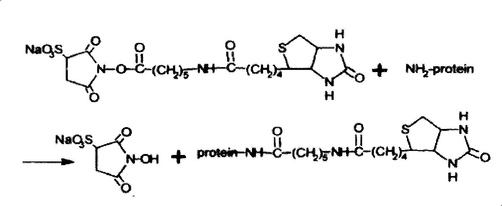
[0057] Experimental procedure: The reaction between asialofetuin and aminobiotinylation reagent was carried out according to the product instructions. Amino biotinylation reagent Sulfo-NHS-lC-Biotin can selectively react with free amino groups in proteins, the reaction formula is as follows figure 1 shown.
[0058] After the reaction, the reaction solution was transferred into a centrifugal ultrafiltration tube, and centrifuged at 25° C. at 4000 g for 20 min. Then add double-distilled water and L-lysine to the centrifuge tube to make the total volume 3ml, the final concentration of L-lysine is 0.08mg / ml, and place it at room temperature for 1h. Then centrifuge the centrifugal ultrafil...
Embodiment 2
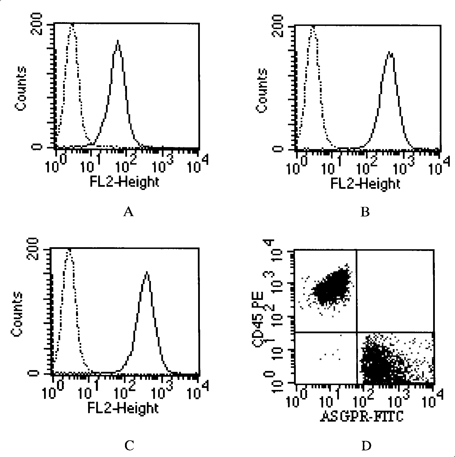
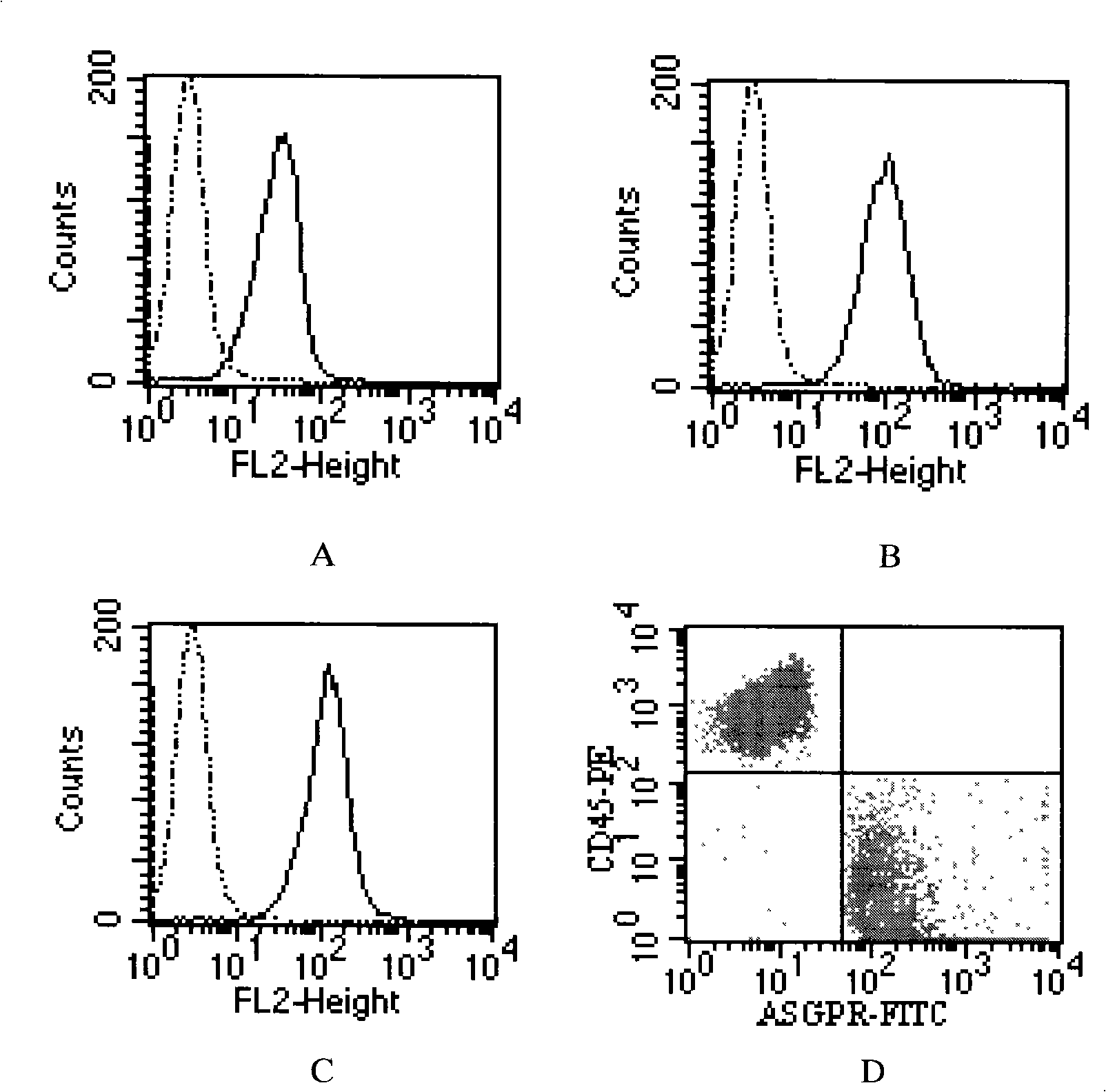
[0059] Example 2 Determining the ideal flow concentration of ASGPR ligands and antibodies
[0060] 1. The following experiments use flow cytometry to verify the reactivity of biotinylated asialofetuin with ASGPR, and determine the ideal concentration of biotinylated asialofetuin for flow cytometry.
[0061] Main materials: PE-labeled mouse anti-human CD45 monoclonal antibody (IgG1), PE-labeled mouse isotype monoclonal antibody (IgG1), FITC-labeled streptavidin, all purchased from Jingmei Company. Biotinylated asialofetuin, homemade. PBS washing solution (PBS+1%FBS), self-made.
[0062] Experimental steps:
[0063] 1) Count 5 million Hep3B liver cancer cells (Cell Bank of the Typical Culture Collection Committee of the Chinese Academy of Sciences), centrifuge at 20°C, 300g for 5min, discard the supernatant, resuspend in 550μl serum-free DMEM culture medium, mix well, 100μl / tube , divided into 5 EP tubes, numbered 1-5 in turn. Tube No. 1 is a blank control tube, Tube No. 2 i...
Embodiment 3
[0102] Example 3 Isolation and Identification of Disseminated Hepatocellular Carcinoma Cells Using ASGPR Ligands
[0103] Main materials: 5ml of peripheral blood from liver cancer patient volunteers, anticoagulated with heparin. Ficoll-Paque Plus, available from GE Healthcare. Magnetic separator, anti-biotin magnetic beads, MS magnetic separation column, 30 μm filter, and CK3-6H5 antibody (tumor cell marker antibody derived from epithelial tissue) were purchased from Miltenyi, Germany. 400R desktop centrifuge (with cytocentrifuge smear attachment), purchased from Heraeus, Germany. Polylysine slides were purchased from Fujian Maixin Company. PV9000 two-step immunohistochemistry kit was purchased from GBI, USA. Biotinylated asialofetuin, homemade. PBS washing solution (containing 0.5% BSA, 80U / ml heparin), prepared by yourself.
[0104] Reagent 1: PBS washing solution (containing 0.5% BSA, 80U / ml heparin)
[0105] Reagent 2: Serum-free DMEM medium
[0106] Reagent 3: Bio...
PUM
| Property | Measurement | Unit |
|---|---|---|
| Concentration | aaaaa | aaaaa |
| Concentration | aaaaa | aaaaa |
Abstract
Description
Claims
Application Information